Download presentation
Presentation is loading. Please wait.

1
TOKSIKOKINETIKA RACUN
Oleh Drs.Sudrajat,S.U. Materi Kuliah FMIPA UNMUL Samarinda Tahun 2011

2
PEMAJANAN RACUN-RACUN YANG UMUM DI SEKITAR KITA
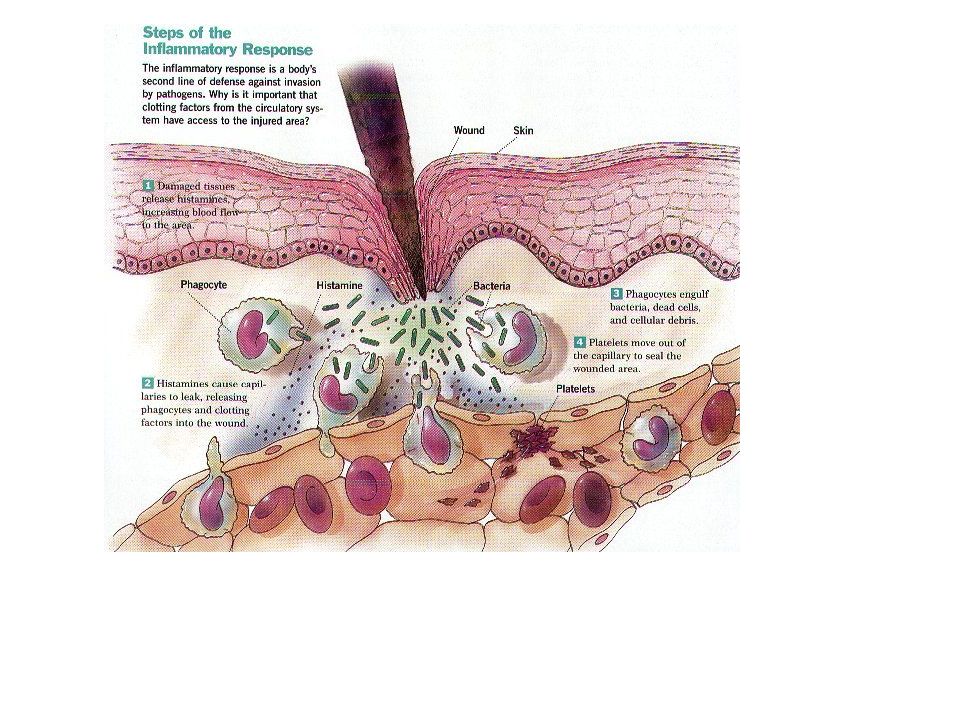
UV radiation PM mercury indoor air pesticides & toxics biologicals lead ozone asbestos ? Nearly 430,000 American children between the ages of one to five had elevated blood lead levels that can cause irreversible disabilities such as lower IQ and neurological damage. Today, children’s exposure to lead is mostly due to the ingestion of contaminated dust, paint, and soil. According to the American Cancer Society, every year, about 130 new childhood cancers are diagnosed for every 1 million children. As such, cancer is the second leading cause of death in children under 14 years of age. The types of cancers that develop in children are different from the types that develop in adults.

3
A Hazard to the Environment

5
Ignitability

6
Corrosivity

7
Toxicity

8
Common Exposures Personal Medications Outdoor Air Pollution
Indoor Air Pollution Industrial Exposures Agricultural Hazards Natural Toxins Radiation Injury Physical Injury

9
This is actually not a bad diagram, fairly logical.

10
Mekanisme kerja suatu racun zat terhadap suatu organ sasaran pada umumnya melewati suatu rantai reaksi yang dapat dibedakan menjadi 3 fase utama : a) Fase Eksposisi b) Fase Toksokinetik c) Fase Toksodinamik ( Lihat gambar ).
 Fase Eksposisi. b) Fase Toksokinetik. c) Fase Toksodinamik ( Lihat gambar ).")
11
Skema dampak pencemaran polutan terhadap makhluk hidup
( dimodifikasi dari : Holdgate, 1979)
")
12
EKSPOSISI TOKSIKOKINETIK TOKSIKODINAMIK
BAHAN KIMIA ABSORPSI INTERAKSI AN- DI AMBIEN : DISTRIBUSI TARA TOKSON - GAS / UAP PENYIMPANAN DENGAN RESEP - DEBU METABOLISME TOR DALAM - KABUT EKSKRESI ORGAN - FUME FASE FASE FASE EKSPOSISI TOKSIKOKINETIK TOKSIKODINAMIK

13
FASE EKSPOSISI

14
1). Fase Eksposisi : Merupakan ketersediaan biologis suatu polutan di lingkungan dan hal ini erat kaitannya dengan perubahan sifat-sifat fisikokimianya. Selama fase eksposisi, zat beracun dapat diubah melalui berbagai reaksi kimia/fisika menjadi senyawa yang lebih toksis atau lebih kurang toksis.

15
Faktor-faktor yang mempengaruhi sifat polutan tersebut adalah atmosfer, air dan biota. Transportasi dan transformasi zat/polutan di lingkungan berhubungan erat dengan sifat-sifat fisikokimia polutan; proses transportasi polutan di lingkungan dan transformasi polutan yang terjadi di lingkungan. Pemaparan bahan polutan ke lingkungan akan mengalami berbagai proses transformasi tergantung atas media transportasinya antara lain air, udara, tanah dan biota ( Connel Des. W . and Gregory J. Miller, 1984).
..")
16
Gas dan Uap Gas : - Zat tanpa bentuk, mengisi slrh ruang pada
kondisi normal (1 atmosfir, suhu kamar) - Mempunyai dimensi tekanan, volume dan suhu - Dapat berubah wujud dengan merubah ke tiga dimensi tsb : - LPG (liquified petroleum gas) - Amoniak cair - CO2 padat (es kering)
 - Mempunyai dimensi tekanan, volume dan suhu. - Dapat berubah wujud dengan merubah ke tiga. dimensi tsb : - LPG (liquified petroleum gas) - Amoniak cair. - CO2 padat (es kering)")
17
Uap : Adalah gas yang pada keadaan normal berupa
cairan atau padatan : - Volatile organic compound (VOC) : - Uap air, dsb. Efek toksik akibat paparan gas / uap pada saluran pernapasan karena 2 hal : 1. paparan gas/uap irritan 2. paparan gas asfiksian
 : - Uap air, dsb. Efek toksik akibat paparan gas / uap pada saluran pernapasan karena 2 hal : 1. paparan gas/uap irritan. 2. paparan gas asfiksian.")
18
GAS / UAP IRITAN - Menyebabkan iritasi - korosi
- Contoh : NH3, formaldehid, ozon, NOx, SOx, H2S, HCl, Cl2, kromium, dll. - Sangat larut air efek pada sal. napas atas - Kurang larut air efek pada saluran napas bawah - Efek : - inflamasi - akut / kronis

19
GAS ASFIKSIAN 1. ASFIKSIAN SEDERHANA
Menyebabkan asfiksia (gagal napas) : 1. ASFIKSIAN SEDERHANA - Gas CO2, NH4, Asetilin, gas inert - Sering pada confined space - Penyebab : tekanan parsial oksigen turun - Udara : 79% N2, 20 % O2, 1% lain-lain - Tekanan oksigen < 16% fatal, kematian sangat cepat
 : 1. ASFIKSIAN SEDERHANA. - Gas CO2, NH4, Asetilin, gas inert. - Sering pada confined space. - Penyebab : tekanan parsial oksigen turun. - Udara : 79% N2, 20 % O2, 1% lain-lain. - Tekanan oksigen < 16% fatal, kematian sangat cepat.")
20
2. ASFIKSIAN KEMIKAL a. Gas CO kegagalan transpor O2 oleh Hb
CO mempunyai afinitas terhadap Hb 300 x darpada O2 b. Gas sianida inhibisi sistem enzim sitokrom oksidase (siklus Kreb), kegagalan pembentukan ATP
, kegagalan. pembentukan ATP.")
21
2. Debu Partikel padat, melayang di udara, organik/anorganik
Bentuk : debu, serat Ukuran : - debu respirable (< 10 mikron) - debu nonrespirable (> 10 mikron) Inhalasi debu deposit pada saluran pernapasan s.d. Alveoli Di mana debu akan terdeposit ? tergantung : ukuran densitas debu pola pernapasan struktur saluran pernapasan
 - debu nonrespirable (> 10 mikron) Inhalasi debu deposit pada saluran. pernapasan s.d. Alveoli. Di mana debu akan terdeposit tergantung : ukuran. densitas debu. pola pernapasan. struktur saluran pernapasan.")
22
Jumlah dan lamanya deposisi akan mempengaruhi besar kecilnya efek Proses pembersihan debu (lung clearence): - mekanis (batuk, bersin) - mucocilliary escalator - fagositosis (by alveolar macrophag) Asap rokok, alkohol dan bahan kimia tertentu melemahkan fungsi tersebut
: - mekanis (batuk, bersin) - mucocilliary escalator - fagositosis (by alveolar macrophag) Asap rokok, alkohol dan bahan kimia tertentu melemahkan fungsi tersebut")
23
3. Kabut 4. Fume Partikel cair berasal dari proses spraying dsb.
Tergantung sifat cairan : mudah larut / sukar larut 4. Fume Partikel padat, berasal dari kondensasi uap metal dengan oksigen oksida logam Ukuran : < 1 mikron Efek : bergantung sifat metalnya Contoh : Pb oksida, Seng oksida, dsb.

24
2.1.2.Media Transpor racun Media transpor dapat berupa : Udara Air Tanah Makanan Organisme Rantai Makanan Dll

26
Gb. Interaksi xenobiotik dengan berbagai faktor di lingkungan ( Sumber : McKinney, 1981).
.")
30
2.1.4. PROSES PERUBAHAN BENTUK
Proses perubahan bentuk polutan di perairan meliputi - hidrolisis, - fotolisis, - degradasi secara mikroorganisme dan - oksidasi.

31
PROSES BIODEGRADASI Biotransformasi ( perubahan bentuk biologis) dan biodegradasi polutan oleh mikroorganisme ( bakteri, jamur, protozoa dan ganggang) merupakan proses pembuangan dan perubahan yang penting dalam air, sedimen dan tanah. Reaksi mencakup : Oksidasi reduksi, hidrolisis dan terkadang penataan ulang dan dipengaruhi oleh bangun molekul dan kepekatan zat polutan, sifat alamiah mikroorganisme, keadaan lingkungan dan suhu.
 dan biodegradasi polutan oleh mikroorganisme ( bakteri, jamur, protozoa dan ganggang) merupakan proses pembuangan dan perubahan yang penting dalam air, sedimen dan tanah. Reaksi mencakup : Oksidasi. reduksi, hidrolisis dan. terkadang penataan ulang dan dipengaruhi oleh bangun molekul dan kepekatan zat polutan, sifat alamiah mikroorganisme, keadaan lingkungan dan suhu.")
32
FASE TOKS0KINETIK

33
2). Fase Toksokinetik : Hanya sebagian dari jumlah zat yang diabsorpsi mencapai organ target suatu zat toksis di dalam tubuh organisme , yakni di lokasi jaringan/molekul yang sesuai. Dibedakan atas proses -proses : - Absorbsi dan distribusi ( Invasi) - Biotransformasi (Perubahan metabolik) - Akumulasi - Ekskresi
 - Biotransformasi (Perubahan metabolik) - Akumulasi. - Ekskresi.")
34
Toxicokinetics Toxicokinetics (Determines the no. molecules that can reach the receptors) Uptake Transport Metabolism & transformation Sequestration Excretion

35
Uptake routes Ingestion (toxicity may be modified by enzymes, pH and microbes) Respiration (Air borne toxicants) Body surface (Lipid soluble toxicants such as carbon terta chloride and organophosphate)
")
36
Exposure Site (skin, Gastro Intestinum, respiratory, placenta)
D e L I V E R y Absorption Distribution Reabsorption Toxication Presystemic Elimination Distribution Away from Target Excretion Detoxication Ultimate Target Target molecule (protein, lipid, DNA, RNA)
")
37
Masuknya racun ke dalam tubuh

38
Uptake and Elimination
K1 Biological System K2 Elimination Uptake K1 > K2 : Accumulation & Toxic effect

39
Efek lokal Bioaktivasi
Lebih toksik Efek lokal Bioaktivasi Pemapar Absorpsi Distribusi Biotransformasi Metabolit fisika Pernapas antar sel fase 1 kimia Kulit sirkulasi fase 2 konsentr Pencern Bioinaktivasi Penyimpanan Efek Ekskresi Ekskresi

40
Uptake Barriers Cell membrane Cell wall/cuticles/stomata
Epithelial cells of GI tract Respiratory surface (lung, gill tracheae) Body surface
 Body surface.")
41
External to Internal environment
..a hint of integration?

42
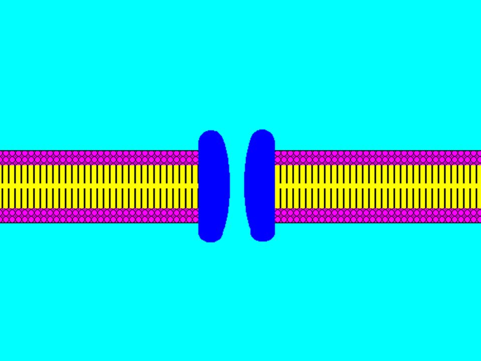
Review the structure and function of the plasma membrane

44
Parts of the membrane Vary depending on cell type
Phospholipids and proteins Vary depending on cell type Bilayer Numerous functions of proteins Structural, carrier, enzyme, channel Unsaturated vs saturated fatty acids Influence fluidity, cholesterol, carbohydrates Structure determines function Selectively permeable

45
Physiochemical characteristics of substances
Size Small cross easier than large Lipid solubility Diffuse easily through lipids Endogenous compound Hormone, protein Polarity/charge Nonpolar (lipid) Polar (protein)
 Polar (protein)")
47
Foreign substances cross
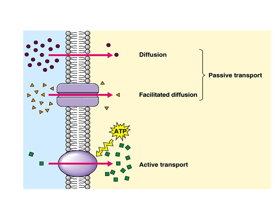
Filtration through pores Passive diffusion through membrane phospholipids Active transport Facilitated diffusion Phagocytosis Pinocytosis

48
Uptake of Toxicants Passive diffusion Facilitated transport Active transport Pinocytosis

49
Uptake by Passive diffusion
Uncharged molecules may diffuse along conc. gradient until equilibrium is reached Not substrate specific Small molecules of < 0.4 nm (e.g. CO, N20, HCN) can move through cell pores Lipophilic chemicals may diffuse through the lipid bilayer
 can move through cell pores. Lipophilic chemicals may diffuse through the lipid bilayer.")
50
Uptake by Passive diffusion
First order rate process, depends on: Concentration gradient Surface area (aveoli = 25 x body surface) Thickness (fluid mosaic phospholipid bi-layer ca. 7 nm) Lipid solubility & ionization(dissolved before transport, polar chemicals have limited diffusion rate) Molecular size (membrane pore size = 4-40 A, allowing MW of ,000 to pass through)
 Thickness (fluid mosaic phospholipid bi-layer ca. 7 nm) Lipid solubility & ionization(dissolved before transport, polar chemicals have limited diffusion rate) Molecular size (membrane pore size = 4-40 A, allowing MW of ,000 to pass through)")
52
Passive diffusion Most important mechanism of absorption for foreign and toxic compounds Conditions required: Concentration gradient Lipid soluble Non-ionized

53
Role of Blood Flow and ionization in absorption
Cell membrane Blood flow H+ + A- HA HA H+ + A- Blood Stream Lumen of Gut

54
Uptake by Facilitated Transport
Carried by trans-membrane carrier along concentration gradient Energy independent May enhance transport up to 50,000 folds Example: Calmodulin for facilitated transport of Ca

55
Uptake by Active Transport
Independent of or against conc. gradient Require energy Substrate –specific Rate limited by no. of carriers Example: P-glycoprotein pump for xenobiotics (e.g. OC) Ca-pump (Ca2+ -ATPase)
 Ca-pump (Ca2+ -ATPase)")
56
Active Transport Specific carrier required Metabolic energy (ATP)
Inhibited Saturated by high concentrations Against concentration gradient Competition of uptake Uniports, symports, antiports

57
Active transport Using an endogenous pathway Drug fluorouracil
Analog for uracil and lead ions absorbed in gut

60
Uptake by Pinocytosis For large molecules ( ca 1 um)
Outside: Infolding of cell membrane Inside: release of molecules Example: Airborne toxicants across alveoli cells Carrageenan accross intestine

62
Distribution to and Away from Target
Exit blood and enter the extracellular space Affect the surface or interior of a tissue cell

63
Mechanisms Opposing Distribution
Binding to Plasma Proteins Most toxins much dissociate from protein to reach target cell Specialized Barriers Tight junctions Not effective against lipophilic substances Distribution to Storage Sites Accumulate in tissues (fat) Association with intracellular binding proteins Nontarget intracellular site (metallothionein) Export from cells Transported back to extracellular space
 Association with intracellular binding proteins. Nontarget intracellular site (metallothionein) Export from cells. Transported back to extracellular space.")
64
Mechanisms Facilitating Distribution to a target
Porosity of the Capillary Endothelium Large fenestrae Passage of protein-bound xenobiotics Accumulate in liver and kidneys Specialized Transport Across the Plasma Membrane Specialized ion channels and membrane transporters Na/K ATP pump, voltage-gated channels, endocytosis Accumulation in Cell Organelles Amphipathic xenobiotics (protonatable amine group and lipophilic characters (lysosomes and mitochondria) Reversible Intracellular Binding Organic and inorganic cations, PAH Bind to protein (melanin)
 Reversible Intracellular Binding. Organic and inorganic cations, PAH. Bind to protein (melanin)")
65
Sites of Absorption Three major sites of absorption of foreign compounds Skin Rarely a significant site for absorption Lungs Airborne compounds Gastrointestinal tract (GI) GI is the most important in toxicology as most foreign compounds are ingested orally
 GI is the most important in toxicology as most foreign compounds are ingested orally.")
66
Excretion vs Reabsorption
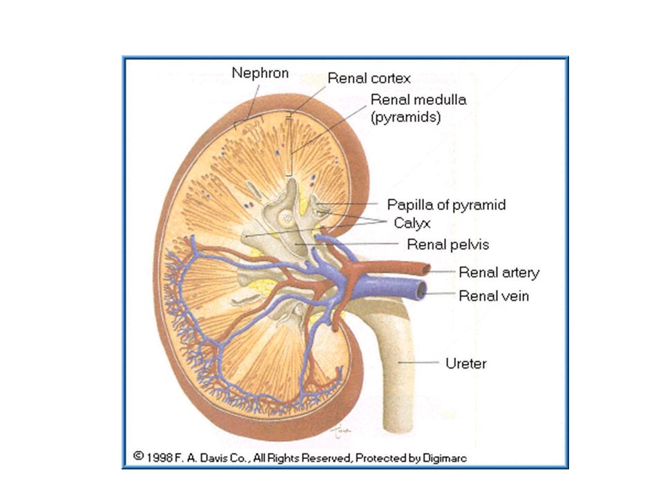
Removal of xenobiotics from the blood and their return to the external environment Physical mechanism Biotransformation is chemical Depends on physiochemical properties of toxicant Major excretory organs – kidney and liver Efficiently remove hydrophilic chemicals (acids, bases) Reabsorption Renal tubules Dependent on lipid solubility GI tract, salivary glands
 Reabsorption. Renal tubules. Dependent on lipid solubility. GI tract, salivary glands.")
67
Transport & Deposition
Blood Lymph, haemolymph Water stream in xylem Cytoplamic strands in phloem Deposition Toxicant Target organs Pb Bone, teeth, brain Cd Kidney, bone, gonad OC, PCB Adipose tissue,milk OP Nervous tissue Aflatoxin Liver

68
Metabolism & Transformation
Evolved to deal with metabolites and naturally occurring toxicants Principle of detoxification: Convert toxicants into more water soluble form (more polar & hydrophilic) Dissolve in aqueous/gas phases and eliminate by excretion (urine/sweat) of exhalation Sequestrate in inactive tissues (e.g bone, fat)
 Dissolve in aqueous/gas phases and eliminate by excretion (urine/sweat) of exhalation. Sequestrate in inactive tissues (e.g bone, fat)")
69
P450 system A heme-containing cytochrome protein located in ER, and is involved in electron transport. Highly conservative, occur in most plants & animals Two phases of transformation May increase or decrease toxicity of toxicants after transformation (e.g turn Benzo[a]pyrene into benzo[a]pyrene diol epoxide, and nitroamines into methyl radicals) Inducible by toxicants
 Inducible by toxicants.")
70
Induction of P450 Toxicant Aryl Hydrocarbon Receptor Toxicant-Receptor
Complex Bind at Specific site hours Translocating protein m-RNA for CYP1A

71
BIOTRANSFORMASI Tujuan utama : detoksifikasi
Lipofil hidrofil (polar) ekskresi Reaksi enzimatik : enzim, ko enzim Di semua sel, terutama sel hati Hasil : metabolit Bioaktivasi metabolit yang lebih aktif Bioinaktivasi metabolit kurang aktif Reaksi fase I : degradasi (oksidasi, reduksi, hidrolisis) Reaksi fase II : konjugasi polar
 ekskresi. Reaksi enzimatik : enzim, ko enzim. Di semua sel, terutama sel hati. Hasil : metabolit. Bioaktivasi metabolit yang lebih aktif. Bioinaktivasi metabolit kurang aktif. Reaksi fase I : degradasi (oksidasi, reduksi, hidrolisis) Reaksi fase II : konjugasi polar.")
72
Oksidasi : Reaksi di mana substrat kehilangan elektron dalam reaksi : oksigenasi, dehidrogenasi atau transfer elektron Enzim : enzim oksidase (mis. Sitokrom) Mikrosomal atau non mikrosomal oksidasi seny. alifatik oksidasi seny. aromatik epoksidasi N-dealkilasi oksidasi amin desulfurisasi, dll
 Mikrosomal atau non mikrosomal. oksidasi seny. alifatik. oksidasi seny. aromatik. epoksidasi. N-dealkilasi. oksidasi amin. desulfurisasi, dll.")
73
Illustration of oxidation :

74
REDUKSI Reaksi kimia di mana substrat mendapat elektron
Biasanya pada bahan yang memiliki atom oksigen sangat sedikit, misalnya golongan azo (N-N dengan ikatan rangkap) atau senyawa nitro (NO2), amino, dll. Amino metabolit aktif Karbon tetraklorida senyawa radikal
 atau senyawa nitro (NO2), amino, dll. Amino metabolit aktif. Karbon tetraklorida senyawa radikal.")
75
HIDROLISIS Terutama untuk golongan ester : asetilkolin (asetilkolin esterase) amida : amidase fosfat : fosfatase
 amida : amidase fosfat : fosfatase")
76
KONJUGASI Oleh senyawa endogen konjugat Mekanisme ; 1. Glukoronid
2. Sulfat 3. Metilasi 4. Asetilasi 5. Glutation Hal ini akan menyebabkan terjadinya mekanisme kejenuhan

77
Reaksi konjugasi :

78
Sequestration Animals may store toxicants in inert tissues (e.g. bone, fat, hair, nail) to reduce toxicity Plants may store toxicants in bark, leaves, vacuoles for shedding later on Lipophilic toxicants (e.g. DDT, PCBs) may be stored in milk at high conc and pass to the young Metallothionein (MT) or phytochelatin may be used to bind metals
 may be stored in milk at high conc and pass to the young. Metallothionein (MT) or phytochelatin may be used to bind metals.")
79
PENYIMPANAN Terutama bahan lipofilik dan yang tidak dibiotransformasi
Tempat : jar. Lemak, tulang, hemoglobin, gusi, hati, ginjal, kuku, rambut, dll. Jar. Lemak : DDT hati-2 pada kondisi kelaparan atau trauma jaringan redistribusi efek toksik Penting dalam rantai trofik makanan kasus penyakit Minamata karena pajanan Merkuri organik Hati & ginjal : tempat penyimpanan sekaligus tempat biotransformasi

80
Excretion Gas (e.g. ammonia) and volatile (e.g. alcohol) toxicants may be excreted from the gill or lung by simple diffusion Water soluble toxicants (molecular wt. < 70,000) may be excreted through the kidney by active or passive transport Conjugates with high molecular wt. (>300) may be excreted into bile through active transport Lipid soluble and non-ionised toxicants may be reabsorbed (systematic toxicity)
 may be excreted through the kidney by active or passive transport. Conjugates with high molecular wt. (>300) may be excreted into bile through active transport. Lipid soluble and non-ionised toxicants may be reabsorbed (systematic toxicity)")
81
EKSKRESI Organ ekskretor utama : ginjal, saluran pencernaan, paru
Lainnya : kulit, air susu, air mata Ginjal : organ utama, bahan hidrofil filtrasi glomeruli diffusi tubuler sekresi tubuler Paru : bahan-bahan volatil

84
PENYEBARAN RACUN LINGK. DI DALAM TUBUH MANUSIA :
- Protein plasma mengikat senyawa asing HATI DAN GINJAL - Bertugas untuk mengeluarkan senyawa asing. Hati berkapasitas merubah senyawa racun ( biotrans-formasi) menjadi tidak aktif). - LEMAK Tempat penyimpanan penting bagi senyawa yang larut dalam lemak ( mis. DDT, PCB, ) - TULANG Berfungsi sebagai penyimpan senyawa Flour, Pb, Strontium.
 menjadi tidak aktif). - LEMAK. Tempat penyimpanan penting bagi senyawa yang larut dalam lemak ( mis. DDT, PCB, ) - TULANG. Berfungsi sebagai penyimpan senyawa Flour, Pb, Strontium.")
85
Elimination Biotransformation Excretion Exhalation
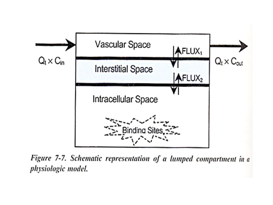
Usually as first order process(one compartment): the rate of elimination at any time is proportional to the amount of the chemical in the body at that time (below saturation level for elimination)
: the rate of elimination at any time is proportional to the amount of the chemical in the body at that time (below saturation level for elimination)")
86
Gb Skematik Mekanisme metabolik zat racun di luar hati dan di dalam hati

87
Gb Skematik Mekanisme metabolik zat racun di luar hati dan di dalam hati

88
FASE TOKS0DINAMIK

89
3). Fase Toksodinamik : Suatu kerja zat toksis pada umumnya adalah hasil interaksi dari sejumlah proses yang sangat rumit dan kompleks.

92
Induction of toxic effects
Toxicodynamics Toxicodynamics (Determines the no. of receptors that can interact with toxicants) Binding Interaction Induction of toxic effects
 Binding. Interaction. Induction of toxic effects.")
94
Reaction of the Ultimate toxicant with target molecule
Noncovalent binding Reversible Hydrogen and ionic bonds Covalent binding Irreversible Permanently alters endogenous molecules Free radicals can bind covalently to molecules Hydrogen Abstraction Remove hydgogen and convert to double bonds Electron Transfer Exchange electrons to oxidize or reduce other molecules Enzymatic Reaction Affect normal reaction

95
Cellular Dysfunction and Toxicities
Gene expression Transcription DNA to RNA Signal Transduction Signals from cell surface receptors to control various cellular activities Cellular activity Influence excitable cells ( neurons, cardiac cells) Neurotransmitters Cell death Depletion of ATP Mitochondria
 Neurotransmitters. Cell death. Depletion of ATP. Mitochondria.")
96
Step 3 – Cellular Dysfunction and Toxicities
Gene expression Transcription DNA to RNA Signal Transduction Signals from cell surface receptors to control various cellular activities Cellular activity Influence excitable cells ( neurons, cardiac cells) Neurotransmitters Cell death Depletion of ATP Mitochondria
 Neurotransmitters. Cell death. Depletion of ATP. Mitochondria.")
97
Step 4 – Repair and Dysrepair
Many toxicants alter macromolecules that may lead to damage of cell, tissue, or complete organism Protein Repair Repair of Lipids Direct Repair Excision Repair Cellular Repair Inflammation Repair fails Tissue necrosis Fibrosis

98
Different types of Repair
Repair of Proteins Oxidation of proteins Repair by using reductants - NADPH Molecular chaperones – refold altered proteins Proteolytic degradation- remove damaged proteins Repair of Lipids Oxidation of lipids Reductants – glutathione reductase Repair of DNA Nuclear DNA is stable. Various repair mechanism (chromatin) Mitochondria DNA – lacks histones and repair mechanisms
 Mitochondria DNA – lacks histones and repair mechanisms.")
99
Different types of Repair
Direct Repair Certain covalent DNA modification can be reversed DNA photolyase Excision Repair Base excision and nucleotide excision Remove damaged bases from DNA

100
Cellular Repair Repair of damaged neurons
Axonal damage is repaired if cell body is intact

101
Tissue Repair Apoptosis Initiated by cell injury Cell shrinks
Nuclear and cytoplasmic materials condense Membrane fragments Eliminating cells that can become cancerous

102
Apoptosis

103
Proliferation and Replacements
Regeneration of Tissue Replacement of Lost Cells by Mitosis Cells adjacent to injury enter cell division Replacement of Extracellular Matrix Growth factors- proteins, collagens, etc. Inflammation Alteration of microcirculation and accumulation of inflammatory cells Produce reactive oxygen and nitrogen species

105
When Repair Fails Repair fails most typically when the damage overwhelms the repair mechanisms Toxicant could affect the repair process Some types of injuries can not be repaired – covalent bonding Sometimes repair may contribute to toxicity Over consumption of NADPH

106
Toxicity resulting from dysrepair
Molecular, Cellular, and Tissue Levels Enzymes or tissue necrosis

107
Tissue Necrosis

108
Tissue Necrosis Cell death
Injury overwhelms and disables repair mechanisms No repair of damaged molecules No elimination of damaged cells by apoptosis No replacement of lost cells by cell division

109
Fibrosis

110
Fibrosis A pathological condition that is characterized by excessive deposition of an extracellular matrix that is abnormal Increase in collagen, laminin, growth factors

111
Carcinogensis Insufficient function of various repair mechanisms
Failure of DNA repair Toxins may cause DNA adducts, strand breakage, oxidation Mutation of proto-oncogenes Proteins needed to control cell cycle Failure of apoptosis Promotion of mutation and continued growth of damaged cells Failure to terminate cell proliferation Tumor formation

112
Cellular Dysfunction and Toxicities
Gene expression Transcription DNA to RNA Signal Transduction Signals from cell surface receptors to control various cellular activities Cellular activity Influence excitable cells ( neurons, cardiac cells) Neurotransmitters Cell death Depletion of ATP Mitochondria
 Neurotransmitters. Cell death. Depletion of ATP. Mitochondria.")
113
Repair and Dysrepair Many toxicants alter macromolecules that may lead to damage of cell, tissue, or complete organism Protein Repair Repair of Lipids Direct Repair Excision Repair Cellular Repair Inflammation Repair fails Tissue necrosis Fibrosis

114
Different types of Repair
Repair of Proteins Oxidation of proteins Repair by using reductants - NADPH Molecular chaperones – refold altered proteins Proteolytic degradation- remove damaged proteins Repair of Lipids Oxidation of lipids Reductants – glutathione reductase Repair of DNA Nuclear DNA is stable. Various repair mechanism (chromatin) Mitochondria DNA – lacks histones and repair mechanisms
 Mitochondria DNA – lacks histones and repair mechanisms.")
115
Different types of Repair
Direct Repair Certain covalent DNA modification can be reversed DNA photolyase Excision Repair Base excision and nucleotide excision Remove damaged bases from DNA

116
Cellular Repair Repair of damaged neurons
Axonal damage is repaired if cell body is intact

117
Tissue Repair Apoptosis Initiated by cell injury Cell shrinks
Nuclear and cytoplasmic materials condense Membrane fragments Eliminating cells that can become cancerous

118
Apoptosis

119
Proliferation and Replacements
Regeneration of Tissue Replacement of Lost Cells by Mitosis Cells adjacent to injury enter cell division Replacement of Extracellular Matrix Growth factors- proteins, collagens, etc. Inflammation Alteration of microcirculation and accumulation of inflammatory cells Produce reactive oxygen and nitrogen species

121
When Repair Fails Repair fails most typically when the damage overwhelms the repair mechanisms Toxicant could affect the repair process Some types of injuries can not be repaired – covalent bonding Sometimes repair may contribute to toxicity Over consumption of NADPH

122
Toxicity resulting from dysrepair
Molecular, Cellular, and Tissue Levels Enzymes or tissue necrosis

123
Tissue Necrosis

124
Tissue Necrosis Cell death
Injury overwhelms and disables repair mechanisms No repair of damaged molecules No elimination of damaged cells by apoptosis No replacement of lost cells by cell division

125
Fibrosis

126
Fibrosis A pathological condition that is characterized by excessive deposition of an extracellular matrix that is abnormal Increase in collagen, laminin, growth factors

127
Carcinogensis Insufficient function of various repair mechanisms
Failure of DNA repair Toxins may cause DNA adducts, strand breakage, oxidation Mutation of proto-oncogenes Proteins needed to control cell cycle Failure of apoptosis Promotion of mutation and continued growth of damaged cells Failure to terminate cell proliferation Tumor formation

128
Toxication vs Detoxication
Biotransformation to harmful products is called toxication or metabolic activation Electrophiles Positively charged Free radicals Unpaired electrons Nucleophiles Negatively charged Redox-active reactants Can donate or accept electrons

129
continued Detoxification
Biotransformations that eliminate the ultimate toxicant or prevent its formation May be competing with toxication Adding a functional group

130
MEKANISME KERJA POLUTAN THDP BAGIAN TUBUH ORGANISME
- Interaksi dengan sistem enzim : Inhibisi enzim tak bolak balik Inhibisi enzim secara reversible Pemutusan Reaksi Biokimia Sintesis Zat mematikan Pengambilan ion logam yang penting untuk kerja enzim Inhibisi penghantaran elektron dalam rantai respirasi

131
MEKANISME KERJA POLUTAN THDP BAGIAN TUBUH ORGANISME
Inhibisi pada transpor oksigen karena gangguan pada hemoglobin Keracunan karbon monoksida Pembentukan Metheglobin dan Sulfahemoglobin Proses Hemolitik

132
Pengaruh Penghantaran Rangsang Neurohumor
- Interaksi dengan Fungsi Umum Sel Kerja Narkose Pengaruh Penghantaran Rangsang Neurohumor - Gangguan pada sintesis DNA dan RNA Kerja Sitostatika Kerja Imunsupresiva Kerja Mutagenik Kerja Karsinogenik

133
- Kerja Teratogenik - Reaksi Hipersensitif ( Reaksi alergi) Reaksi fotoalergik Sensibilisasi cahaya Reaksi fototoksis

134
Kerusakan kulit akibat zat kimia Gas yang merangsang Gas air mata
Iritasi Kimia langsung pada Jaringan Kerusakan kulit akibat zat kimia Gas yang merangsang Gas air mata Zat yang berbau - Toksisitas pada Jaringan - Penimbunan ( Sekuestrasi) Zat asing Penimbunan dalam jaringan lemak Penimbunan dalam Tulang Pneumokoniosis
 Zat asing. Penimbunan dalam jaringan lemak. Penimbunan dalam Tulang. Pneumokoniosis.")
136
Sasaran Proses yang Terganggu
Tabel 1. Rangkuman beberapa pengaruh biokimia dan fisiologis penting dari suatu zat beracun. No Sasaran Proses yang Terganggu 1. Membran sel Perubahan atau modifikasi permeabilitas memberan b. Pengacauan sistem transportasi membran sel 2. Enzim Inhibisi dapat balik atau tidak balik dari enzim (koenzim, subtrat atau pengaktif logam), oleh zat kimia 3. Metabolisme Lemak Pengacauan metabolisme lemak dapat menyebabkan kegagalan fungsi hati, termasuk akumulasi lemak patologis dalam hati dan kapasitas lemak untuk mengsintesis kolesterol dapat digagalkan.
, oleh zat kimia. 3. Metabolisme. Lemak. Pengacauan metabolisme lemak dapat menyebabkan kegagalan fungsi hati, termasuk akumulasi lemak patologis dalam hati dan kapasitas lemak untuk mengsintesis kolesterol dapat digagalkan.")
137
Tabel 1. Rangkuman beberapa pengaruh biokimia dan fisiologis penting dari
suatu zat beracun. No Sasaran Proses yang Terganggu 4. Biositensis Protein Sintesis zat protein dapat dipengaruhi oleh sejumlah besar zat eksogenus, terutama melalui penekanan kapasitas protein untuk mensintesis yang bertempat di dalam reticulkum kasar endoplasmik (r ER) di dalam sitoplasma. Dalam beberapa kasus, salah satu pengaruh dapat merangsang timbulnya pertambahan sintesis protein mikrosomal. 5. Sistem enzim Mikrosomal Pergantian dalam fungsi enzim mikrosomal-rangsangan atau inhibisi yang diinduksi oleh banyak zat kimia di lingkungan. 6. Proses Pengaturan dan Pertumbuhan Struktur atau kegiatan enzim pengatur dapat diubah dan sintesis, penyimpanan, pelepasan atau pengasingan hormon dapat digagalkan oleh zat beracun dalam berbagai cara, penurunan laju pertumbuhan dapat mengikuti gangguan kimiawi jalur dan laju metabolisme.
 di dalam sitoplasma. Dalam beberapa kasus, salah satu pengaruh dapat merangsang timbulnya pertambahan sintesis protein mikrosomal. 5. Sistem enzim Mikrosomal. Pergantian dalam fungsi enzim mikrosomal-rangsangan atau inhibisi yang diinduksi oleh banyak zat kimia di lingkungan. 6. Proses Pengaturan dan Pertumbuhan. Struktur atau kegiatan enzim pengatur dapat diubah dan sintesis, penyimpanan, pelepasan atau pengasingan hormon dapat digagalkan oleh zat beracun dalam berbagai cara, penurunan laju pertumbuhan dapat mengikuti gangguan kimiawi jalur dan laju metabolisme.")
138
EFEK BIOLOGIS MERUPAKAN RESULTANTE AKHIR DARI SEJUMLAH PROSES YANG SANGAT KOMPLEKS, YAKNI INTERAKSI ANTARA FUNGSI HOMEOSTASISNYA DENGAN XENOBIOTIK. Jika proses homeostasis gagal, karena berbagai hal misalnya dosis terlalu tinggi, paparan konsentrasi terlalu pekat dan kontinyu, keadaan gizi kurang, dstnya maka akan terjadi efek biologis yang diekspresikan bermacam-macam .

139
Toxicity = tissue necrosis, cancer, fibrosis, disease
Toxicant Delivery Step 1 Interaction with Target molecule Alteration of biological environment Step 2 Cellular dysfunction Injury Dysrepair Step 3 TOXICITY Step 4 Toxicity = tissue necrosis, cancer, fibrosis, disease

140
More Symptoms A significant effect of Minamata is the onset of symptoms similar to those of cerebral palsy Fetal Minamata Disease A pregnant mother ingests toxic fish and the methylmercury concetrates inside the placenta. Harms the fetus while the mother is relatively unaffected

141
These are all children with congenital (fetal) Minamata Disease due to intrauterine methyl mercury poisoning (Harda 1986).
 Minamata Disease due to intrauterine methyl mercury poisoning (Harda 1986).")
142
Examples of chemicals in food, air, water linked to birth defects Cross placenta to embryo
Defects of brain, nerves, heart Defects of skeleton (often limbs) Blindness, deafness Spasticity Mental retardation Defects of heart, brain Decreased fetal growth Defects of face (cleft palate/lip) Emotional & learning problems
 Blindness, deafness. Spasticity. Mental retardation. Defects of heart, brain. Decreased fetal growth. Defects of face (cleft palate/lip) Emotional & learning problems.")
143
Sasaran Proses yang Terganggu
Tabel . Rangkuman beberapa pengaruh biokimia dan fisiologis penting dari suatu zat beracun. No Sasaran Proses yang Terganggu 1. Membran sel Perubahan atau modifikasi permeabilitas memberan b. Pengacauan sistem transportasi membran sel 2. Enzim Inhibisi dapat balik atau tidak balik dari enzim (koenzim, subtrat atau pengaktif logam), oleh zat kimia 3. Metabolisme Lemak Pengacauan metabolisme lemak dapat menyebabkan kegagalan fungsi hati, termasuk akumulasi lemak patologis dalam hati dan kapasitas lemak untuk mengsintesis kolesterol dapat digagalkan.
, oleh zat kimia. 3. Metabolisme Lemak. Pengacauan metabolisme lemak dapat menyebabkan kegagalan fungsi hati, termasuk akumulasi lemak patologis dalam hati dan kapasitas lemak untuk mengsintesis kolesterol dapat digagalkan.")
144
Tabel . Rangkuman beberapa pengaruh biokimia dan fisiologis penting dari
suatu zat beracun. No Sasaran Proses yang Terganggu 4. Biositensis Protein Sintesis zat protein dapat dipengaruhi oleh sejumlah besar zat eksogenus, terutama melalui penekanan kapasitas protein untuk mensintesis yang bertempat di dalam reticulkum kasar endoplasmik (r ER) di dalam sitoplasma. Dalam beberapa kasus, salah satu pengaruh dapat merangsang timbulnya pertambahan sintesis protein mikrosomal. 5. Sistem enzim Mikrosomal Pergantian dalam fungsi enzim mikrosomal-rangsangan atau inhibisi yang diinduksi oleh banyak zat kimia di lingkungan. 6. Proses Pengaturan dan Pertumbuhan Struktur atau kegiatan enzim pengatur dapat diubah dan sintesis, penyimpanan, pelepasan atau pengasingan hormon dapat digagalkan oleh zat beracun dalam berbagai cara, penurunan laju pertumbuhan dapat mengikuti gangguan kimiawi jalur dan laju metabolisme.
 di dalam sitoplasma. Dalam beberapa kasus, salah satu pengaruh dapat merangsang timbulnya pertambahan sintesis protein mikrosomal. 5. Sistem enzim Mikrosomal. Pergantian dalam fungsi enzim mikrosomal-rangsangan atau inhibisi yang diinduksi oleh banyak zat kimia di lingkungan. 6. Proses Pengaturan dan Pertumbuhan. Struktur atau kegiatan enzim pengatur dapat diubah dan sintesis, penyimpanan, pelepasan atau pengasingan hormon dapat digagalkan oleh zat beracun dalam berbagai cara, penurunan laju pertumbuhan dapat mengikuti gangguan kimiawi jalur dan laju metabolisme.")
145
Reversible – irreversible Segera – tertunda
SPEKTRUM EFEK : Akut - kronik Lokal – sistemik Reversible – irreversible Segera – tertunda Perubahan morfologi-fungsi-biokimiawi

146
ORGAN TARGET DAN KERACUNANNYA:
Hepatotoksik Nefrotoksik Neurotoksik Hematotoksik Pulmotoksik Dll.

147
Factors influencing toxicity :
Form and innate chemical activity Dosage, especially dose-time relationship Exposure route Species Age Sex Ability to be absorbed Metabolisme Distribution within the body Excretion Presence of other chemicals

148
Mechanisms of Toxicity
Threshold effect Absorption at portals of entry ingestion inhalation skin contact Distribution within the body Metabolism and Excretion Toxic effects

149
Type of interaction :

151
EFEK GENETIK RACUN Yakni dapat menyebabkan gangguan struktur dan jumlah kromosom a). Aneuploidisasi Susunan kromosom di dalam sel kelamin ( spermatozoa atau sel telur) tidak memisah. Sehingga jika terjadi fertilisasi, maka anak yang lahir memiliki susunan kromosom kurang atau lebih. Anak lahir cacat/ abnormal. b). Klastogenesis DNA rusak, misalnya akibat sinar ultraviolet atau bahan kimia. Anak lahir cacat/ abnormal. c). Mutagenesis Terjadi perubahan struktur gen, sehingga anak yang lahir akan cacat/ abnormal.
 tidak memisah. Sehingga jika terjadi fertilisasi, maka anak yang lahir memiliki susunan kromosom kurang atau lebih. Anak lahir cacat/ abnormal. b). Klastogenesis. DNA rusak, misalnya akibat sinar ultraviolet atau bahan kimia. Anak lahir cacat/ abnormal. c). Mutagenesis. Terjadi perubahan struktur gen, sehingga anak yang lahir akan cacat/ abnormal.")
152
KARSINOGEN a). DNA REAKTIF KARSINOGEN Direct acting karsinogen
Prokarsionogen Inorganik b). EPIGENETIK KARSINOGEN Promotor Cytotoxic Hormon Modifying Imunopressor Solid c). UNCLASSIFIED KARSINOGEN
. EPIGENETIK KARSINOGEN. Promotor. Cytotoxic. Hormon Modifying. Imunopressor. Solid. c). UNCLASSIFIED KARSINOGEN.")
153
DNA REAKTIF KARSINOGEN
Direct acting karsinogen Bekerja langsung terhadap molekul-molekul nukleotida Tidak dijumpai di alam. Mis. Halogen ether, Nitrosoamida Prokarsinogen Reaksinya tidak langsung menyebabkan kanker Kanker muncul setelah terjadi aktivasi metabolis Dari alam atau sintetik. Mis. PAH, Aromatic Heterosiklik Amine, Senyawa azo, Senyawa N-Nitroso, Aldehyda, Hexamethylphosphoramida, Karbamat, Ethyonine, HC terhalogenisasi, mikroba karsinogenik, senyawa dari tumbuhan karsinogenik Inorganik Termasuk kelompok ini adalah Uranium, Polonium, Radium, Titanium, Nikel, kromium dan Cobalt Bersifat sinergestik dengan debu/ partikulat dan meningkatkan risiko kanker paru, saluran napas.

154
EPIGENETIK KARSINOGEN
Kelompok ini tidak langsung hasil reaksi dengan materi genetik. a. Promotor Merupakan agen yang memberikan sarana pada dorman neoplastic untuk tumbuh menjadi tumor. Misalnya Tetradecanoyl Phorbol Acetate ( TPA), Phenolbarbital, Hydrocarbon terklorinasi, Saccharin, Butylated Hydroxyanisole dan Butylated Hydroxytoluol. b. Cytotoxic Merupakan penyebab kanker sebagai implikasi dari iritasi kronis, kemudian menjadi agen yang mengakibatkan sel menjadi degeratif sebagai konpensasi proliferasi yang menaikkan risiko kanker. Misalnya. Nitrolotriacetic Acid ( NTA)
, Phenolbarbital, Hydrocarbon terklorinasi, Saccharin, Butylated Hydroxyanisole dan Butylated Hydroxytoluol. b. Cytotoxic. Merupakan penyebab kanker sebagai implikasi dari iritasi kronis, kemudian menjadi agen yang mengakibatkan sel menjadi degeratif sebagai konpensasi proliferasi yang menaikkan risiko kanker. Misalnya. Nitrolotriacetic Acid ( NTA)")
155
c. Hormon Modifying Pada Usia di atas 40 tahun, hormon terumata estrogen dapat menyebabkan kanker. Misalnya. Estrogen, Androgen, 3-Aminotriazole, Ethylenethiourea Imunopressor Proses pemberian kekebalan kadang-kadang dapat menimbulkan kanker. Misalnya pemberian serum azathioprine pada hewan dapat menaikkan risiko leukemia dan pemberian obat 6-mercaptopurine dapat menaikkan risiko lymphoma. d. Solid Bahan padat dapat mendukung untuk proliferasi sel, sehingga menjadi kanker.Misalnya. Asbestos, Kompleks Iron-Carbohidrat

Similar presentations


 Absorption Distribution Metabolism Excretion Toxicology.>")


>")


