Download presentation
Presentation is loading. Please wait.

1
Early Development Gametes

2
Sperm

3
Egg Lecithal = Yolk

4
Microlecithal egg Small amount of yolk Amphioxus Eutherians

5
Mesolecithal egg Medium amount of yolk Amphibians

6
Macrolecithal egg Large amount of yolk Bird and reptiles Most fish

7
Isolecithal Even yolk distribution In microlecithal eggs

8
Telolecithal Uneven yolk distribution
Macrolecithal and Mesolecithal eggs Vegetal Pole – yolk region Animal Pole – relatively yolk-free, high metabolic activity/embryo

9
Amniote eggs Amnion Allantoic cavity Amnionic cavity Allantois Albumin
Chorion Yolk

10
Layers around egg Vitelline membrane Jelly Capsule Shell Albumin
Corona Radiata Zona Pellucida

11
Oviparous

12
Viviparous

13
Ovoviviparous

14
Fertilization

15
Internal Fertilization
Apodans Urodeles Amniotes

16
External Fertilization
Fish Frogs

17
Zygote

18
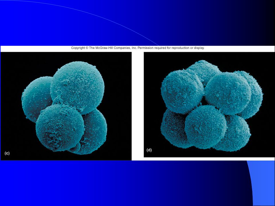
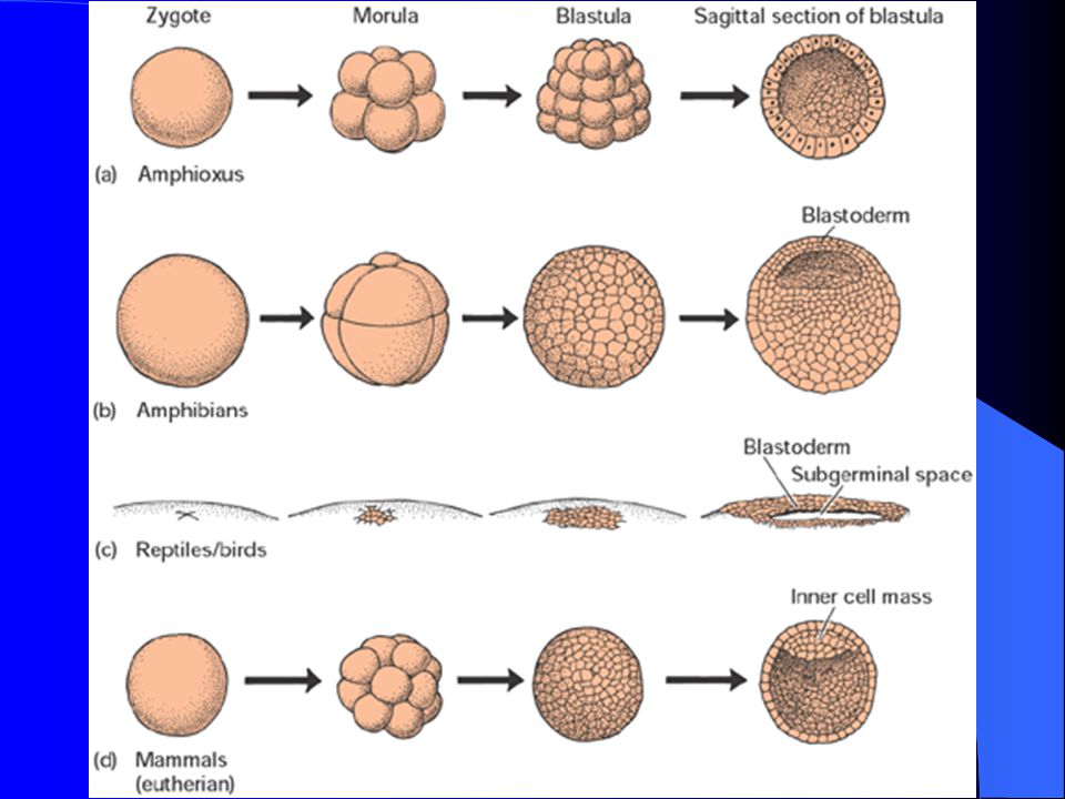
Cleavage & Blastula Microlecithal Eggs
Cleavage – Mitosis divisions Blastomeres Blastocoel

21
Cleavage & Blastula Blastocyst in mammals Inner cell mass in mammals
Trophoblast cells in mammals

24
Cleavage & Blastula Mesolecithal eggs

27
Cleavage & Blastula Macrolecithal Eggs
Blastoderm Blastocoel

30
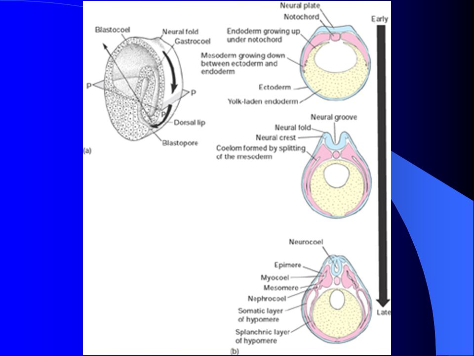
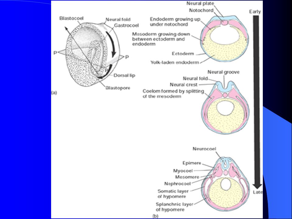
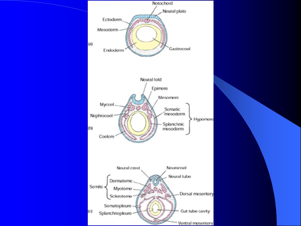
Gastrulation Germ layers form from which ALL future organs form
Notochord forms Bilateral symmetry established

31
Gastrulation/Microlecithal eggs
Involution Blastopore Archenteron

33
Germ layers Ectoderm –outer layer Mesoderm – middle layer
Forms notochord Splits to form coelom Endoderm – inner layer around archenteron

34
Gastrulation Mesolecithal eggs
Epiboly

36
Ectoderm Nervous System Sensory structures
Neural crest cells that become melanocytes, adrenal gland… Epidermis of skin Epithelium of mouth/nose and anus

39
Endoderm Lungs & Swim bladders Digestive viscera

42
Mesoderm Chordomesoderm becomes notochord

44
Mesoderm Dorsal Mesoderm = Epimere Segmented bands called somites
Divides into Dermatome Myotome Sclerotome

48
Mesoderm Lateral plate mesoderm = hypomere
Splits into Somatic and Splanchnic layers Coelom between these layers

50
Hypomere Somatic Mesoderm plus Ectoderm = Somatopleure
Splanchnic Mesoderm plus Endoderm = Splanchnopleure

52
Mesoderm Intermediate mesoderm = Mesomere
Kidney tubules and associated ducts

55
Key Points Which germ layer (ectoderm, endoderm, mesoderm) gives rise to the following structures: Heart Liver Lung Biceps muscle Notochord Brain Kidney Spinal cord Skin

56
Gastrulation in Macrolecithal eggs
Delamination Blastoderm forms upper sheet of cells called Epiblast and Lower sheet of cells called Hypoblast Epiblast becomes Ectoderm Hypoblast becomes Endoderm

59
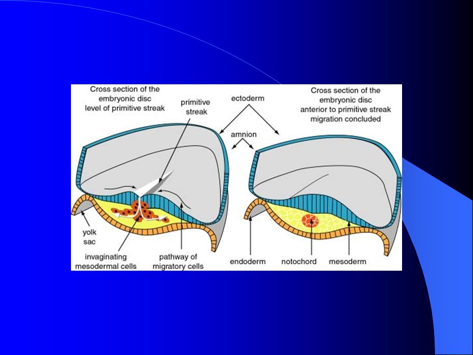
Gastrulation in Macrolecithal eggs
Mesoderm forms by Primitive Streak Cells stream inward from posterior to anterior Gives rise to notochord

64
Gastrulation in macrolecithal eggs
Some mesoderm is unorganized and migrates, called MESENCHYME

65
Gastrulation in macrolecithal eggs
Body stalk Connection from body to yolk

68
Gastrulation in mammal
Blastoderm Delamination to form hypoblast & epiblast Primitive streak forms mesoderm Notochord Mesenchyme Coelom from splitting of lateral plate mesoderm

71
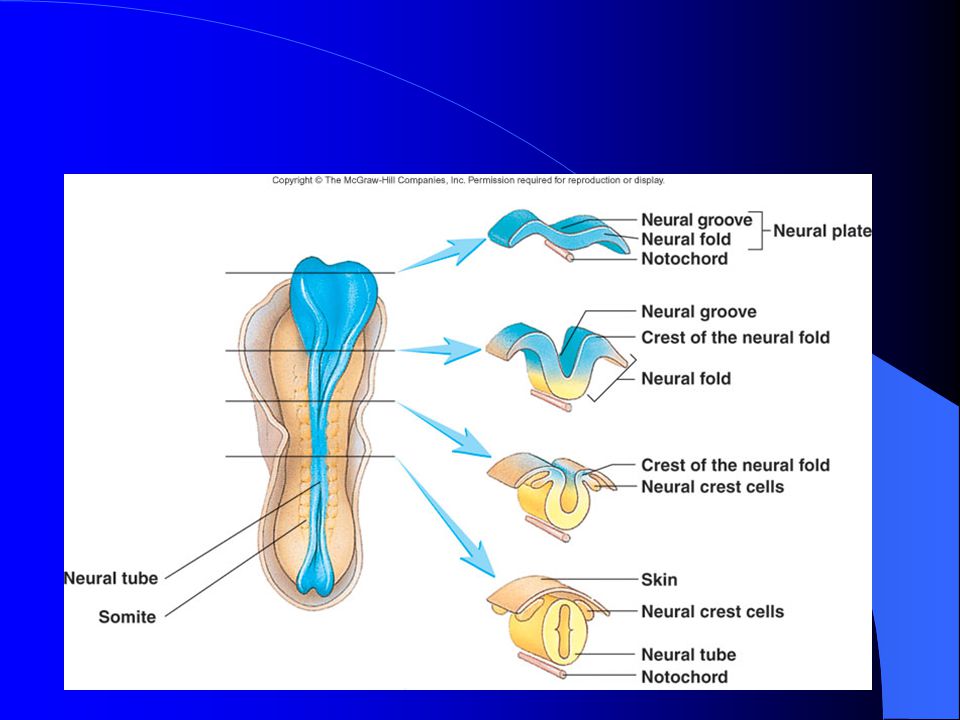
Neurulation Dorsal hollow nerve cord Neural crest cells branch off

74
Organogenesis Beginning of all major organs of the body

75
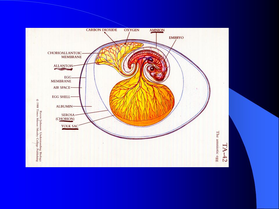
Extraembryonic membranes
Fish Body stalk Yolk sac

76
Amphibians

77
Extraembryonic membranes
AMNIOTES Yolk sac (from splanchnopleure)
")
80
Amniotes Amnion From somatopleure Amniotic Fluid

83
Amniotes Chorion From somatopleure
For communication with oxygen source Helps form placenta in mammals Against shell in birds

85
Amniotes Allantois From splanchnopleure Gas exchange in reptiles
Waste receptacle in eutherians

88
Placenta in Eutherians
Excellent waste removal & nutrient uptake Mom’s uterus plus baby’s extraembryonic membrane Attaches to baby via umbilical cord Yolk sac functions as placenta in marsupials

Similar presentations


 Ectoderm: Outside skin, nerves Mesoderm: Blood, Muscle, some.>")


 How do oogenesis and spermatogenesis differ? (Ch. 46) How do these hormones affect the menstrual cycle? LH FSH Estrogen Progesterone.>")
>")

 of a monoploid sperm nucleus (n) with a monoploid egg nucleus (n). During.>")

