Download presentation
Presentation is loading. Please wait.

1
Complete and Partial Edentulism
April 2, 2004 ICD-9 C & M Meeting Baltimore, MD

2
525 Other diseases and conditions of the teeth and supporting structures
525.1 Classification of edentulism based on the etiology of tooth loss - Trauma - Extraction - Periodontal Disease

3
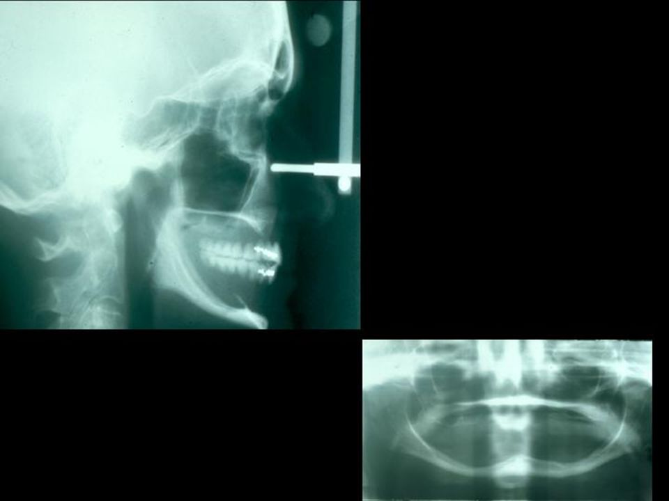
Complete Edentulism

4
Complete Edentulism

5
Complete Edentulism Edentulism, defined as total tooth loss, is more prevalent among persons with less than a high school education, those without dental insurance, non-Hispanic blacks, and current everyday smokers (CDC, 1999) Between the 1950s and the early 1990s the prevalence of edentulism in the United States decreased from 50% to 42% among people aged 65 and older, from 28% to 11% for 45- to 64-year-olds, and from 5% to 2% for persons 18 to 44 years old (Oliver & Brown, 1993) 1998 National Health Interview Survey, National Center for Health Statistics, and the 1999 Behavioral Risk Factor Surveillance System, CDC
 Between the 1950s and the early 1990s the prevalence of edentulism in the United States decreased from 50% to 42% among people aged 65 and older, from 28% to 11% for 45- to 64-year-olds, and from 5% to 2% for persons 18 to 44 years old (Oliver & Brown, 1993) 1998 National Health Interview Survey, National Center for Health Statistics, and the 1999 Behavioral Risk Factor Surveillance System, CDC.")
6
525 Other diseases and conditions of the teeth and supporting structures
525.4 Classification of complete edentulism based on the severity of the completely edentulous predicament

7
Complete Edentulism Classification System for Complete Edentulism
McGarry TJ, Nimmo A, Skiba JF, Ahlstrom RH, Smith CR, Koumjian JH J Prosthodont Mar;8(1):27-39
:")
8
Classification System for the Completely Edentulous Patient
Ideal or minimally compromised Class II Moderately compromised Diagnostic Criteria Bone height--mandibular Maxillomandibular relationship Residual ridge morphology- maxilla Muscle attachments Class III Substantially compromised Class IV Severely compromised

9
Diagnostic Criteria Bone height--mandibular
Maxillomandibular relationship Residual ridge morphology- maxilla Muscle attachments

10
1. Bone Height Mandibular

11
Type I Residual bone height of 21mm or greater measured at the least vertical height of the mandible.

12
Type IV Residual vertical bone height of 10 mm or less measured at the least vertical height of the mandible

14
2. Residual Ridge Morphology
Maxilla

15
Type A Anterior labial and posterior buccal vestibular depth that resists vertical and horizontal movement of the denture base Palatal morphology that resists vertical and horizontal movement of the denture base Sufficient tuberosity definition that resists vertical and horizontal movement of the denture base Hamular notch is well defined to establish the posterior extension of the denture base Absence of tori or exostoses

16
Type D Loss of anterior labial and posterior buccal vestibules
Maxillary palatal and/or lateral tori-rounded or undercut- that interferes with the posterior border of the denture Hyperplastic, redundant anterior ridge Palatal vault morphology that does not resist vertical or horizontal movement of the denture base Prominent anterior nasal spine

17
3. Maxillomandibular Relationship

18
Class I Maxillomandibular relationship allows tooth position that has normal articulation with the teeth supported by the residual ridge.

19
Class III Maxillomandibular relationship requires tooth position outside the normal ridge relation in order to attain phonetics and articulation; i.e., crossbite—anterior or posterior, tooth position not supported by the residual ridge.

20
4. Muscle Attachments

21
Type A Adequate attached mucosal base without undue muscular impingement during normal function in all regions.

22
Type D Adequate attached mucosal base only in the posterior lingual region All other regions are detached

23
Diagnostic Classification of Complete Edentulism

24
Class I All four of the diagnostic criteria are favorable.
This classification level describes the stage of edentulism that is most apt to be successfully treated by conventional prosthodontic techniques with complete denture prosthesis. All four of the diagnostic criteria are favorable.

25
Class I Residual bone height of 21 mm or greater measured at the least vertical height of the mandible Class I maxillomandibular relationship

26
Class II This classification level distinguishes itself with the noted continuation of the physical degradation of the denture supporting structures and in addition is characterized with the early onset of systemic disease interactions, localized soft tissue factors and patient management/lifestyle considerations.

27
Residual bone height of 16-20 mm measured at the least vertical height of the mandible
Class I maxillomandibular relationship Residual ridge morphology that resists horizontal and vertical movement of the denture base—Type A, B--Maxilla Class II

28
Class III This classification level is characterized by the need for surgical revision of denture supporting structures to allow for adequate prosthodontic function. Additional factors now play a significant role in treatment outcomes.

29
Class III Residual bone height of mm measured at the least vertical height of the mandible Class I, II and III maxillomandibular relationship Residual ridge morphology has minimum influence to resist horizontal or vertical movement of the denture base—Type C—Maxilla Location of muscle attachments with moderate influence on denture base stability and retention—Type C--Mandible

30
Class IV This classification level depicts the most debilitated edentulous condition Surgical reconstruction is almost always indicated but can not always be accomplished due to the patient’s health, desires, past dental history and financial considerations When surgical revision is not selected, prosthodontic techniques of a specialized nature must be used in order to achieve an adequate treatment outcome

31
Class IV Residual bone height of least vertical height of the mandible
Class I, II and III maxillomandibular relationships Residual ridge offers no resistance to horizontal or vertical movement – Type D—Maxilla Location of muscle attachments with significant influence on denture base stability and retention— Type D and E--Mandible

33
Completely Dentate

34
Partial Edentulism

35
Partial Edentulism

36
Partial Edentulism

37
525 Other diseases and conditions of the teeth and supporting structures
525.5 Classification of partial edentulism based on the severity of the partially edentulous predicament

38
Partial Edentulism Classification System for Partial Edentulism
McGarry TJ, Nimmo A, Skiba JF, Ahlstrom RH, Smith CR, Koumjian JH, Arbree NS J Prosthodont Sep;11(3):181-93
:")
39
Classification System for the Partially Edentulous Patient
Ideal or minimally compromised Class II Moderately compromised Diagnostic Criteria 1. Location and extent of the edentulous area(s) 2. Condition of the abutment teeth 3. Occlusal scheme 4. Residual ridge Class III Substantially compromised Class IV Severely compromised
 2. Condition of the abutment teeth. 3. Occlusal scheme. 4. Residual ridge. Class III. Substantially. compromised. Class IV. Severely. compromised.")
40
DIAGNOSTIC CRITERIA Location and extent of the edentulous area(s)
Condition of the abutment teeth Occlusal scheme Residual ridge

42
Partial Edentulism

43
Committed to developing a dental educational curriculum that is diagnosis driven
The only dental school in the third largest city in the US providing service to more than 100,000 patient visits per year Need for clinical studies that have a common, transparent and systematic diagnosis. Achieved by employing the evidence-based process to assemble, organize and synthesize clinical research in a rigorous and transparent fashion. This body of evidence, coupled with clinical expertise, will lead to the creation of guidelines designed to enhance clinical judgment and decision-making

44
Concluding Remarks The codes being proposed are part of normal diagnostic data collection that occurs for all patients, meeting with the existing standard of care in dentistry The proposed new codes are within the scope and conventions of the existing classification By adopting these codes into the public domain, dental educators, researchers and clinicians will be able to contribute significantly to the body of evidence

45
Acknowledgements Dr. Stephen Campbell UIC COD
Dr. Kent Knoernschild UIC COD Dr. John Zarb UIC COD Dr. Thomas McGarry ACP Dr. Barry Shipman ACP Dr. Rosemary Walker UIC SBHI Ms. Teri Jorwic UIC SBHI Dr. Bruce Graham UIC COD Ms. Lea Alexander UIC COD

Similar presentations