Download presentation
Presentation is loading. Please wait.

1
Functional Human Physiology for the Exercise and Sport Sciences The Respiratory System
Jennifer L. Doherty, MS, ATC Department of Health, Physical Education, and Recreation Florida International University

2
Overview of Respiratory Function
Respiration = the process of gas exchange Two levels of respiration: Internal respiration (cellular respiration) The use of O2 with mitochondria to generate ATP by oxidative phosphorylation CO2 is the waste product External respiration (ventilation) The exchange of O2 and CO2 between the atmosphere and body tissues
 The use of O2 with mitochondria to generate ATP by oxidative phosphorylation. CO2 is the waste product. External respiration (ventilation) The exchange of O2 and CO2 between the atmosphere and body tissues.")
4
Internal respiration (cellular respiration)
Involves gas exchange between capillaries and body tissues cells Tissue cells continuously use O2 and produce CO2 during metabolism Partial pressure (P) The PO2 is always higher in arterial blood than in the tissues The PCO2 is always higher in the tissues than in arterial blood O2 and CO2 move down their partial pressure gradients O2 moves out of the capillary into the tissues CO2 moves out of the tissues into the capillary
 The PO2 is always higher in arterial blood than in the tissues. The PCO2 is always higher in the tissues than in arterial blood. O2 and CO2 move down their partial pressure gradients. O2 moves out of the capillary into the tissues. CO2 moves out of the tissues into the capillary.")
6
External respiration (ventilation)
4 Processes: Pulmonary Ventilation Movement of air into the lungs (inspiration) and out of the lungs (expiration) Exchange of O2 and CO2 between lung air spaces and blood Transportation of O2 and CO2 between the lungs and body tissues Exchange of O2 and CO2 between the blood and tissues
 and out of the lungs (expiration) Exchange of O2 and CO2 between lung air spaces and blood. Transportation of O2 and CO2 between the lungs and body tissues. Exchange of O2 and CO2 between the blood and tissues.")
8
Overview of Pulmonary Circulation
Deoxygenated blood Under resting conditions, 5 liters of deoxygenated blood are pumped to the lungs each minute from the right ventricle CO2 blood concentration is higher than O2 blood concentration in: Systemic veins Right atrium Right ventricle Pulmonary arteries

10
Overview of Pulmonary Circulation
Oxygenated blood Transported from the pulmonary capillaries → pulmonary veins → left atrium → left ventricle → aorta → systemic arterial circulation O2 blood concentration is higher than CO2 blood concentration in: Alveoli Pulmonary capillaries Pulmonary veins Left atrium Left ventricle Systemic arteries

12
Anatomy of the Respiratory Zone
Gas exchange occurs between the air and the blood within the alveoli

13
Anatomy of the Respiratory Zone
Alveoli (singular is alveolus) Tiny air sacs clustered at the distal ends of the alveolar ducts Alveoli have a thin respiratory membrane separating the air from blood in pulmonary capillaries
 Tiny air sacs clustered at the distal ends of the alveolar ducts. Alveoli have a thin respiratory membrane separating the air from blood in pulmonary capillaries.")
15
Respiratory Membrane The thin alveolar wall consists of:
The fused alveolar and capillary walls Alveolar epithelial cells Capillary endothelial cells The basement membrane Sandwiched between the alveolar epithelial cells and the endothelial cells of the capillary

17
Respiratory Membrane Gas exchanges occurs across the respiratory membrane It is < 0.1 μm thick Lends to very efficient diffusion It is the site of external respiration and diffusion of gases between the inhaled air and the blood Occurs in the pulmonary capillaries

18
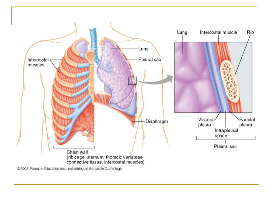
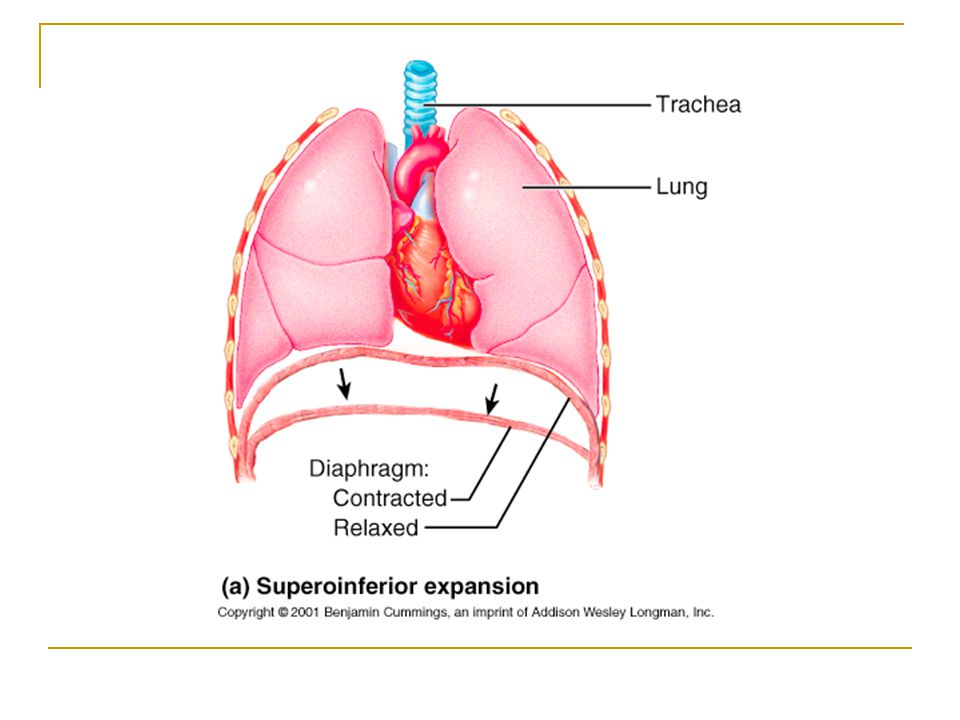
Structures of the Thoracic Cavity
A container with a single opening, the trachea Volume of the container changes Diaphragm moves up and down Muscles move the rib cage in and out Volume of the thoracic cavity increases by enlarging all diameters ↑ diameter = ↑ volume

20
Boyle’s Law Volume and pressure are inversely related
↑ volume = ↓ pressure Air always flows from an area of higher pressure to an area of lower pressure Decreased pressure in the thoracic cavity in relation to atmospheric pressure causes air to flow into the lungs The process of inspiration

21
Structures of the Thoracic Cavity
Pleura Parietal pleura: A membrane that lines the interior surface of the chest wall Visceral pleura: A membrane that lines the exterior surface of the lungs Intrapleural space A thin compartment between the two pleurae filled with intrapleural fluid

23
Pulmonary Pressures Pressure gradient 4 Pulmonary Pressures
The difference between intrapulmonary and atmospheric pressures 4 Pulmonary Pressures Atmospheric pressure Intra-alveolar (Intrapulmonary) pressure Intrapleural pressure Transpulmonary pressure
 pressure. Intrapleural pressure. Transpulmonary pressure.")
24
Pulmonary Pressures Atmospheric pressure
The pressure exerted by the weight of the air in the atmosphere (~ 760 mmHg at sea level) Intra-alveolar (Intrapulmonary) pressure The pressure inside the lungs Intrapleural pressure The pressure inside the pleural space Transpulmonary pressure The difference between the intrapleural and intra-alveolar pressure
 Intra-alveolar (Intrapulmonary) pressure. The pressure inside the lungs. Intrapleural pressure. The pressure inside the pleural space. Transpulmonary pressure. The difference between the intrapleural and intra-alveolar pressure.")
25
Pleural Pressures Intrapleural pressure Surface tension
The pressure inside the pleural space or cavity This cavity contains intrapleural fluid, necessary for surface tension Surface tension The force that holds moist membranes together due to an attraction that water molecules have for one another Responsible for keeping lungs patent

26
Surface Tension The force of attraction between liquid molecules
Type II alveolar cells secrete surfactant Creates a thin fluid film in the alveoli Surfactant (a phospholipoprotein) reduces the surface tension in the alveoli It interferes with the attraction between fluid molecules Decreasing surface tension reduces the amount of energy required to expand the lungs
 reduces the surface tension in the alveoli. It interferes with the attraction between fluid molecules. Decreasing surface tension reduces the amount of energy required to expand the lungs.")
29
Inspiration Drawing or pulling air into the lungs
Atmospheric pressure forces air into the lungs The diaphragm moves downward, decreasing intra-alveolar pressure The thoracic rib cage moves upward and outward, increasing the volume of the thoracic cavity Surface tension Holds the pleural membranes together, which assists with lung expansion Surfactant reduces surface tension within the alveoli

33
Inspiration During inspiration, forces are generated that attempt to pull the lungs away from the thoracic wall Surface tension of the intraplueral fluid hold the lungs against the thoracic wall, preventing collapse

36
Expiration Pushing air out of the lungs
Results due to the elastic recoil of tissues and due to the surface tension within the alveoli Expiration can be aided by: Thoracic and abdominal wall muscles that pull the thoracic cage downward and inward, decreasing intra-alveolar pressure This compresses the abdominal organs upward and inward, decreasing the volume of the thoracic cavity

38
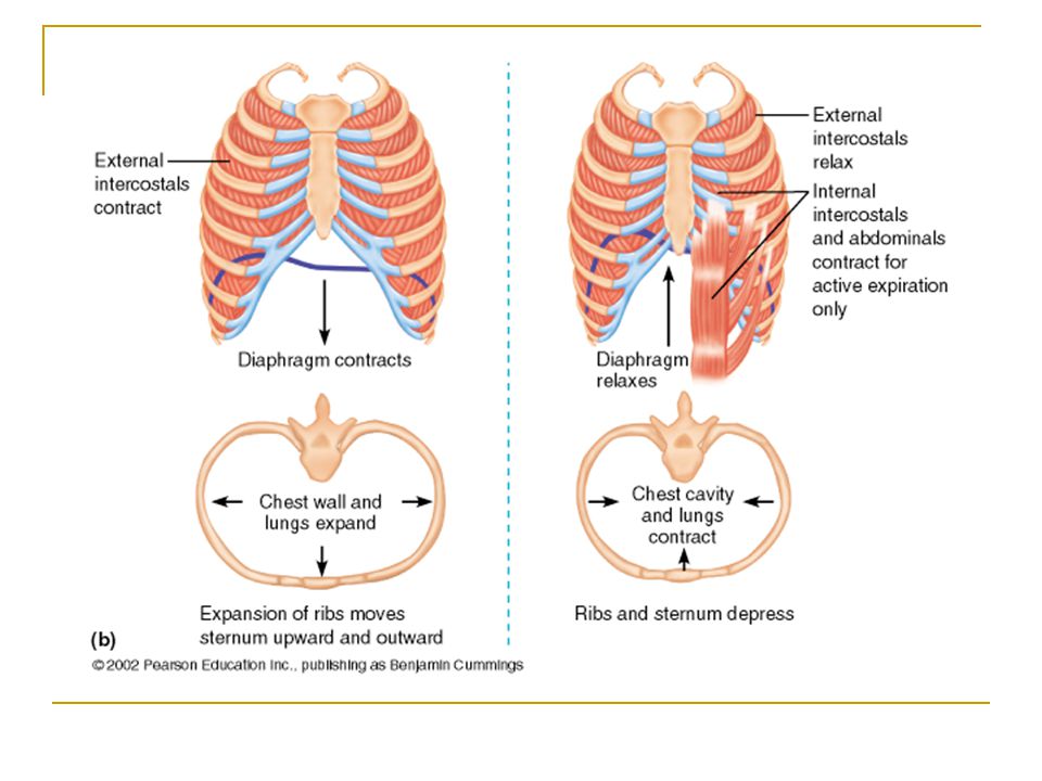
Muscles of Breathing - Inspiration
Quiet Breathing Muscles include: External intercostals Diaphragm Contract to expand the rib cage and stretch the lungs = ↑ volume of the thoracic cavity ↑ intrapulmonary volume ↓ intrapulmonary pressure (relative to atmospheric pressure) Decreased pressure inside the lungs pulls air into the lungs down its pressure gradient until intrapulmonary pressure equals atmospheric pressure
 Decreased pressure inside the lungs pulls air into the lungs down its pressure gradient until intrapulmonary pressure equals atmospheric pressure.")
41
Muscles of Breathing - Inspiration
Forced or Deep Inspiration Involves several accessory muscles: Sternocleidomastoid Pectoralis minor Scalenes (neck muscles) Maximal ↑ in thoracic volume Greater ↓ in intrapulmonary pressure More air moves into the lungs At the end of inspiration, the intrapulmonary pressure equals atmospheric pressure
 Maximal ↑ in thoracic volume. Greater ↓ in intrapulmonary pressure. More air moves into the lungs. At the end of inspiration, the intrapulmonary pressure equals atmospheric pressure.")
42
Muscles of Breathing - Expiration
Quiet Breathing Passive process Depends on the elasticity of the lungs Muscles of inspiration relax The rib cage descends The lungs recoil ↓ intrapulmonary volume ↑ intrapulmonary pressure Alveoli are compressed, thus forcing air out of the lungs

43
Muscles of Breathing - Expiration
Forced Expiration It is an active process Occurs in activities such as blowing up a balloon, exercising, or yelling Abdominal wall muscles are involved in forced expiration Function to ↑ the pressure in the abdominal cavity forcing the abdominal organs upward against the diaphragm ↓ volume of the thoracic cavity ↑ pressure in the thoracic cavity Air is forced out of the lungs

45
Factors Affecting Pulmonary Ventilation
Lung compliance The ease with which the lungs may be expanded, stretched, or inflated Depends primarily on the elasticity of the lung tissue Elasticity refers to the ability of the lung to recoil after it has been inflated

46
Factors Affecting Pulmonary Ventilation
Lung and thoracic cavity tissue has a natural tendency to recoil, or become smaller Lung recoil is essential for normal expiration and depends on the fibroelastic qualities of lung tissue In normal lungs there is a balance between compliance and elasticity

47
Factors Affecting Pulmonary Ventilation
Recoil pressure is inversely proportional to compliance Increased compliance results in decreased recoil Example: Emphysema Results in difficulty resuming the shape of the lung during exhalation Decreased compliance results in increased recoil Example: Cysitc fibrosis Results in difficulty expanding the lung because of increased fibrous tissue and mucous

48
Factors Affecting Pulmonary Ventilation
Airway Resistance Opposition to air flow in the respiratory passageways Resistance and air flow are inversely related ↑ airway resistance = ↓ air flow (and vice versa) Airway resistance is most affected by changes in the diameter of the bronchioles ↓ diameter of the bronchioles = ↑ airway resistance Examples: Asthma Bronchiospasm during an allergic reaction A high resistance to air flow produces a greater energy cost of breathing
 Airway resistance is most affected by changes in the diameter of the bronchioles. ↓ diameter of the bronchioles = ↑ airway resistance. Examples: Asthma. Bronchiospasm during an allergic reaction. A high resistance to air flow produces a greater energy cost of breathing.")
49
The Respiratory System: Gas Exchange and Regulation of Breathing
Jennifer L. Doherty, MS, ATC Department of Health, Physical Education, and Recreation Florida International University

50
Diffusion of Gases Partial Pressure of Gases (Pgas)
Concentration of gases in a mixture (air) Gases move from areas of high partial pressure to areas of low partial pressure Movement of gases also occurs between cells and the blood in the capillaries Movement of gases occurs between blood in the pulmonary capillaries and the air within the alveoli Movement of gasses is by diffusion across the respiratory membrane of the alveoli
 Gases move from areas of high partial pressure to areas of low partial pressure. Movement of gases also occurs between cells and the blood in the capillaries. Movement of gases occurs between blood in the pulmonary capillaries and the air within the alveoli. Movement of gasses is by diffusion across the respiratory membrane of the alveoli.")
52
Dalton’s Law of Partial Pressure
Each gas in a mixture (air) tends to diffuse independently of all other gases Oxygen does not interfere with carbon dioxide diffusion or vice versa Each gas diffuses at a rate proportional to its partial pressure gradient until it reaches equilibrium This allows for two-way traffic of gases in the lungs and in the body tissues The total pressure exerted by a mixture of gases is the same as the sum of the pressure exerted by each individual gas in the mixture Pair = PN2 + PO2 + PH2O
 tends to diffuse independently of all other gases. Oxygen does not interfere with carbon dioxide diffusion or vice versa. Each gas diffuses at a rate proportional to its partial pressure gradient until it reaches equilibrium. This allows for two-way traffic of gases in the lungs and in the body tissues. The total pressure exerted by a mixture of gases is the same as the sum of the pressure exerted by each individual gas in the mixture. Pair = PN2 + PO2 + PH2O.")
53
Partial Pressure: Atmospheric Air
The partial pressure of a gas is the pressure exerted by each gas in a mixture and is directly proportional to its percentage in the total gas mixture Example: Atmospheric Air At sea level, atmospheric pressure is 760 mmHg Air is ~78% Nitrogen The partial pressure of nitrogen (PN2) is: 0.78 x 760 mmHg = PN2 = 593 mmHg Air is ~ 21% Oxygen The partial pressure of oxygen (PO2) is: 0.21 x 760 mmHg = PO2 = 160 mmHg Air is ~ 0.04% carbon dioxide The partial pressure of carbon dioxide (PCO2) is: x 760 mmHg = PCO2 = 0.3 mmHg.
 is: 0.78 x 760 mmHg = PN2 = 593 mmHg. Air is ~ 21% Oxygen. The partial pressure of oxygen (PO2) is: 0.21 x 760 mmHg = PO2 = 160 mmHg. Air is ~ 0.04% carbon dioxide. The partial pressure of carbon dioxide (PCO2) is: x 760 mmHg = PCO2 = 0.3 mmHg.")
54
Partial Pressure: Alveolar Air
Composition of the partial pressures of oxygen and carbon dioxide in the pulmonary capillaries and alveolar air: Pulmonary arterial capillary blood PCO2 of pulmonary capillary blood is 45 mmHg PO2 of pulmonary capillary blood is 40 mmHg Alveolar air: PCO2 of alveolar air is 40 mmHg PO2 of alveolar air is 104 mmHg

55
Solubility of Gases in a Liquid
The ability of a gas to dissolve in water Important because O2 and CO2 are exchanged between air in the alveoli and blood (which is mostly water) Even when dissolved in water, gases exert a partial pressure Gases diffuse from regions of higher partial pressure toward regions of lower partial pressure
 Even when dissolved in water, gases exert a partial pressure. Gases diffuse from regions of higher partial pressure toward regions of lower partial pressure.")
56
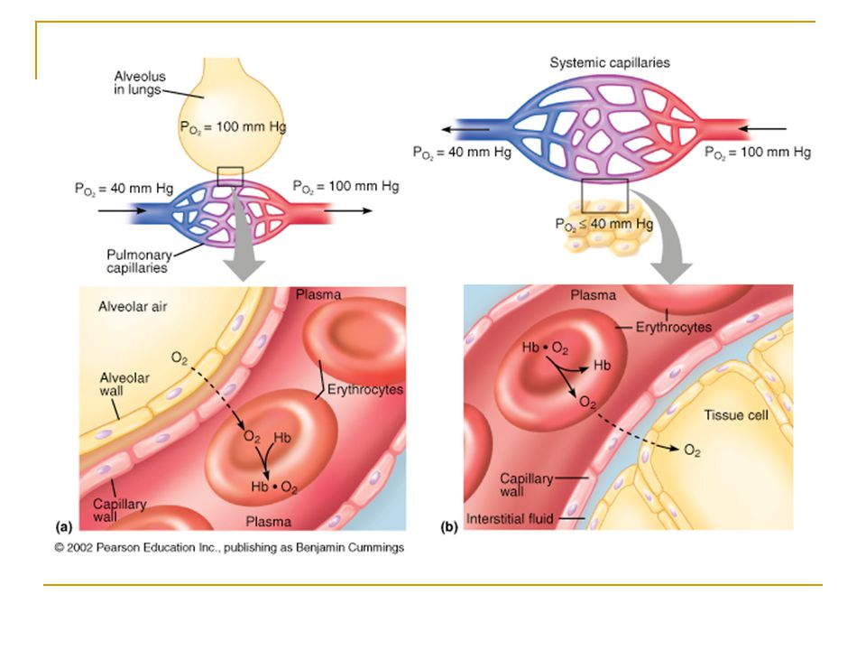
Gas Exchange in the Lungs
Gas exchange occurs by diffusion across the respiratory membrane in the alveoli Oxygen diffuses from the alveolar air into the blood Alveolar air PO2 = 104 mmHg Pulmonary capillaries PO2 = 40 mmHg Carbon dioxide diffuses from the pulmonary capillary blood into the alveolar air Pulmonary capillaries PCO2 = 46 mmHg Alveolar air PCO2 = 40 mmHg

59
Gas Exchange in Respiring Tissue
Gas partial pressures in systemic capillaries depends on the metabolic activity of the tissue Oxygen concentrations Systemic arteries PO2 = 100 mmHg Systemic veins PO2 = 40 mmHg Carbon dioxide concentrations Systemic arteries PCO2 = 40 mmHg Systemic veins PCO2 = 46 mmHg

61
Transport of Gases in the Blood: O2
98% of O2 is transported in combination with hemoglobin molecules (98%) 2% of O2 is dissolved and transported in the plasma Hemoglobin (Hb) A protein found in RBCs O2 binds loosely to Hb due to its molecular structure Hemoglobin consists of four polypeptide chains Consists of 4 globin molecules, each of which is bound to a heme group The heme group contains an iron molecule, which is the site of O2 binding Each Hb molecule is able to carry 4 molecules of O2
 2% of O2 is dissolved and transported in the plasma. Hemoglobin (Hb) A protein found in RBCs. O2 binds loosely to Hb due to its molecular structure. Hemoglobin consists of four polypeptide chains. Consists of 4 globin molecules, each of which is bound to a heme group. The heme group contains an iron molecule, which is the site of O2 binding. Each Hb molecule is able to carry 4 molecules of O2.")
62
Transport of Gases in the Blood: O2
O2 binds temporarily, or reversibly, to Hb Oxyhemoglobin (HbO2) Hb + O2 = HbO2 Hb attached to four O2 molecules is saturated Saturated Hb is relatively unstable and easily releases O2 in regions where the PO2 is low Deoxyhemoglobin (HHb) HHb = Hb + O2
 Hb + O2 = HbO2. Hb attached to four O2 molecules is saturated. Saturated Hb is relatively unstable and easily releases O2 in regions where the PO2 is low. Deoxyhemoglobin (HHb) HHb = Hb + O2.")
63
The Hemoglobin-Oxygen Dissociation Curve
Describes the relationship between the aterial PO2 and Hb saturation The Hb- O2 Dissociation Curve plots the percent saturation of Hb as a function of the PO2

65
The Hemoglobin-Oxygen Dissociation Curve
Hb Saturation Full saturation All four heme groups of the Hb molecule in the blood are bound to O2 Partial saturation Not all of the heme groups are bound to O2 Hb saturation is largely determined by the PO2 in the blood At normal alveolar PO2 (104 mm Hg), Hb is % saturated
, Hb is % saturated.")
67
The Hemoglobin-Oxygen Dissociation Curve
Hb Unloading of O2 Factors that increase O2 unloading from hemoglobin at the tissues: Increased body temperature Decreases Hb affinity for O2 Decreased blood pH (the Bohr effect) H+ ions bind to Hb Increased arterial PCO2 (the Carbamino effect)
 H+ ions bind to Hb. Increased arterial PCO2 (the Carbamino effect)")
69
The Bohr Effect Based on the fact that when O2 binds to Hb, certain amino acids in the Hb molecule release H+ ions Hb + O2 ↔ HbO2 + H+ An increase in H+ (a decrease in pH) pushes the reaction to the left, causing O2 to dissociate from Hb Hb affinity for O2 is decreased when H+ ions bind to Hb, therefore O2 is unloaded from Hb H+ concentration increases in active tissues, which facilitates O2 unloading from Hb so that it may be utilized by the active tissues
 pushes the reaction to the left, causing O2 to dissociate from Hb. Hb affinity for O2 is decreased when H+ ions bind to Hb, therefore O2 is unloaded from Hb. H+ concentration increases in active tissues, which facilitates O2 unloading from Hb so that it may be utilized by the active tissues.")
71
The Carbamino Effect Based on the fact that CO2 may bind to Hb
Hb + CO2 ↔ HbCO2 An increase in PCO2 pushes the reaction to the right, forming carbaminohemoglobin (HbCO2) HbCO2 decreases Hb affinity for O2 This decreases O2 transport in the blood The carbamino effect is one method of transporting CO2 in the blood
 HbCO2 decreases Hb affinity for O2. This decreases O2 transport in the blood. The carbamino effect is one method of transporting CO2 in the blood.")
73
The Hemoglobin-Oxygen Dissociation Curve
These factors are all present during exercise and enable Hb to release more O2 to meet the metabolic demands of working tissues ↑ body temperature = ↓ Hb affinity for O2 ↑ H+ ions (↓ pH) = ↓ Hb affinity for O2 ↑ arterial PCO2 = ↓ Hb affinity for O2
 = ↓ Hb affinity for O2. ↑ arterial PCO2 = ↓ Hb affinity for O2.")
75
Transport of Gases in the Blood: CO2
CO2 may be transported in the blood by… Dissolving in the plasma Dissolving as bicarbonate Binding to Hb (carbaminohemoglobin)
")
76
Transport of Gases in the Blood: CO2
CO2 Dissolved in Plasma CO2 is very soluble in water ~ 5 - 6% of CO2 in the blood is dissolved in plasma The partial pressure gradient between the tissues and blood allows CO2 to easily diffuse from the tissues into the plasma The amount of CO2 dissolved in the plasma is proportional to the partial pressure of CO2

78
Transport of Gases in the Blood: CO2
CO2 as Bicarbonate (H2CO3) ~ 86 – 90% of CO2 in the blood is transported in the form of bicarbonate ions In water, carbonic acid dissociates to release H+ ions and bicarbonate ions CO2 + H2O ↔ H2CO3 ↔ H+ + HCO3- Catalyzed by carbonic anhydrase This chemical reaction occurs slowly in both plasma and in red blood cells The blood becomes more acidic due to the accumulation of CO2
 ~ 86 – 90% of CO2 in the blood is transported in the form of bicarbonate ions. In water, carbonic acid dissociates to release H+ ions and bicarbonate ions. CO2 + H2O ↔ H2CO3 ↔ H+ + HCO3- Catalyzed by carbonic anhydrase. This chemical reaction occurs slowly in both plasma and in red blood cells. The blood becomes more acidic due to the accumulation of CO2.")
80
Transport of Gases in the Blood: CO2
CO2 bound to Hb (carbaminohemoglobin) Carbaminohemoglobin CO2 attached to a hemoglobin molecule Hb + CO2 ↔ HbCO2 ~ 5 - 8% of CO2 is bound to Hb in RBCs CO2 diffuses into RBCs and binds with the globin component (not the heme component) of Hb for transport to the lungs
 Carbaminohemoglobin. CO2 attached to a hemoglobin molecule. Hb + CO2 ↔ HbCO2. ~ 5 - 8% of CO2 is bound to Hb in RBCs. CO2 diffuses into RBCs and binds with the globin component (not the heme component) of Hb for transport to the lungs.")
82
CO2 Exchange and Transport in Systemic Capillaries and Veins
The Chloride Shift CO2 may be transported as HbCO2 or H2CO3 H+ ions or bicarbonate may accumulate in RBCs Hb functions as a buffer for H+ ions Hb binding to H+ ions forms HHb as a buffer so that RBCs do not become too acidic Hb + H+ ↔ HHb The bicarbonate ion (H2CO3) diffuses out of the RBC into the plasma to be carried to the lungs As bicarbonate ions leave the RBC, Cl- ions enter the RBC
 diffuses out of the RBC into the plasma to be carried to the lungs. As bicarbonate ions leave the RBC, Cl- ions enter the RBC.")
83
The Effect of O2 on CO2 Transport
The Haldane effect Loading/Unloading of CO2 onto Hb is directly related to: 1) The partial pressure of CO2 (PCO2) In areas of high PCO2, carbaminohemoglobin forms This helps unload CO2 from tissues 2) The partial pressure of O2 (PO2 ) In areas of high PO2 (such as in the lungs), the amount of CO2 transported by Hb decreases This helps unload CO2 from the blood 3) The degree of oxygenation of Hb Deoxygenated Hb is able to carry more CO2 than a Hb molecule loaded with O2 The binding of O2 to Hb decreases the affinity of Hb for CO2
 The partial pressure of CO2 (PCO2) In areas of high PCO2, carbaminohemoglobin forms. This helps unload CO2 from tissues. 2) The partial pressure of O2 (PO2 ) In areas of high PO2 (such as in the lungs), the amount of CO2 transported by Hb decreases. This helps unload CO2 from the blood. 3) The degree of oxygenation of Hb. Deoxygenated Hb is able to carry more CO2 than a Hb molecule loaded with O2. The binding of O2 to Hb decreases the affinity of Hb for CO2.")
84
Central Regulation of Ventilation
The purpose of ventilation is to deliver O2 to and remove CO2 from cells at a rate sufficient to keep up with metabolic demands Breathing is under both involuntary and voluntary control Normal breathing is rhythmic and involuntary However, the respiratory muscles may be controlled voluntarily

85
Neural Control of Breathing by Motor Neurons
The brainstem generates breathing rhythm Signals are delivered to the respiratory muscles via somatic motor neurons Phrenic nerve Innervates the diaphragm Intercostal nerves Innervate the internal and external intercostal muscles

86
Generation of the Breathing Rhythm by the Brainstem
Central control of respiration is not completely understood Research indicates that respiratory control centers are located in the brainstem Respiratory control centers include… Medullary Rhythmicity Area of the medulla oblongata Pneumotaxic Area of the pons Apneustic Center of the pons

87
Medullary Rhythmicity Area
Includes two groups of neurons: Dorsal Respiratory Group Ventral Respiratory Group

88
Medullary Rhythmicity Area
The Dorsal Respiratory Group The medullary inspiratory center Functions to generate the basic respiratory rhythm The respiratory cycle is repeated times/minute Dorsal neurons have an intrinsic ability to spontaneously depolarize at a rhythmic rate Quiet breathing - Inhalation The dorsal inspiratory neurons transmit nerve impulses via the phrenic and intercostal nerves to the diaphragm and external intercostal muscles When these muscles contract, the lungs fill with air Quiet breathing - Exhalation When the dorsal inspiratory neurons stop sending impulses, expiration occurs passively as the inspiratory muscles relax and the lungs recoil

89
Medullary Rhythmicity Area
The Ventral Respiratory Group The medullary expiratory center Functions to promote expiration during forceful breathing If the rate and depth of breathing increases above a critical threshold, it stimulates a forceful expiration The ventral expiratory neurons transmit nerve impulses to the muscles of expiration The internal intercostals The abdominal muscles

90
Pneumotaxic Area Includes two groups of neurons:
Pontine Respiratory Group The Central Pattern Generator

91
Pneumotaxic Area The Pontine Respiratory Group
Facilitates the transition between inspiration and expiration Regulates the depth or the extent of inspiration Regulates the frequency of respiration

92
Pneumotaxic Area The Central Pattern Generator
A network of neurons scattered between the pons and the medulla Exact location of these neurons is unknown Coordinates the control centers of the brainstem Regulates the rate of breathing Regulates the length of inspiration Avoid over-inflation of the lungs Regulates the depth of breathing ↑ pneumotaxic output = shallow, rapid breathing ↓ pneumotaxic output = deep, slow breathing

93
Peripheral Input to Respiratory Centers
Receptors and reflexes monitor and respond to stimuli Feed information (input) to the Central Pattern Generator Input received from… Chemoreceptors Pulmonary stretch receptors Detect lung tissue expansion and may protect lungs from over inflation through the Hering-Breuer reflex Irritant receptors Detect inhaled particles (dust, smoke) and trigger coughing, sneezing, or bronchiospasm
 to the Central Pattern Generator. Input received from… Chemoreceptors. Pulmonary stretch receptors. Detect lung tissue expansion and may protect lungs from over inflation through the Hering-Breuer reflex. Irritant receptors. Detect inhaled particles (dust, smoke) and trigger coughing, sneezing, or bronchiospasm.")
94
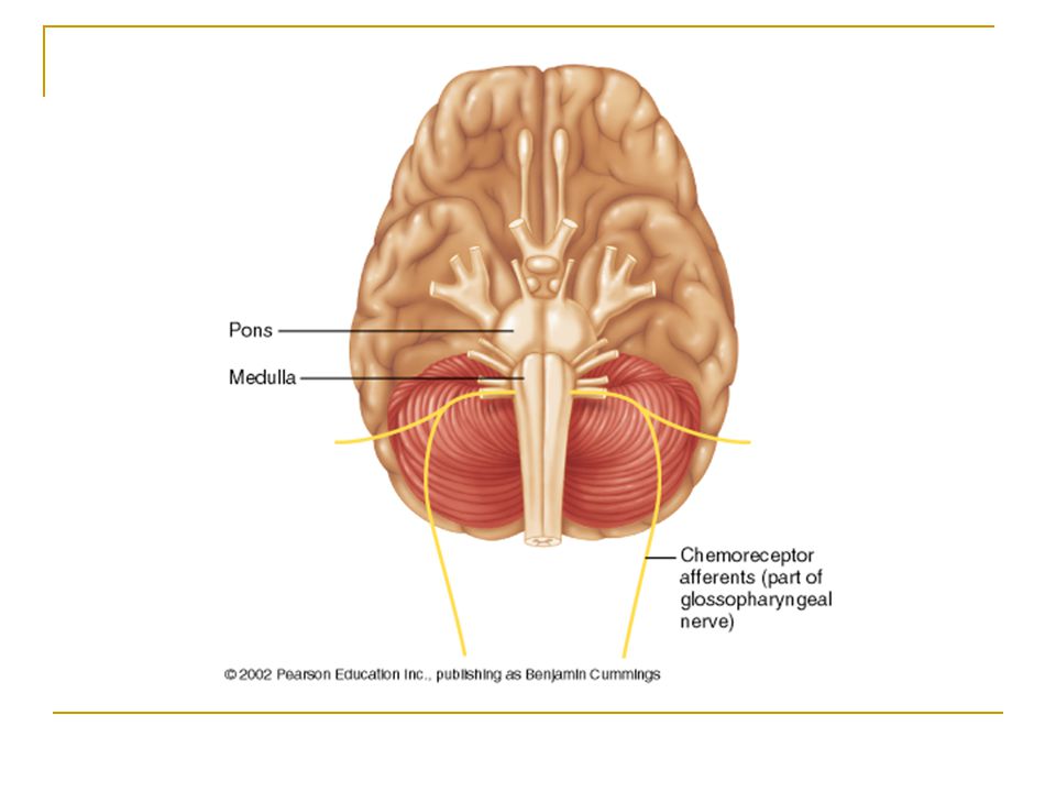
Peripheral Input to Respiratory Centers: Chemoreceptors
Peripheral Chemoreceptors Detect chemical concentration of blood and cerebrospinal fluid Location: Carotid sinus At its bifurcation into the internal and external carotid arteries Connected to medulla by afferent neurons in the glossopharyngeal (CN IX) nerve Chemical concentration of the blood is most important Changing levels of CO2, O2, and pH of the blood Sensitive to low arterial O2 concentrations (below 60 mmHg)
 nerve. Chemical concentration of the blood is most important. Changing levels of CO2, O2, and pH of the blood. Sensitive to low arterial O2 concentrations (below 60 mmHg)")
97
Peripheral Input to Respiratory Centers: Chemoreceptors
Peripheral chemoreceptors are very sensitive to changes in arterial pH ↓ arterial pH (↑ H+ ion concentration) occurs: When arterial CO2 levels increase When lactic acid accumulates in the blood Therefore, ↓ arterial pH is the most powerful stimulant for respiration When O2 concentration is low, ventilation increases
 occurs: When arterial CO2 levels increase. When lactic acid accumulates in the blood. Therefore, ↓ arterial pH is the most powerful stimulant for respiration. When O2 concentration is low, ventilation increases.")
98
Peripheral Input to Respiratory Centers: Chemoreceptors
Central chemoreceptors Sensitive to H+ ion concentration in cerebrospinal fluid Located in the medulla within the blood-brain barrier CO2 is able to diffuse across the blood-brain barrier and combine with water to form carbonic acid This reaction releases H+ ions in the cerebrospinal fluid CO2 then combines with water in cerebrospinal fluid to form carbonic acid Stimulation of these central chemoreceptors increases respiration rate, thus increasing blood pH to homeostatic levels

100
Chemoreceptor reflexes
Chemoreceptors maintain normal levels of arterial CO2 through chemoreceptor reflexes Increased CO2 = increased concentration of H+ ions (↓ pH) This stimulates the chemoreceptors Decreased blood pH can be caused by Exercise and accumulation of lactic acid Breath holding Other metabolic causes ↓ arterial pH causes the respiratory system to attempt to restore normal blood pH by… ↑ ventilation to decrease CO2 levels This results in an increase in pH to normal levels
 This stimulates the chemoreceptors. Decreased blood pH can be caused by. Exercise and accumulation of lactic acid. Breath holding. Other metabolic causes. ↓ arterial pH causes the respiratory system to attempt to restore normal blood pH by… ↑ ventilation to decrease CO2 levels. This results in an increase in pH to normal levels.")
101
Conscious Control of Breathing
Control over respiratory muscles is voluntary Therefore, breathing patterns may be consciously altered Voluntary control is made possible by neural connections between higher brain centers (the cortex) and the brain stem Voluntary control includes… Holding your breath Emotional upset Strong sensory stimulation (irritants) that alter normal breathing patterns
 and the brain stem. Voluntary control includes… Holding your breath. Emotional upset. Strong sensory stimulation (irritants) that alter normal breathing patterns.")
102
Disturbances in Respiration
Hyperpnea An ↑ in the arterial CO2 concentration with a resultant ↓ in CSF fluid pH This condition stimulates the… Central chemoreceptors, and Medullary respiratory centers Stimulates an increase in ventilation Hyperventilation More CO2 is exhaled resulting in ↓ arterial CO2 concentration This returns arterial pH to normal levels

103
The Respiratory System in Acid-Base Homeostasis
Acid-Base Disturbances in Blood The average pH of body fluids is 7.38 This is slightly alkaline, but, acidic compared to blood The pH of arterial blood is 7.4. The pH of venous blood and extracellular fluid is 7.35 The pH of intracellular fluid is 7.0 This reflects the greater amounts of acidic wastes and CO2 that are produced inside cells Acidosis Arterial blood pH less than 7.35 Alkalosis Arterial blood pH greater than 7.45

104
The Respiratory System in Acid-Base Homeostasis
Hydrogen Ion Concentration Regulation Body pH is regulated by the respiratory system through the regulation of H+ ion concentration in the blood Very important because metabolic reactions generally produce more acids than bases Acid-base buffers Bind with H+ ions when fluids become acidic Release H+ ions when fluids become alkaline Convert strong acids into weaker acids Convert strong bases into weaker bases Examples: Hemoglobin Bicarbonate ions

105
The Respiratory System in Acid-Base Homeostasis
Respiratory centers located in the brainstem help regulate pH by controlling the rate and depth of breathing Respiratory responses to changes in pH are not immediate, it requires several minutes to modify pH Respiratory responses to changes in pH are almost twice the buffering power of all the chemical buffers combined

106
Abnormalities of Acid-Base Balance
pH disturbances result due to inadequate or improper functioning of respiratory mechanics Respiratory acidosis The most common type of acid-base imbalance Accumulation of CO2 as the result of shallow breathing, pneumonia, emphysema, or obstructive respiratory diseases Respiratory alkalosis Develops during hyperventilation Excessive loss of CO2 Treatment includes re-breathing air to increase arterial CO2

Similar presentations


 To improve gas absorption Increase surface.>")


