Download presentation
Presentation is loading. Please wait.

1
MGUS (interpreting the test you didnt order) Family Medicine Review Course 2011 Christian Cable, MD, FACP
 Family Medicine Review Course 2011 Christian Cable, MD, FACP")
2
The Case

3
What is the laboratory abnormality? 10-3 = 7 Whats in there?

4
What comprises the blood?

6
Whats in blood... Cellular (bone marrow) – RBCs – Platelets – WBCs Plasma (liver) – Water – Proteins Albumin Antibodies Clotting factors
 – RBCs – Platelets – WBCs Plasma (liver) – Water – Proteins Albumin Antibodies Clotting factors.")
9
Proteins in the Blood?

10
Brainstorm As many globins as you can think of...

11
Tell me more about antibodies

12
What is the correct test?

14
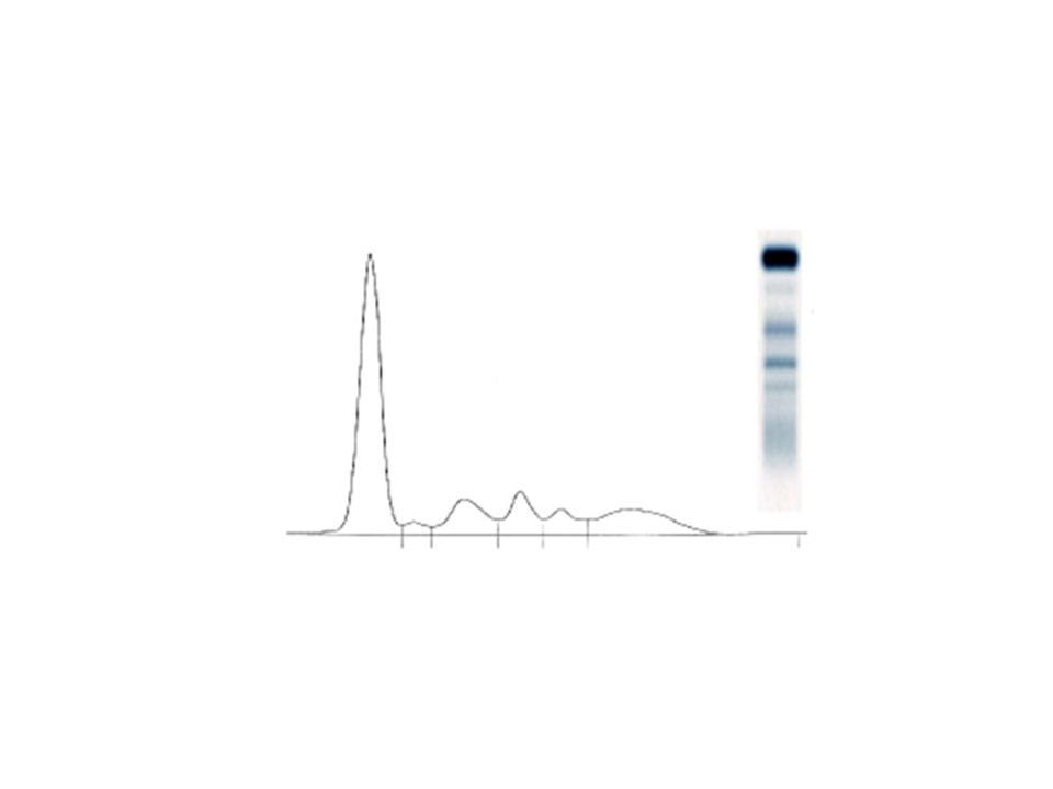
SPEP/SIEP SPEP qualitative (is it there?) SIEP quantitative (how much, which one?)
 SIEP quantitative (how much, which one )")
15
Copyright ©2001 American Society of Hematology. Copyright restrictions may apply. Lazarchick, J. ASH Image Bank 2001;2001:100185 Figure 8. Immunofixation electrophoresis showing a monoclonal IgA lambda light chain restricted band

16
Gammopa-what?

17
Greek to me (I)... Gamma - - region in electrophoretic mobility Pathy - - disease or condition
... Gamma - - region in electrophoretic mobility Pathy - - disease or condition")
18
Greek to me (II)... Clonal - - type Mono - - one Poly - - many (much)
... Clonal - - type Mono - - one Poly - - many (much)")
19
Differentiate Polyclonal from Monoclonal

20
M-spike

21
What is normal?

22
How high?

23
Polyclonal gammopathy - -significance Think of an elevated ESR What could cause that?

25
Is polyclonal gammopathy a plasma cell disorder?

26
Monoclonal gammopathy - - determined significance

27
New Myeloma Classification

28
Copyright ©2002 American Society of Hematology. Copyright restrictions may apply. Schrier, S. ASH Image Bank 2002;2002:100514 Figure 2. This is a bone marrow aspirate from a patient with multiple myeloma showing the abnormal accumulation of malignant plasma cells

29
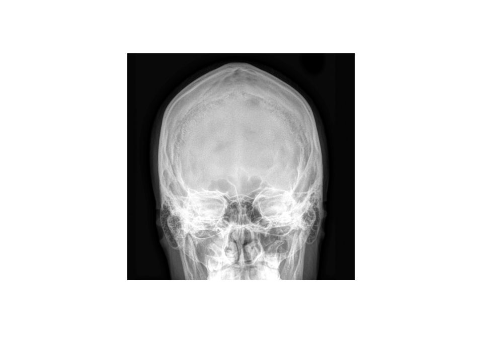
Copyright ©2001 American Society of Hematology. Copyright restrictions may apply. Lazarchick, J. ASH Image Bank 2001;2001:100185 Figure 11. Skull x-ray showing multiple lytic areas

30
Monoclonal gammopathy - - undetermined significance

31
Common?

32
3% of population over 50 twice that prevalence African Americans

33
Defined M-spike < 3 g/dL absence of CRAB symptoms (at least those attributable to MM) - - tricky with pre-existing renal disease! Bone Marrow involvement <10% with clonal plasma cells

34
How to evaluate CBC, Creatinine, Calcium, SPEP/SIEP Skeletal survey (plain films)
")
35
When to refer

37
Higher risk non-Ig G (IgA & Ig M) African American total M spike: >1.5 g/dL
 African American total M spike: >1.5 g/dL")
38
Why follow? Over 20 years: 1% per year turn into either Multiple Myeloma or another blood cancer Double that risk for non-IgG subtypes and African American patients

39
How do you follow it? Id like to help follow higher risk patients. Lower risk: – re-test in 6 months then annually

40
Our Patient

41
SPEP

42
SIEP 1.6 g/dL IgA kappa

44
Recommendations referral bone marrow biopsy

45
ccable@swmail.sw.org

Similar presentations


 Idiopathic Associated with other diseases (autoimmune, infectious, non-heme.>")


BB Topic 3 Autoimmunity Part 8 Immunoproliferative Diseases.>")


.>")