Download presentation
Presentation is loading. Please wait.

1
Soft Tissues Mark Bromley PGY4 November 19, 2009
Lateral Neck X-Ray Soft Tissues Mark Bromley PGY4 November 19, 2009

2
Outline Relevant Anatomy Cases Test Characteristics Epiglottitis
Retropharyngeal Abscess Bacterial Tracheitis Croup FB Test Characteristics

3
Enlarged epiglottis Loss of valecular airspace Thickened aryepiglotic folds Distended hypopharynx Straightening of the cervical spine

4
Normal sagittal MR of an adult and lateral soft
tissue neck radiograph of a child showing normal upper airway structures. Adenoids (none in adult) 1. Adenoids 2. Vallecula 3. Epiglottis 4. Aryepiglottic folds 5. Subglottic trachea 6. Retropharyngeal soft tissues
 1. Adenoids. 2. Vallecula. 3. Epiglottis. 4. Aryepiglottic folds. 5. Subglottic trachea. 6. Retropharyngeal soft tissues.")
5
Case 1 4 year old male Inspiratory stridor Unwell for past 24 hours
Mom found him in his room with increased work of breathing

6
When to order films… Sub-acute presentation Diagnosis is unclear
Impending airway embarrassment something else Friend in the or Triple set-up Value judgement – you should be ashamed of your airway right now

7
Enlarged epiglottis Loss of valecular airspace Thickened aryepiglotic folds Distended hypopharynx Straightening of the cervical spine

9
Normal on left Distended hypopharynx Straightening spine Lulu

11
Epiglottitis Enlarged epiglottis Loss of valecular airspace
Thickened aryepiglotic folds Distended hypopharynx Straightening of the cervical spine Sensitivity (38-88%) Specificity (78%) Longus colli Croup and epiglottitis: a radiologic study. AU Stankiewicz JA; Bowes AK SO Laryngoscope 1985 Oct;95(10): Epiglottitis and croup. AU Sobol SE; Zapata S SO Otolaryngol Clin North Am Jun;41(3):551-66, ix. Adult epiglottitis: the Toronto Hospital experience. AU Solomon P; Weisbrod M; Irish JC; Gullane PJ SO J Otolaryngol Dec;27(6):332-6.
 Specificity (78%) Longus colli. Croup and epiglottitis: a radiologic study. AU Stankiewicz JA; Bowes AK SO Laryngoscope 1985 Oct;95(10): Epiglottitis and croup. AU Sobol SE; Zapata S SO Otolaryngol Clin North Am Jun;41(3):551-66, ix. Adult epiglottitis: the Toronto Hospital experience. AU Solomon P; Weisbrod M; Irish JC; Gullane PJ SO J Otolaryngol Dec;27(6):")
12
Case 1 4 year old male Inspiratory stridor Unwell for past 24 hours
Mom found him in his room with increased work of breathing

13
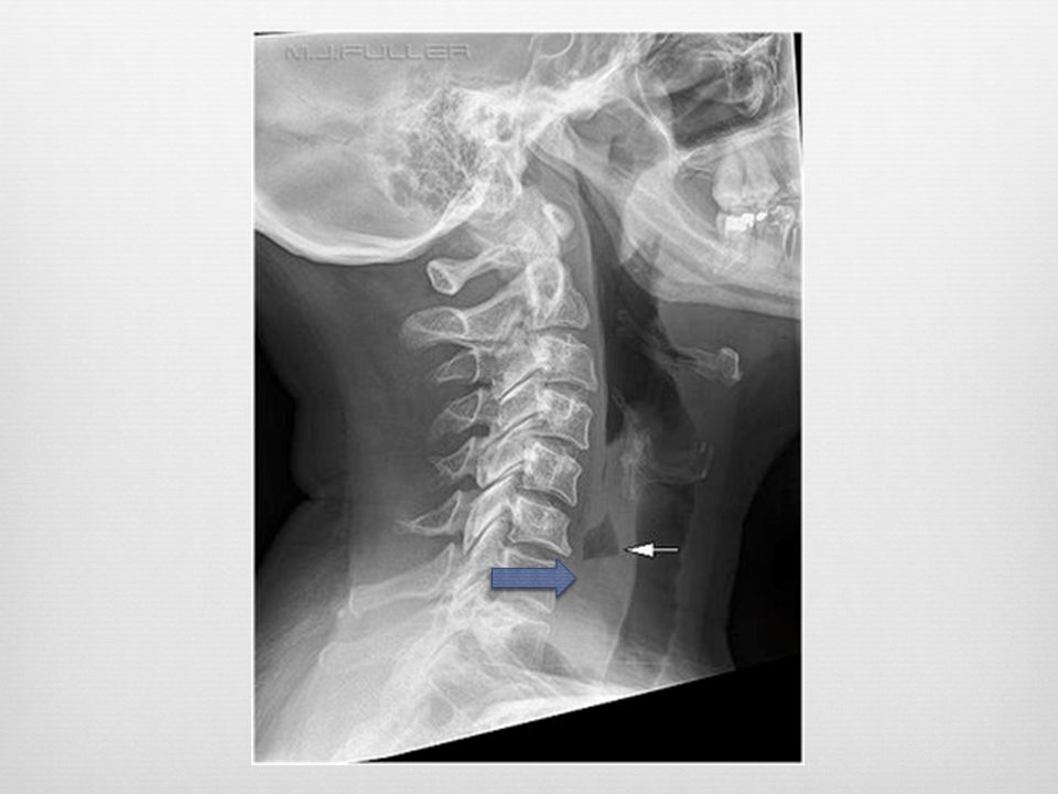
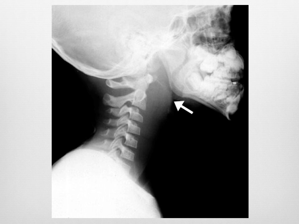
Lateral neck radiograph demonstrating widening of the retropharyngeal space and reversal of the normal cervical spine curvature. The retropharyngeal space is considered widened if it is greater than 7 mm at C2 or 14 mm at C6. The epiglottis and sub-glottic area in this radiograph are normal Longus colli

14
Widened retropharyngeal space
C2 – 7mm or C6 – 14mm (kids) C6 – 22mm (adults) Reversal of normal cervical lordosis Foreign body Air-fluid level Gas
 C6 – 22mm (adults) Reversal of normal cervical lordosis. Foreign body. Air-fluid level. Gas.")
15
Retropharyngeal Infection
Widened retropharyngeal space C2 – 7mm or C6 – 14mm (kids) C6 – 22mm (adults) Reversal of normal cervical lordosis Foreign body Air-fluid level Gas Give me 5 signs of retropharyngeal infection on lateral XR Longus colli
 C6 – 22mm (adults) Reversal of normal cervical lordosis. Foreign body. Air-fluid level. Gas. Give me 5 signs of retropharyngeal infection on lateral XR. Longus colli.")
17
False Pre-vertebral Swelling
Oblique lateral Neck flexion Crying Give me 2 false positive scenarios True lateral Neck in slight extension – neutral expiration

18
Case 1 4 year old male Inspiratory stridor Unwell for past 24 hours
Mom found him in his room with increased work of breathing

20
Tracheal membranes irregularity, clouding
Clinical: toxic fevor stridor Staph aureus

21
Case 1 3 year old male Inspiratory stridor Unwell for past 24 hours
Mom found him in his room with increased work of breathing

22
Barky cough Croup – subglottic narrowing and distended hypopharynx “ballooning” of pharynx Subglottic edema

23
Steeple sign

24
Clinical: 6-24 months Influenza, parainfluenza

25
Case 1 4 year old male Inspiratory stridor Unwell for past 24 hours
Mom found him in his room with increased work of breathing

27
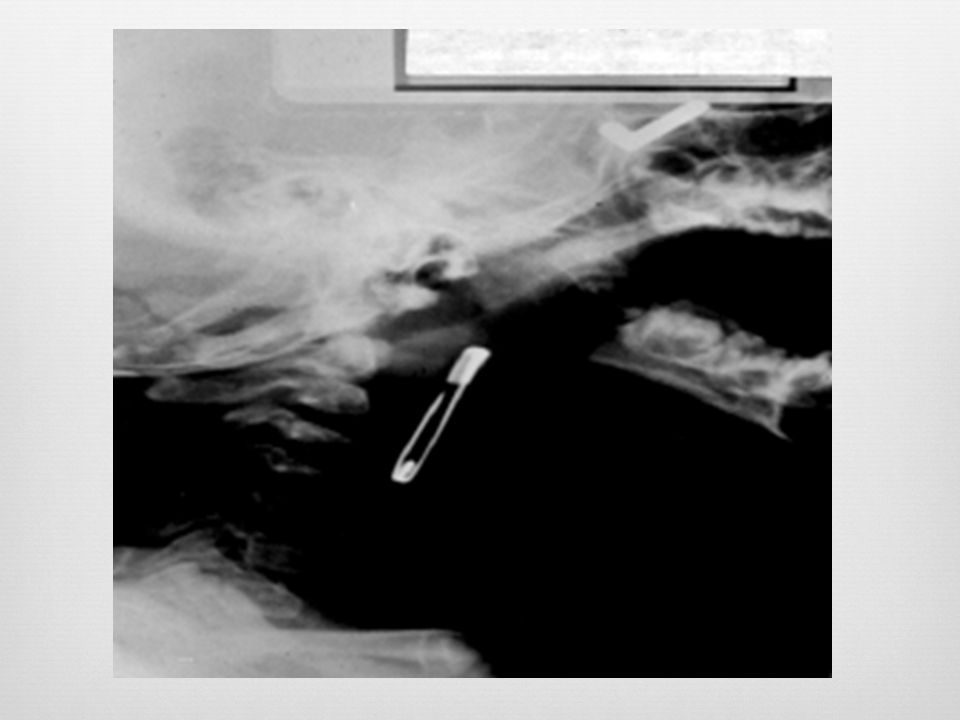
Foreign Body Visualization of radiopaque FB
Widened pre-vertebral shadow Loss of lordosis Location Esophagus – coronal plane Trachea – sagittal plane (best seen on lateral) Longus colli
 Longus colli.")
28
Chickenbone air

31
Test Characteristics Sensitivity Specificity Epiglottitis 33-88% 78%
Retropharyngeal abscess 80% 100% Croup Foreign Body 84%

32
Questions? Thank You!

34
Triple blinded prospective study 100 elective surgery patients
Lateral neck x-ray compared with Mallampati Anesthesiologist gold standard (grade III or IV) Results Mallampati sens: 26% spec: 100% Lateral neck film: sens: 100% spec:100%
 Results. Mallampati sens: 26% spec: 100% Lateral neck film: sens: 100% spec:100%")
35
h – middle of hyoid bone e – highest point of epiglottis t – thyroid cartilage a – posterior surface of arytenoid bone A line from point h and parallel to TA was drawn (HE) and the angle formed between HE and HE’ was called B’. HEE’ is triangle of safety.
 and the angle formed between HE and HE’ was called B’. HEE’ is triangle of safety.")
37
Chicken bone at c7 – retropharyngeal air

39
Croup – steeple sign …may vary with respiration even in normal kids

40
Enlarged epiglottis Loss of valecular airspace Thickened aryepiglotic folds Distended hypopharynx Straightening of the cervical spine

41
Adult Epiglottitis Radiographic Criteria
Epiglottic height-to-width ratio >0.6 Epiglottic to C4 vertebral body width ratio >0.33 Aryepiglottic fold to C3 vertebral body width ratio >0.35 Prevertebral soft-tissue to C4 vertebral body width ratio >0.25 Hypopharyngeal airway to C4 vertebral body width ratio >1.5

Similar presentations


>")


 Motion Controller Design for A Class of Second-order Systems Center for Self-Organizing Intelligent.>")


