Download presentation
Presentation is loading. Please wait.

1
The Lower Limb

2
Lower limb The lower limb has six major parts or regions
Gluteal region Thigh Knee Leg Ankle Foot

6
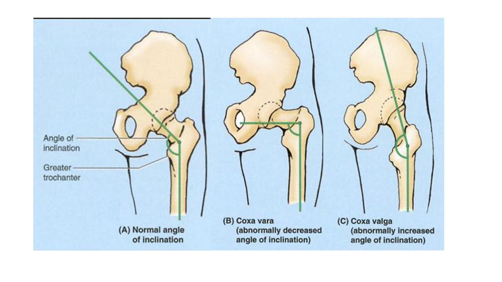
The angle of inclination & torsion
Through evolution and development, our largest bone, the femur, has developed a bend (angle of inclination) and has twisted (medial rotation and torsion so that the knee and all joints inferior to it flex posteriorly) to accommodate our erect posture and to enable bipedal walking and running. This obtuse angle of inclination is greatest (most nearly straight) at birth and gradually diminishes (becomes more acute) until the adult angle is reached. The angle is less in females because of the increased width between the acetabula (a consequence of a wider lesser pelvis) and the greater obliquity of the shaft. The angle of inclination allows greater mobility of the femur at the hip joint because it places the head and neck more perpendicular to the acetabulum in the neutral position.
 and has twisted (medial rotation and torsion so that the knee and all joints inferior to it flex posteriorly) to accommodate our erect posture and to enable bipedal walking and running. This obtuse angle of inclination is greatest (most nearly straight) at birth and gradually diminishes (becomes more acute) until the adult angle is reached. The angle is less in females because of the increased width between the acetabula (a consequence of a wider lesser pelvis) and the greater obliquity of the shaft. The angle of inclination allows greater mobility of the femur at the hip joint because it places the head and neck more perpendicular to the acetabulum in the neutral position.")
7
…torsion & inclination

9
Fascia, Vessels, and Cutaneous Nerves of the Lower Limb
The subcutaneous tissue (superficial fascia) lies deep to the skin and consists of loose connective tissue that contains a variable amount of fat, cutaneous nerves, superficial veins (great and small saphenous veins and their tributaries), lymphatic vessels, and lymph nodes. The deep fascia of the thigh is called fascia lata (L. lata, broad). It is continues inferior to the knee as the deep fascia of the leg The fascia lata is substantial because it encloses the large thigh muscles, especially laterally where it is thickened and strengthened by additional reinforcing longitudinal fibers to form the iliotibial tract
 lies deep to the skin and consists of loose connective tissue that contains a variable amount of fat, cutaneous nerves, superficial veins (great and small saphenous veins and their tributaries), lymphatic vessels, and lymph nodes. The deep fascia of the thigh is called fascia lata (L. lata, broad). It is continues inferior to the knee as the deep fascia of the leg. The fascia lata is substantial because it encloses the large thigh muscles, especially laterally where it is thickened and strengthened by additional reinforcing longitudinal fibers to form the iliotibial tract.")
11
Cutaneous Innervation of the Lower Limb
The areas of skin supplied by the individual spinal nerves, including those contributing to the plexuses, are called dermatomes. The dermatomal (segmental) pattern of skin innervation is retained throughout life but is distorted by limb lengthening and the torsion of the limb that occurs during development
 pattern of skin innervation is retained throughout life but is distorted by limb lengthening and the torsion of the limb that occurs during development.")
14
The Gluteal region

20
The abductor muscles of hip

21
The rotator muscles of hip

22
The flexor muscles of hip
Lumbar plexus via anterior branches of L1-L3 nerves femoral nerve and direct branches from the lumbar plexus

23
Quadriceps femoris Anterior group Sartorius Pectineus Adductor longus Adductor brevis Gracilis Adductor magnus The muscle of thigh Medial group Biceps femoris posterior group Semitendinosus semimembranosus

24
Quadriceps Femoris The quadriceps femoris (L. four-headed femoral muscle) forms the main bulk of the anterior thigh muscles and collectively constitutes the largest and one of the most powerful muscles in the body. It may be three times stronger than its antagonistic muscle group, the hamstrings
 forms the main bulk of the anterior thigh muscles and collectively constitutes the largest and one of the most powerful muscles in the body. It may be three times stronger than its antagonistic muscle group, the hamstrings.")
26
The Hamstring muscles The Hamstring muscles comprised of three separate muscles: the Biceps Femoris, Semitendinosus and Semimembranosus

27
The Hamstring muscles The Hamstring muscles comprised of three separate muscles: the Biceps Femoris, Semitendinosus and Semimembranosus

29
Adductor muscles

31
Femoral Sheath The femoral sheath is a funnel-shaped fascial tube of varying length that passes deep to the inguinal ligament, lining the vascular lacuna of the subinguinal space The sheath is formed by an inferior prolongation of transversalis and iliopsoas fascia from the abdomen/greater pelvis. The compartments of the femoral sheath are the: Lateral compartment for the femoral artery. Intermediate compartment for the femoral vein. Medial compartment, which constitutes the femoral canal.

33
Femoral hernia
34
The femoral triangle & adductor canal
The adductor canal (subsartorial canal; Hunter canal) is a long (approximately 15 cm), narrow passageway in the middle third of the thigh. It extends from the apex of the femoral triangle, where the sartorius crosses over the adductor longus, to the adductor hiatus in the tendon of the adductor magnus. The adductor canal provides an intermuscular passage for the femoral artery and vein, the saphenous nerve, and the nerve to vastus medialis, delivering the femoral vessels to the popliteal fossa where they become the popliteal vessels.
 is a long (approximately 15 cm), narrow passageway in the middle third of the thigh. It extends from the apex of the femoral triangle, where the sartorius crosses over the adductor longus, to the adductor hiatus in the tendon of the adductor magnus. The adductor canal provides an intermuscular passage for the femoral artery and vein, the saphenous nerve, and the nerve to vastus medialis, delivering the femoral vessels to the popliteal fossa where they become the popliteal vessels.")
39
Popliteal fossa & its contents

Similar presentations