Download presentation
Presentation is loading. Please wait.

1
Infectious diseases of Respiratory tract

2
Microbial Diseases of the Upper Respiratory System
Laryngitis: S. pneumoniae, S. pyogenes, viruses Tonsillitis: S. pneumoniae, S. pyogenes, viruses Sinusitis: Bacteria Epiglottitis: H. influenzae

3
Upper respiratory normal microbiota may include pathogens.

4
Streptococcal pharyngitis (Strep throat)
Causative organism: group A streptococci. This group consists solely of Streptococcus pyogenes. The pathogenicity of GAS is enhanced by their resistance to phagocytosis. They are also able to produce special enzymes, called Streptokinases, which lyse fibrin clots, and Streptolysins which are cytotoxic to tissue cells, red blood cells, and protective leukocytes.

5
Streptococcal pharyngitis (Strep throat)
Pharyngitis is characterized by local inflammation and fever . Frequently, tonsillitis occurs, and the lymph nodes in the neck become enlarged and tender. Another frequent complication is otitis media. Transmission: Pharyngitis is now most commonly transmitted by respiratory secretions, but epidemics of streptococcal pharyngitis spread by unpasteurized milk were once frequent.

6
Scarlet fever Causative agent: Streptococcus pyogenes.
When the strain causing streptococcal pharyngitis produces an reddening toxin the resulting infection is called scarlet fever. When the strain produces an erythrogenic (reddening) toxin, it has been lysogenized by a bacteriophage that means the genetic information of a bacteriophage (bacterial virus) has been incorporated into the chromosome of the bacterium, so the characteristics of the bacterium have been altered. The toxin causes a pinkish red skin rash, which is probably the skin's hypersensitivity reaction to the circulating toxin, and a high fever. The tongue has a spotted, strawberry-like appearance and then, as it loses upper membrane, becomes very red and enlarged. Classically, scarlet fever has been considered to be associated with streptococcal pharyngitis, but it might accompany a streptococcal skin infection. The incidence of scarlet fever has varied over time in severity.
 toxin, it has been lysogenized by a bacteriophage that means the genetic information of a bacteriophage (bacterial virus) has been incorporated into the chromosome of the bacterium, so the characteristics of the bacterium have been altered. The toxin causes a pinkish red skin rash, which is probably the skin s hypersensitivity reaction to the circulating toxin, and a high fever. The tongue has a spotted, strawberry-like appearance and then, as it loses upper membrane, becomes very red and enlarged. Classically, scarlet fever has been considered to be associated with streptococcal pharyngitis, but it might accompany a streptococcal skin infection. The incidence of scarlet fever has varied over time in severity.")
7
Scarlet fever

8
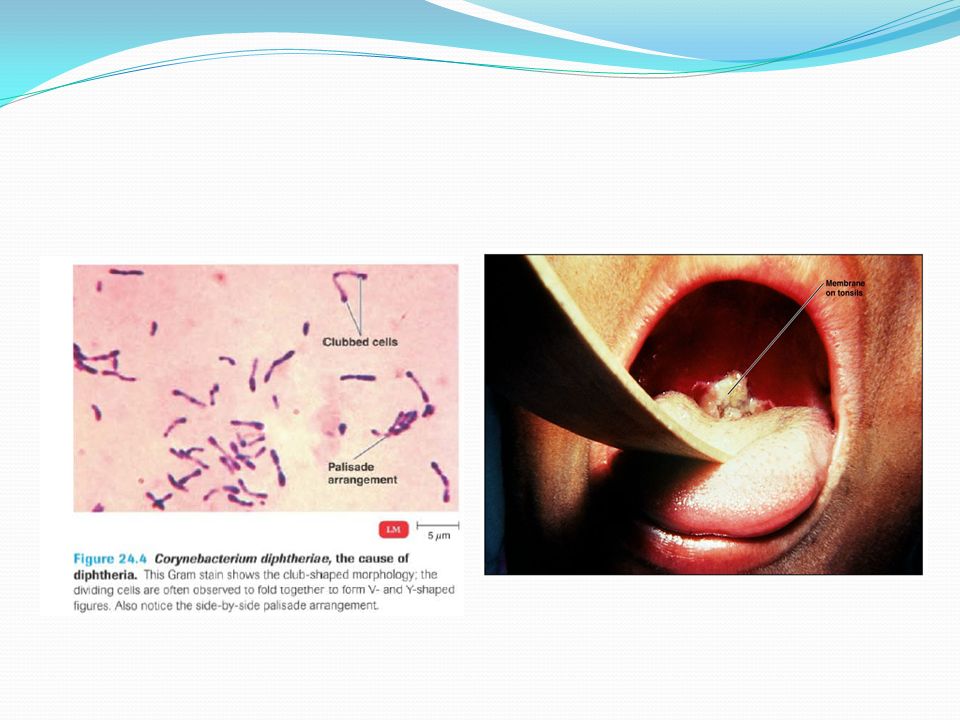
Diptheria Causative agent: Corynebacterium diptheriae
Morphology and cultural characteristics: They are Gram-ve, rods Non motile and non spore forming Characteristically they possess irregular swelling at one end that give them a characteristic ‘Club-shaped” appearance They are aerobic and pleomorphic in microscopy.

10
In nature, Corynebacterium diptheriae occurs in the respiratory tract, or on the skin, in wounds of infected persons. It is spread by droplets or by contact to susceptible individuals. The bacilli then grow on mucus membranes or in the skin abrasions and those that are toxigenic start producing toxin. Although the bacteria do not invade tissues, those that have been lysogenized by a phage, produce a powerful exotoxin.

11
Diptheria toxin This toxin is a heat-labile polypeptide (MW 62000) that can be lethal in a dose of 0.1µg/kg. If the disulfide bond is broken, the molecule can be split into two fragments. Fragment B (MW 38000) has no independent activity but is required for the transport of fragment A into the cell. Fragment A inhibits polypeptide chain Elongation –provided NAD is present-by inactivating elongation factor EF-2. This factor is required for translocation of polypeptidyl-transfer RNA from the acceptor to the donor site on the eukaryotic ribosome. Toxin fragment A inactivates EF-2 by catalyzing a reaction that yields free Nicotinamide plus an adenine diphosphate-ribose-EF-2 complex. It is assumed that abrupt arrest of protein synthesis is responsible for the necrotizing and neurotoxic effects of diptheria toxin.
 has no independent activity but is required for the transport of fragment A into the cell. Fragment A inhibits polypeptide chain Elongation –provided NAD is present-by inactivating elongation factor EF-2. This factor is required for translocation of polypeptidyl-transfer RNA from the acceptor to the donor site on the eukaryotic ribosome. Toxin fragment A inactivates EF-2 by catalyzing a reaction that yields free Nicotinamide plus an adenine diphosphate-ribose-EF-2 complex. It is assumed that abrupt arrest of protein synthesis is responsible for the necrotizing and neurotoxic effects of diptheria toxin.")
12
Diptheria toxin (AB toxin)
B subunit: Binds to host cell - Delivers A subunit to cytoplasm - Often five B subunits form a pore for A entry. Figure 25.17A A subunit: Has toxic activity - ADP-ribosyltransferase - Diphtheria toxin - Cholera toxin Figure 25.17B 12

14
Diptheria toxin mechanism

15
Pathology Diptheria toxin absorbed into mucus membrane and causes destruction of epithelium and a superficial inflammatory response. The necrotic epithelium becomes embedde in exuding fibrin and redand white cells, so that a grayish “ Pseudomembrane” is formed commonly over tonsils, pharynx or larynx. Any attempt to remove the Pseudomembrane exposes and tear the capillaries and thus result in bleeding.

16
Clinical findings of diptheria
When diptheria inflammation begins in the respiratory tract, sore throat and fever usually develops. Pseudomembrane that forms in the throat can cause the obstruction of the passage of air to the lungs. This obstruction may even cause suffocation if not promptly relieved by intubation or tracheostomy. Irregularities of cardiac rhythm indicate damage to the heart. Later, there may be difficulties in swallowing, speech.

17
Diptheria Transmission:
It is spread by droplets or by contact to susceptible individuals. Prevention: Active immunization in childhood with diptheria toxoid. Part of the normal immunization program for children in the United States is the DTaP vaccine. The D stands for diphtheria toxoid, an inactivated toxin that causes the body to produce antibodies against the diphtheria toxin . Treatment: Antimicrobial drugs Antibiotics e.g. penicillin control the growth of bacteria, they do not neutralize the toxin. Thus antibiotics should be used only in conjunction with antitoxin.

18
Lower respiratory tract of Human

19
Bacterial diseases of Lower rerspiratory tract:
i) Tuberculosis ii) Pertussis iii) Pneumonia
 Tuberculosis. ii) Pertussis. iii) Pneumonia.")
20
Tuberculosis Causative agent: Mycobacterium tuberculosis
Morphological and cultural properties: In tissue, tubercle bacilli are thin straight rods measuring about .4 X3 µm The rods grow slowly (20 hours or longer generation time). Sometimes form filamentous or tend to grow in clumps. Can not be classified as either G+ve or G-ve. They are called acid fast bacilli. They are obligate aerobes and derive energy from the oxidation of many simple carbon compounds. tubercle bacilli are resistant to drying and survive for long periods in dried sputum.
. Sometimes form filamentous or tend to grow in clumps. Can not be classified as either G+ve or G-ve. They are called acid fast bacilli. They are obligate aerobes and derive energy from the oxidation of many simple carbon compounds. tubercle bacilli are resistant to drying and survive for long periods in dried sputum.")
22
Pathogenesis of tuberculosis

23
Pathogenesis of tuberculosis
If the infection progresses, the host isolates the pathogens in a walled off lesion called a tubercle (meaning a lump or knob), a disease that gives the disease its name. When the disease is arrested at this point, the lesions slowly heal, becoming calcified. These show up clearly on X-ray films and are called Ghon complexes. If the body’s defense fail at this stage, the tubercle breaks down and releases virulent bacilli into the airways of the lungs and then the cardiovascular and lymphatic systems.
, a disease that gives the disease its name. When the disease is arrested at this point, the lesions slowly heal, becoming calcified. These show up clearly on X-ray films and are called Ghon complexes. If the body’s defense fail at this stage, the tubercle breaks down and releases virulent bacilli into the airways of the lungs and then the cardiovascular and lymphatic systems.")
24
Clinical finding: Fatigue, weakness, weight loss and fever Pulmonary involvement giving rise to chronic cough and splitting of blood usually is associated with far-advanced lesions. Bloodstream dissemination leads to miliary tuberculosis with lesions in many organs and high a mortality rate.

25
Prevention of Tuberculosis:
The BCG vaccine is a live culture of Mycobacterium bovis (pathogen mainly of cattle, cause of bovine tuberculosis) that has been made avirulent by long cultivation on artificial media. Mycobacterium bovis is transmitted to humans via contaminated milk or food. BCG stands for bacillus of Calmette and Guerin, the people who originally isolated the strain. The BCG vaccine has been available since the 1920s and is one of the most widely used vaccines in the world. In 1990, it was estimated that 70% of the world's school children received it.
 that has been made avirulent by long cultivation on artificial media. Mycobacterium bovis is transmitted to humans via contaminated milk or food. BCG stands for bacillus of Calmette and Guerin, the people who originally isolated the strain. The BCG vaccine has been available since the 1920s and is one of the most widely used vaccines in the world. In 1990, it was estimated that 70% of the world s school children received it.")
26
Pertussis (Whooping Cough)
Bordetella pertussis: Gram-negative coccobacillus Virulent strains posses a capsule Tracheal cytotoxin of cell wall damaged ciliated cells Pertussis toxin Prevented by DTaP vaccine (acellular Pertussis cell fragments) Figure 24.8
 Figure")
27
Pertussis (Whooping Cough)
The bacteria attach specifically to ciliated cells in the trachea, first impeding their ciliary action and then progressively destroying the cells. This prevents the ciliary escalator system from moving mucus. B. pertussis produces several toxins. Tracheal cytotoxin, a fixed cell wall fraction of the bacterium, is responsible for damage to the ciliated cells, and pertussis toxin enters the bloodstream and is associated with systemic symptoms of the disease.

28
Primarily a childhood disease, pertussis can be quite severe.
The initial stage, called the catarrhal stage, resembles a common cold. Prolonged sieges of coughing characterize the paroxysmal stage, or second stage. (The name pertussis is derived from the Latin per, meaning thoroughly, and tussis, meaning cough.) When ciliary action is compromised, mucus accumulates, and the infected person desperately attempts to cough up these mucus accumulations. The violence of the coughing in small children can actually result in broken ribs. Gasping for air between coughs causes a whooping sound, hence the informal name of the disease. Coughing episodes occur several times a day for 1 to 6 weeks The convalescence stage, the third stage, may last for months.
 When ciliary action is compromised, mucus accumulates, and the infected person desperately attempts to cough up these mucus accumulations. The violence of the coughing in small children can actually result in broken ribs. Gasping for air between coughs causes a whooping sound, hence the informal name of the disease. Coughing episodes occur several times a day for 1 to 6 weeks. The convalescence stage, the third stage, may last for months.")
29
Bacterial pneumonia The term Pneumonia is applied to most of which are caused by bacteria. Pneumonia caused by Streptococcus pneumoniae is the most common, about two-thirds of cases, and is therefore referred to Typical pneumonia. Pneumonias caused by other microorganisms, which can include fungi, protozoa, viruses, and other bacteria, are termed atypical pneumonia

30
Pnemococcal Pneumonia
Pneumonias also are named after the portions of the lower respiratory tract they affect. For example, if the lobes of the lungs are infected, it is called lobar pneumonia. pneumonias caused by S. pneumoniae are usually of this type. Bronchopneumonia indicated the alveoli of the lungs adjacent to the bronchi are infected. Pleurisy is often a complication of various pneumonias, in which the pleural membranes become painfully inflamed.

31
Pnemococcal Pneumonia
Pneumonia caused by S.pneumoniae is called pneumococcal pneumonia. S. pneumoniae is a gram-positive, ovoid bacterium. The cell pairs are surrounded by a dense capsule that makes the pathogen resistant to phagocytosis.

32
Pneumococcal Pnumonia
Symptoms: High fever, breathing difficulty, and chest pain. (Atypical pneumonias usually have a slower onset and less fever and chest pain.) The lungs have a reddish appearance because blood vessels are dilated. In response to the infection, alveoli fill with some red blood cells, neutrophils and fluid from surrounding tissues. The sputum is often rust-colored from blood coughed up from the lungs. Pneumococci can invade the blood stream, the pleural cavity surrounding the lung, and occasionally the meninges.
 The lungs have a reddish appearance because blood vessels are dilated. In response to the infection, alveoli fill with some red blood cells, neutrophils and fluid from surrounding tissues. The sputum is often rust-colored from blood coughed up from the lungs. Pneumococci can invade the blood stream, the pleural cavity surrounding the lung, and occasionally the meninges.")
33
Rust-colored sputum

34
Pneumococcal Pnumonia
Diagnosis: A presumptive diagnosis can be made by isolating the pneumococci from the throat, sputum, and other fluids. Pneumococci can be distinguished from other alpha-hemolytic streptococci by observing the inhibition of growth next to a disk of optochin (ethylhydrocuprcine hydrochloride). They can also be serologically typed.
. They can also be serologically typed.")
35
Viral disease of Lower respiratory tract
Viral Pneumonia Influenza

36
Viral Pneumonia Viral pneumonia as a complication of influenza, measles, chickenpox Viral etiology suspected if no cause determined Respiratory Syncytial Virus (RSV) Common in infants; 4500 deaths annually Symptoms: coughing Diagnosis by serologic test for viruses and antibodies Treatment: Ribavirin
 Common in infants; 4500 deaths annually. Symptoms: coughing. Diagnosis by serologic test for viruses and antibodies. Treatment: Ribavirin.")
37
Influenza Viruses in the genus Influenza consist of eight separate RNA segments of differing lengths enclosed by an inner layer of protein and an outer lipid bilayer. Embedded in the lipid bilayer are numerous projections that characterize the virus. There are two types of projections: Hemagglutinin (H) spikes used for attachment to host cells. Neuraminidase (N) spikes used to release virus from cell.
 spikes used for attachment to host cells. Neuraminidase (N) spikes used to release virus from cell.")
38
Hemagglutinin spikes The HA spikes, of which there are about 500 on each virus, allow the virus to recognize and attach to body cells before infecting them. Antibodies against the influenza virus are directed mainly at these spikes. The term Hemagglutinin refers to the agglutination of red blood cells (hemagglutination) that occurs when the viruses are mixed with them.
 that occurs when the viruses are mixed with them.")
39
Neuraminidase Spikes The NA spikes, of which there are about 100 per virus, differ from the HA spikes in appearance and function. Apparently they enzymatically help the virus separate from the infected cell as the virus exits after intracellular reproduction. NA spikes also stimulate the formation of antibodies, but these are less important in the body's resistance to the disease than those produced in response to the HA spikes.

40
Influenza

41
Influenza Antigenic Shift Antigenic drift

42
Influenza Influenza viruses are also classified into groups according to the antigens of their protein coats. These groups are A, B, and (rarely) C. The A-type viruses are responsible for the major pandemics. The B-type virus also circulates and mutates, but it is usually responsible for more geographically limited and milder infections.
 C. The A-type viruses are responsible for the major pandemics. The B-type virus also circulates and mutates, but it is usually responsible for more geographically limited and milder infections.")
43
Avian Influenza

44
Influenza A Virus Negative sense RNA Single stranded Segmented
16 Hemagglutinin subtypes 9 Neuraminidase Subtypes

46
Influenza Subtypes 16 Hemagglutinin subtypes 9 Neuraminidase subtypes
2 Nonstructural subtypes Can occur in any combination Useful for epidemiology

47
Avian Influenza: Infection and Disease
Infection may cause a wide range of clinical signs from no disease (asymptomatic), respiratory disease, to severe disease with high mortality Localized Infection-mild to moderate disease Intestinal-wild ducks and shorebirds, poultry Respiratory-humans, swine, horses, poultry, domestic ducks, seal, mink Systemic Infection-high mortality chickens, turkeys, other gallinaceous birds
, respiratory disease, to severe disease with high mortality. Localized Infection-mild to moderate disease. Intestinal-wild ducks and shorebirds, poultry. Respiratory-humans, swine, horses, poultry, domestic ducks, seal, mink. Systemic Infection-high mortality. chickens, turkeys, other gallinaceous birds.")
48
Highly Pathogenic Avian Influenza
Systemic, rapidly fatal disease of poultry Only H5 and H7 subtypes are recognized to cause HPAI OIE List A Disease-outbreaks are reportable HA cleavage site critical virulence factor Low pathogenic H5 and H7 AI viruses can mutate into the highly pathogenic form of the virus

49
H5 Hemagglutinin Cleavage Site
For H5 LPAI waterfowl viruses, the consensus cleavage site sequence is Arg Glu Thr Arg/ Gly Most H5 HPAI viruses have additional basic amino acids at cleavage site Mexico Arg Lys Arg Lys Thr Arg/ Gly Hong Kong 1997 Arg Glu Arg Arg Arg Lys Lys Arg/Gly The loss of a glycosylation site was also important in the emergence of HPAI in Pennsylvania in 1983 LPAI PA/83 Lys Lys Lys Arg/ Gly + glycosylation at 11-13 HPAI PA/83 Lys Lys Lys Arg/ Gly - glycosylation at 11-13

50
Methods of Control Stamping out-identify infected flocks and destroy them to prevent spread to other flocks Vaccination in conjunction with stamping out Vaccination only

Similar presentations


. The genus haemophilus organisms.>")


 Virus>")


