Download presentation
Presentation is loading. Please wait.

1
Left Ventricular Filling Pressure by Doppler Echocardiography in Patients With End-Stage Renal Disease Angela Y-M Wang, Mei Wang, Christopher W-K Lam, Iris H-S Chan, Yan Zhang, John E. Sanderson Hypertension 2008 ; July : 107-114 R2 손주웅

2
Background Cardiovascular disease leading cause of death in ESRD patients accelerated atherosclerosis and LVH Starting long-term dialysis therapy LVH was already present in three quarters LV dilatation and systolic dysfunction. important predictors of outcome

3
Background LV filling pressure noninvasively using echocardiography important value for prognostication The ratio of early transmitral flow velocity (E) to early diastolic mitral annular velocity (EM) : E/Em ratio using tissue doppler imaging more accurate than other methods for estimating LV filling pressure
 to early diastolic mitral annular velocity (EM) : E/Em ratio using tissue doppler imaging more accurate than other methods for estimating LV filling pressure")
4
Background The objective of this study The estimation of the E/Em ratio using tissue doppler imaging adds significant long-term prognostic value Standard clinical, biochemical, dialysis, echocardiographic parameters

5
Methods Study design and subjects 4-year prospective study 220 stable patients with ESRD on CAPD Continuous PD for > 3 months Exclusion criteria Severe valvular disease Hypertrophic cardiomyopathy Underlying malignancy Chronic liver disease SLE Chronic rheumatic heart disease CHF

6
Methods Clinical and Demographic Data Atherosclerotic vascular disease Presence of coronary artery disease History of angina, previous MI CVA peripheral vascular disease Systolic and diastolic blood pressures Measured on every follow-up

7
Methods Echocardiography and Tissue Doppler Imaging LV ejection fraction Midwall fractional shortening LV mass Mitral inflow velocities Myocardial velocities

8
Methods Outcome Measures Death from all causes Cardiovascular death Definite myocardial ischemic event Heart failure Cerebrovascular event Arrhythmia Peripheral vascular accident

9
Results

12
During the 48 month follow-up 84 patients had died 53 deaths(63%) were of cardiovascular causes 31 deaths(37%) were of noncardiovascular causes E/Em ratio was highest among patients who died from cardiovascular causes.
 were of cardiovascular causes 31 deaths(37%) were of noncardiovascular causes E/Em ratio was highest among patients who died from cardiovascular causes.")
13
Results

16
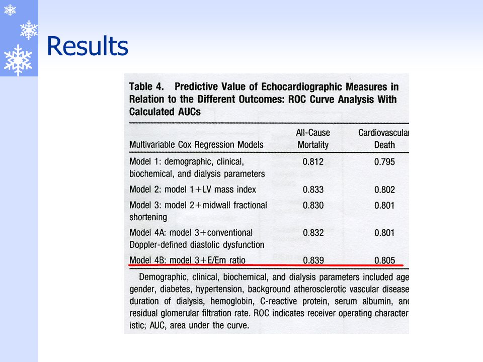
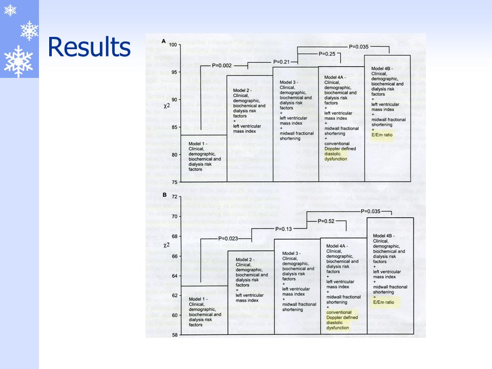
Discussion In this study >60% of our patients with ESRD had elevated LV filling pressure E/Em independent and additional prognostic value for long-term mortality and cardiovascular death single best Doppler predictor of elevated LV filling pressure powerful independent predictor of all-cause mortality

17
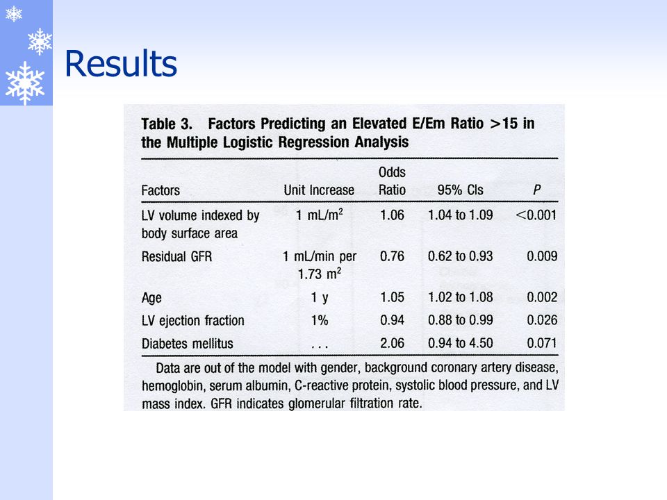
Discussion E/Em ratio of > 15 predicting LV end-diastolic pressure >15 mmHg sensitivity : 82% specificity : 88% reliable marker of increased LV filling pressure

18
Discussion E/Em ratio more significant predictor of long-term mortality and cardiovascular death compare with mwFS Higher E/Em ratio indicating a higher LV diastolic filling pressure associated with greater LV hypertrophy and dilatation and more systolic dysfunction

19
Discussion E/Em ratio > 15 greater prevalence of diabetes, atherosclerotic vascular disease and heart failure, higher systolic blood pressure, greater inflammation, more anemia and hypoalbuminemia and lower residual renal function LV hypertrophy, reduced aortic distensibility, diabetes, and coronary artery disease all contribute to an increased LV filling pressure in ESRD Higher E/Em ratio was associated with a worse echocardiographic profile E/Em ratio as surrogate marker of LV filling pressure

20
Discussion Limitation only prevalent dialysis patients were included,which may introduce survival bias all of the parameters were measured at a single time point and did not reflect changes over time invasive measurement of LV filling pressure was not performed volume status was not directly assessed

21
Conclusion the incremental value of measuring E/Em ratio an estimate of LV filling pressure standard clinical, biochemical, and dialysis risk factors and conventional echocardiographic and Doppler-derived measures LV filling pressure should be estimated during echocardiographic examination for additional prognostication in patients with ESRD.

Similar presentations


 For Broker/Dealer Use Only.>")

