Download presentation
Presentation is loading. Please wait.

1
DEMO - IV DEMO - IV (Thigh and Gluteal Regions) Ali Jassim Alhashli Year IV – Unit VII – Musculoskeletal System
 Ali Jassim Alhashli Year IV – Unit VII – Musculoskeletal System")
2
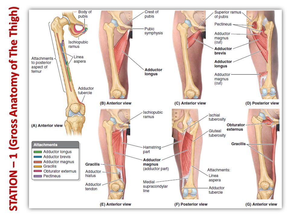
STATION – 1 (Gross Anatomy of The Thigh) Muscles of the thigh: Muscles of the thigh: – Anterior thigh muscles (all of them are innervated by the femoral nerve L2-L4 except for psoas major which is innervated by the lumbar nerve): Pectineus: extending from the superior ramus of the pubis to the pectineal line on the femur. It causes: adduction, flexion and medial rotation of the thigh. Sartorius ( العضلة الخياطة ): extending from the anterior superior iliac spine to the supero-medial aspect of the tibia. It causes abduction, flexion and lateral rotation of the thigh. Iliopsoas muscle which is composed of: – Psoas major: extending from T12-L5 vertebrae to the lesser trochanter of the femur. – Iliacus: extending from iliac crest and iliac fossa and joining the tendon of psoas major to the lesser trochanter of the femur. Note: both of them will cause flexion of the thigh and maintain posture. Psoas major will cause lumbar lordosis and compensatory thoracic kyphosis of the vertebral column during standing. Pectineus
: extending from the anterior superior iliac spine to the supero-medial aspect of the tibia. It causes abduction, flexion and lateral rotation of the thigh. Iliopsoas muscle which is composed of: – Psoas major: extending from T12-L5 vertebrae to the lesser trochanter of the femur. – Iliacus: extending from iliac crest and iliac fossa and joining the tendon of psoas major to the lesser trochanter of the femur. Note: both of them will cause flexion of the thigh and maintain posture. Psoas major will cause lumbar lordosis and compensatory thoracic kyphosis of the vertebral column during standing. Pectineus.")
3
Muscles of the thigh: Muscles of the thigh: – Anterior thigh muscles (all of them are innervated by the femoral nerve L2-L4 except for psoas major which is innervated by the lumbar nerve): Quadriceps femoris: – Rectus femoris: originating from the anterior inferior iliac spine. It causes flexion of the thigh and extension of the leg at knee joint. Its ability to extend the knee is limited when the thigh is flexed. – Vastus lateralis: originating from the greater trochanter and lateral lip of linea aspera. – Vastus medialis: originating from intertrochanteric line and medial lip of linea aspera. – Vastus intermedius: originating from anterior and lateral aspects of the femur bone. – Note: all of these 4 muscles will merge to form the quadriceps tendon in which the patella will be embedded and then will continue as the patellar ligament to inset in the tibial tuberosity. STATION – 1 (Gross Anatomy of The Thigh)
.")
4
Muscles of the thigh: Muscles of the thigh: – Medial thigh muscles (they are adductor muscles causing adduction of the thigh. All of them are innervated by obturator nerve except for the hamstring part of adductor magnus): Adductor longus: extending from body of pubis to the middle third of linea aspera of the femur. Adductor brevis: found deep to pectineus and adductor longus muscles. Extending from the body and inferior ramus of pubis to the pectineal line (same insertion of pectineus muscle). Adductor magnus: with 2 parts (adductor part which is innervated by obturator nerve and hamstring part which is innervated by the tibial division of sciatic nerve). Note: the hamstring part is inserted in the adductor tubercle in the distal end of the femur. Gracilis: because of its minor function it can be removed and used in transplantation surgeries. It extends from the body and inferior ramus of pubis to the supero-medial aspect of the tibia (same insertion of sartorius muscle). Obturator externus. STATION – 1 (Gross Anatomy of The Thigh)
: Adductor longus: extending from body of pubis to the middle third of linea aspera of the femur. Adductor brevis: found deep to pectineus and adductor longus muscles. Extending from the body and inferior ramus of pubis to the pectineal line (same insertion of pectineus muscle). Adductor magnus: with 2 parts (adductor part which is innervated by obturator nerve and hamstring part which is innervated by the tibial division of sciatic nerve). Note: the hamstring part is inserted in the adductor tubercle in the distal end of the femur. Gracilis: because of its minor function it can be removed and used in transplantation surgeries. It extends from the body and inferior ramus of pubis to the supero-medial aspect of the tibia (same insertion of sartorius muscle). Obturator externus. STATION – 1 (Gross Anatomy of The Thigh).")
6
Muscles of the thigh: Muscles of the thigh: – Posterior thigh muscles (their main function is to extend the thigh and flex the knee. All of them are innervated by the tibial division of sciatic nerve): Biceps femoris: with its long and short heads. Note: the short head of biceps femoris is not considered a hamstring muscle because it only crosses the knee joint and it is innervated by the common fibular division of sciatic nerve. Semi-tendinosus. Semi-membranosus: has a groove for semi-tendinosus. Note: the ability of these muscles to flex the knee is limited when the thigh is fully extended (the opposite is true). Hamstring muscles originate from ischial tuberosity except for the short head of biceps femoris which originates from linea aspera and lateral supracondylar line of the femur. Femoral triangle: Femoral triangle: – Boundaries: Superior: inguinal ligament. Lateral: medial border of sartorius muscle. Medial: lateral border of adductor longus muscle. – Contents: Femoral artery: a continuation of external iliac artery → passing through the femoral triangle lateral to the femoral vein and surrounded by femoral sheath → giving a branch named “profunda artery of femur”. Note: femoral artery is best exposed in adductor canal in the middle third of the thigh. Femoral vein: passing through femoral triangle and surrounded by femoral sheath. Note: femoral canal is medial to femoral vein allowing it to expand when there is increased venous return. Femoral nerve (L2-L4): passing through femoral triangle outside the femoral sheath.
: Biceps femoris: with its long and short heads. Note: the short head of biceps femoris is not considered a hamstring muscle because it only crosses the knee joint and it is innervated by the common fibular division of sciatic nerve. Semi-tendinosus. Semi-membranosus: has a groove for semi-tendinosus. Note: the ability of these muscles to flex the knee is limited when the thigh is fully extended (the opposite is true). Hamstring muscles originate from ischial tuberosity except for the short head of biceps femoris which originates from linea aspera and lateral supracondylar line of the femur. Femoral triangle: Femoral triangle: – Boundaries: Superior: inguinal ligament. Lateral: medial border of sartorius muscle. Medial: lateral border of adductor longus muscle. – Contents: Femoral artery: a continuation of external iliac artery → passing through the femoral triangle lateral to the femoral vein and surrounded by femoral sheath → giving a branch named profunda artery of femur . Note: femoral artery is best exposed in adductor canal in the middle third of the thigh. Femoral vein: passing through femoral triangle and surrounded by femoral sheath. Note: femoral canal is medial to femoral vein allowing it to expand when there is increased venous return. Femoral nerve (L2-L4): passing through femoral triangle outside the femoral sheath..")
7
STATION – 1 (Gross Anatomy of The Thigh)
")
9
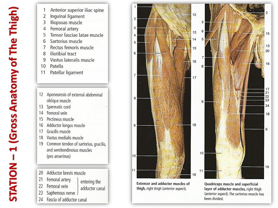
Psoas major Iliacus Inguinal ligament Femoral vein, artery & nerve Pectineus muscle Adductor longus Sartorius Vastus medialis Rectus femoris Vastus lateralis Quadriceps tendon Patella Patellar ligament inserting in tibial tuberosity Adductor longus Gracilis Adductor magnus Vastus medialis

10
STATION – 1 (Gross Anatomy of The Thigh) Biceps femoris Semitendinosus semimembranosus
 Biceps femoris Semitendinosus semimembranosus")
11
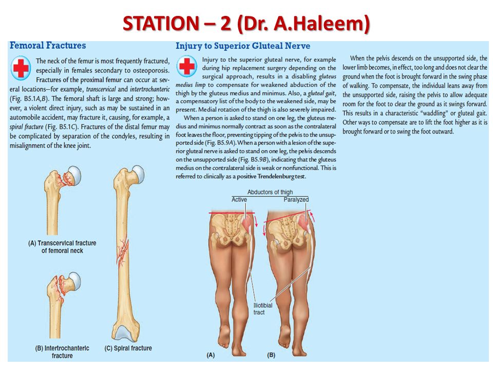
STATION – 2 (Dr. A.Haleem) Anterior superior iliac spine Anterior inferior iliac spine Pubic tubercle Pubic symphysis Ischial spine Greater sciatic notch Lesser sciatic notch Iliac crest Obturator foramen Iliac fossa Posterior superior iliac spine Posterior inferior iliac spine HeadFoveaNeck Greater trochanter Intertrochanteric line Lesser trochanter Shaft of femur Lateral condyle Medial condyle The sacrotuberous and sacrospinous ligaments convert the sciatic notches in the hip bones into the greater and lesser sciatic foramina.
 Anterior superior iliac spine Anterior inferior iliac spine Pubic tubercle Pubic symphysis Ischial spine Greater sciatic notch Lesser sciatic notch Iliac crest Obturator foramen Iliac fossa Posterior superior iliac spine Posterior inferior iliac spine HeadFoveaNeck Greater trochanter Intertrochanteric line Lesser trochanter Shaft of femur Lateral condyle Medial condyle The sacrotuberous and sacrospinous ligaments convert the sciatic notches in the hip bones into the greater and lesser sciatic foramina..")
12
The pulsation of femoral artery The pulsation of femoral artery can be felt in the midway of inguinal ligament (which is extending from the anterior superior iliac spine to the pubic tubercle). Femoral artery Femoral artery: it is the continuation of external iliac artery. It passes through the femoral triangle surrounded by femoral sheath and then continues in the adductor canal (middle third of the thigh) until it passes through the adductor hiatus (after which it will be called popliteal artery in the popliteal fossa). Femoral triangle: Femoral triangle: – Boundaries: Superior: inguinal ligament. Lateral: medial border of sartorius muscle. Medial: lateral border of adductor longus muscle. – Contents: Femoral artery. Femoral vein. Femoral nerve (L2-L4): passing through femoral triangle outside the femoral sheath. Sciatic nerve Sciatic nerve is coming from the sacral plexus (L4, L5, S1, S2, S3). It is exiting the pelvis through the greater sciatic foramen and passing posteriorly in the mid-point between greater trochanter and ischial tuberosity. It lies on adductor magnus during its passage. When it reaches the distal third of the thigh it will bifurcate to tibial and common fibular divisions. To know the course of sciatic nerve: – Draw a line from posterior superior iliac spine to ischial tuberosity and mark the midpoint. – Draw another line from greater trochanter to ischial tuberosity and mark the midpoint. Connect both points to get a curved line representing the course of sciatic nerve. STATION – 2 (Dr. A.Haleem)
 until it passes through the adductor hiatus (after which it will be called popliteal artery in the popliteal fossa). Femoral triangle: Femoral triangle: – Boundaries: Superior: inguinal ligament. Lateral: medial border of sartorius muscle. Medial: lateral border of adductor longus muscle. – Contents: Femoral artery. Femoral vein. Femoral nerve (L2-L4): passing through femoral triangle outside the femoral sheath. Sciatic nerve Sciatic nerve is coming from the sacral plexus (L4, L5, S1, S2, S3). It is exiting the pelvis through the greater sciatic foramen and passing posteriorly in the mid-point between greater trochanter and ischial tuberosity. It lies on adductor magnus during its passage. When it reaches the distal third of the thigh it will bifurcate to tibial and common fibular divisions. To know the course of sciatic nerve: – Draw a line from posterior superior iliac spine to ischial tuberosity and mark the midpoint. – Draw another line from greater trochanter to ischial tuberosity and mark the midpoint. Connect both points to get a curved line representing the course of sciatic nerve. STATION – 2 (Dr. A.Haleem).")
13
Movements of the hip joint: Movements of the hip joint: – Flexion: by anterior thigh muscles Iliopsoas (psoas major and iliacus). Pectineus and sartorius. Rectus femoris. – Extension: Gluteus maximus (acting to extend the hip joint against resistance). Hamstrings. – Abduction and medial rotation: Gluteus medius and gluteus minimus. – Adduction: by medial thigh muscles (adductors) Adductor longus. Adductor brevis. Adductor magnus. Gracilis. Obturator externus. – Lateral rotation: by deep gluteal muscles Piriformis. Obturator internus. Superior and inferior gemelli. Quadratus femoris. STATION – 2 (Dr. A.Haleem)
. Hamstrings. – Abduction and medial rotation: Gluteus medius and gluteus minimus. – Adduction: by medial thigh muscles (adductors) Adductor longus. Adductor brevis. Adductor magnus. Gracilis. Obturator externus. – Lateral rotation: by deep gluteal muscles Piriformis. Obturator internus. Superior and inferior gemelli. Quadratus femoris. STATION – 2 (Dr. A.Haleem).")
16
Muscles of the gluteal region: classified to: Muscles of the gluteal region: classified to: – Superficial muscles which include: Gluteus maximus: causing extension of the thigh and lateral rotation. Its outer-lateral quadrant is a preferred site for intramuscular injections (large area for distribution of the drug). This muscle is innervated by inferior gluteal nerve. Gluteus medius and gluteus minimus: they cause abduction and medial rotation of the thigh. These two muscles are innervated by superior gluteal nerve. – Deep muscles which cause lateral rotation of the thigh and include: Piriformis. Obturator internus. Superior and inferior gemelli. Quadratus femoris. STATION – 3 (Muscles of Gluteal Region)
. This muscle is innervated by inferior gluteal nerve. Gluteus medius and gluteus minimus: they cause abduction and medial rotation of the thigh. These two muscles are innervated by superior gluteal nerve. – Deep muscles which cause lateral rotation of the thigh and include: Piriformis. Obturator internus. Superior and inferior gemelli. Quadratus femoris. STATION – 3 (Muscles of Gluteal Region).")
17
Gluteus medius Gluteus maximus Piriformis Superior gemellus Obturator internus Inferior gemillus Quadratus femoris Sciatic nerve

18
STATION – 3 (Muscles of Gluteal Region)
")
19
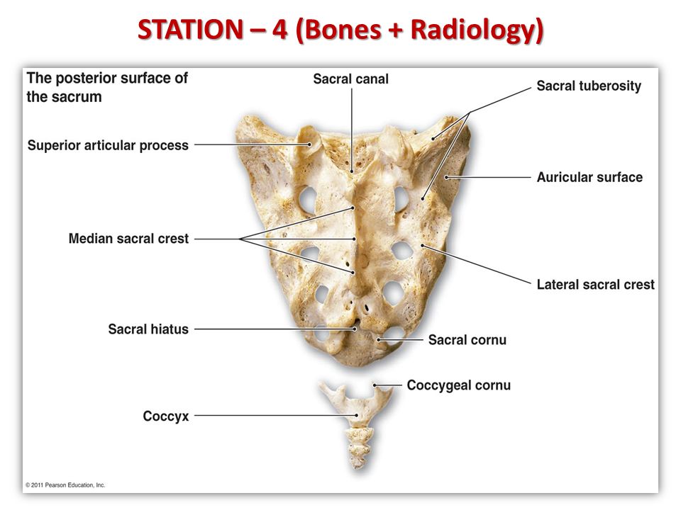
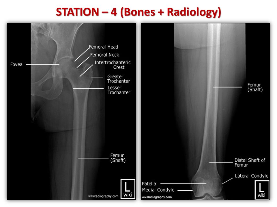
STATION – 4 (Bones + Radiology)
")
23
Good Luck!

Similar presentations