Download presentation
Presentation is loading. Please wait.

1
Approach to arthritis DR.AFSAR SAYEEDA MRCP,FRCP(UK)
CONSULTANT & HEAD,CTU DEPT OF MEDICINE, KKUH

2
Outline Rheumatological history and clinical examination
Inflammatory /non-inflammatory arthritis Mono/ Oligo arthritis Polyarthritis Soft tissue rheumatism Lab investigations Synovial fluid analysis Imaging

3
Normal Joint..

4
Evaluation of a patient with arthritis in rheumatology opd
Articular or non articular Inflammatory or non inflammatory Acute or chronic Monoarticular or polyarticular Extra articular signs

5
ARTICULAR - Deep or diffuse pain. NONARTICLAR localised pain
Painful or limited range of movemnt - both active and passive Swelling of joint Crepitation. Joint instability. Locking of joint. Deformity. NONARTICLAR localised pain Point or local tenderness Painful active movements but not on passive Physical findings are remote from joint capsule. swelling,crepitation,joint instability, deformity are rare.

6
arthritis Cardinal signs Systemic symptoms Stiffness- >1 hr
inflammatory Non- inflammatory Cardinal signs Systemic symptoms Stiffness- >1 hr - prolon. rest Lab evidences ESR CRP NO signs Stiffness-<60 mnt - intermittent -brief rest Trauma,rept.use, Degenerative,tumor

7
The Rheumatologic History
h/o presenting complaints - Onset - progression - distribution of disease - stiffness - aggravating or relieving factor - diurnal variation - other systemic feature - functional disability General systematic medical history. Past medical and surgical history. Family history. Drug history.

8
Rheumatic disease signs
Swelling Posture of joint Deformity Warmth Redness Tenderness Limitation of joint movement Crepitus Stability Function

9
Extra articular signs & symptoms
Constitutional symptoms Skin rashes Mucous membrane lesions Ocular Nails Raynauds Serositis

10
Chronology of complaints
ONSET acute- < 6 wks eg.infectious arthritis crystal arthropathy reactive arthritis. Chronic - >6 wks eg. Non inflamatory arthritis (OA) Inflammatory arthritis(RA) Fibromyalgia. EVOLUTION – chronic eg.OA intermittent eg. Crystal / lymes arthritis migratory arthritis eg.Rheumaticfever,
 Inflammatory arthritis(RA) Fibromyalgia. EVOLUTION – chronic eg.OA. intermittent eg. Crystal / lymes arthritis. migratory arthritis eg.Rheumaticfever,")
11
Chronology of complaints
C. Extent of articular involvement - Monoarticular (one joint involved) - Oligo or pauciarticular (two or three joint) - Polyarticular (> 3 joints) D. Distribution of joint involvement -symmetrical- upper and lower limb eg. RA, SLE -Asymmetrical-eg. psoriatic arthritis, spondyloarthropathy, gout -Involvement of axial skeletal-eg AS, OA, RA(only cervical spine)
 - Oligo or pauciarticular (two or three joint) - Polyarticular (> 3 joints) D. Distribution of joint involvement. -symmetrical- upper and lower limb eg. RA, SLE. -Asymmetrical-eg. psoriatic arthritis, spondyloarthropathy, gout. -Involvement of axial skeletal-eg AS, OA, RA(only cervical spine)")
12
History and physical examination
no Trauma/fracture Soft tissue rheumatism Is it articular No yes Infectious arthritris Crystal induced Reactive arthritis Acute > 6 weeks yes Chronic yes Chronic inflammatory arthritis no Chronic noninflamatory arthritis Signs of inflammation yes Joints involved DIP, CMC1,Hip ,Knee joint osteoarthritis 1-3 >3 yes no no Psoriatic Pauci JA PCP,MCP/MTP symmetrical Osteonecrosis Charcots joint no yes Psoriatic Reactive SLE/Scleroderma Rheumatoid

13
CAUSES:Mono/ oligo arthritis
Septic Arthritis–Bacteria,fungal,parasitic arthritis Internal derangement or trauma –Meniscus Injury –Ligament tears - hemarthrosis crystal-induced arthritis Charcot joint Psoariatic arthritis Juvenile Rheumatoid Arthritis(pauci articular) Mono art.presentation of c/c arthritis Ischemic bone (avascular necrosis Neoplasms –Villonodularsynovitis
 Mono art.presentation of c/c arthritis. Ischemic bone (avascular necrosis. Neoplasms –Villonodularsynovitis.")
14
Septic Arthritis: Risk Factors
Prosthetic hip joint. Prosthetic knee joint. Skin Infection. Joint surgery. Rheumatoid Arthritis. Elderly patients over age 80 years old. Diabetes Mellitus. Intravenous drug use (unusual joints affected). Large vein catheterization (unusual joints affected).
. Large vein catheterization (unusual joints affected).")
15
CAUSES OF SEPTIC ARTHRITIS
Young sexually active adults –Neisseria gonorrhoeae (most common) More common in women –Staphylococcus aureus –Streptococcus Older adults –Staphylococcus aureus(50%) –Streptococcus species -Gram Negative Bacilli
 More common in women. –Staphylococcus aureus. –Streptococcus. Older adults. –Staphylococcus aureus(50%) –Streptococcus species. -Gram Negative Bacilli.")
16
Signs and Symptoms Rapid onset monoarticular joint inflammation
Joints affected in bacterial infection –Septic Knee (50% of cases),hip (children), ankle, - shoulder Joints affected with intravenous Drug Abuse –SI joint, SC joint.pubic symphysis,vertebral spaces
,hip (children), ankle, - shoulder. Joints affected with intravenous Drug Abuse. –SI joint, SC joint.pubic symphysis,vertebral spaces.")
17
Gout: Uric Acid Crystals
RISK FACTOR -Obesity -Diabetes Mellitus -Hyperlipidemia -Hypertension -Atherosclerosis -Alcohol use -Thiazide Diuretics -Renal insufficiency -Myeloproliferativedisease

18
GOUT :SIGN AND SYMPTOMS
Acute onset of lower extremity joint pain –First Metatarsophalangeal joint (great toe) - Affected in 50% of first gout attacks Fever and chills Joint Inflammation - Asymmetric joint involvement - May only involve one side with the first attack
 - Affected in 50% of first gout attacks. Fever and chills. Joint Inflammation - Asymmetric joint involvement. - May only involve one side with the first attack.")
19
gouty arthritis

20
Gout SynovialFluid Polarizing Microscopy
Negatively birefringent Needle shaped Uric Acid crystals Gram Stain and Culture Rule out Septic Arthritis

21
Polyarthritis

22
Polyarthritis Acute Polyarthritis - Viral Chronic Polyarthritis:
- < 6 wks - Viral - Borrelia burgdorferi Chronic Polyarthritis: - >6 weeks <60yrs age : RA, SLE, psoriatic arthritis, spondyloarthropathies >60yrs age : crystal induced, OA

23
Osteoarthritis Most common form of arthritis. Associated functional.
Impairment increases with age. Prevalence directly increases with age

24
PATHOPHYSIOLOGY Primary lesion resides in the articular cartilage
–Abnormal cartilage repair and remodeling –Chondrocytes proteolytic enzymes destroy cartilage subchondral subchondral sclerosis cysts Marginal osteophytes

25
osteoarthritis

26
Sign and Symptoms Pain on motion that worsens with increasing joint usage Slowly progressive deformity and possibly pain No systemic manifestations Associated muscle spasm, contractures and atrophy Symptoms uncommon before age 40 Morning stiffness of short duration (<30 minutes)
")
27
Distribution of Osteoarthritis
Joints spared –Wrist –Metacarpal-phalangeal (except thumb) –Elbow –Ankle Joints commonly involved knee hip foot hand –DIP (Heberden'sNodes) –PIP (Bouchard's Nodes) –First CMC jt(thumb) Cervical and lumbar spine
 –Elbow. –Ankle. Joints commonly involved. knee. hip. foot. hand –DIP (Heberden sNodes) –PIP (Bouchard s Nodes) –First CMC jt(thumb) Cervical and lumbar spine.")
28
Rheumatoid Arthritis Affects all ethnic groups
Peak incidence 4-6th decades Most widely used criteria ACR Diagnosis is based on the clinical criterion and cant be made until symptoms present for several weeks positive RF supports Diagnosis (20% are seronegative)
")
29
ACR Rheumatoid Arthritis Criteria Need to have 4 of 7
Morning stiffness:-in and around the joint lasting 1 hr before maximal improvement. Arthritis of 3 or more joint area observed by the physician. 14 possible joint area involved are rt < PIP,MCP, wrist, elbow, knee, ankle and MTP joint. Arthritis of hand joints- wrist,mcp &pip joint. Symmetrical arthritis. Rheumatoid nodule. Serum Rheumatoid factor. Radiographic changes – erosion or bony decalcification in or adjacent to involved joints. Criteria 1 to 5 must be present for at least 6 wks Criteria 2 to 5 must be observed by physician

30
New ACR/EULAR RA Criteria
RA can be classifiable or diagnosed with a score ≥6: Joints (0–5) 1 large joint 0 2–10 large joints 1 1–3 small joints (large jts excluded) 2 4–10 small joints (large jts excluded) 3 >10 joints (at least 1 small joint) 5 Serology (0–3) Negative RF and negative anti-CCP 0 Low positive RF or anti-CCP 2 High positive RF or anti-CCP 3 Symptom duration (0–1) <6 weeks 0 ≥6 weeks 1 Acute phase reactants (APR) (0–1) Normal CRP and ESR 0 Abnormal CRP or ESR 1 Goal Establish new criteria for classifying early RA Phase I RA patient data from cohorts. Analyzed at start of MTX. Factor analysis developed 4 discreet domains: serology, acute phase reactants, joint involvement and distribution, duration of arthritis Phase 2: 32 European and USA Rheumatologists, used consensus methodology to refine factors / approach from phase I, using actual patient vignettes. Criteria: 2 considered essential evidence of synovitis lack of other disease to explain findings Patients with typical erosions would be classified as having RA. 4 other factors can also help lead to a diagnosis of RA: pattern of involved joints (number, size, symmetry – 6 different groupings) serology (either RF / anti-CCP; discussions ongoing about titer and cutoffs) duration of arthritis (6 weeks as key cutoff point) acute phase reactants (ESR / CRP > normal -local lab) Final weighted scoring version presented at ACR2009 Aletaha D. Ann Rheum Dis 2010 30
 1 large joint 0. 2–10 large joints 1. 1–3 small joints (large jts excluded) 2. 4–10 small joints (large jts excluded) 3. >10 joints (at least 1 small joint) 5. Serology (0–3) Negative RF and negative anti-CCP 0. Low positive RF or anti-CCP 2. High positive RF or anti-CCP 3. Symptom duration (0–1) <6 weeks 0. ≥6 weeks 1. Acute phase reactants (APR) (0–1) Normal CRP and ESR 0. Abnormal CRP or ESR 1. Goal Establish new criteria for classifying early RA Phase I RA patient data from. cohorts. Analyzed at start of MTX. Factor analysis developed 4 discreet domains: serology, acute phase reactants, joint involvement and distribution, duration of. arthritis. Phase 2: 32 European and USA Rheumatologists, used consensus methodology to refine. factors / approach from phase I, using actual patient vignettes. Criteria: 2 considered essential. evidence of synovitis. lack of other disease to explain findings. Patients with typical erosions would be classified as having RA. 4 other factors can also help lead to a diagnosis of RA: pattern of involved joints (number, size, symmetry – 6 different groupings) serology (either RF / anti-CCP; discussions ongoing about titer and cutoffs) duration of arthritis (6 weeks as key cutoff point) acute phase reactants (ESR / CRP > normal -local lab) Final weighted scoring version presented at ACR2009. Aletaha D. Ann Rheum Dis")
31
Guidelines for classification
Four of the seven criteri are required to classify a pts is having RA. Pts with two or more clinical diagnoses are not excluded.

32
Distribution of Rheumatoid Arthritis
Affects small and large joints Typically patient has symmetrical inflammation in the wrists and/or MCP joints Spares DIP Morning stiffness, inactivity stiffness. Acute, severe onset %; subacute 20%

33
Acute Polyarthritis - RA

34
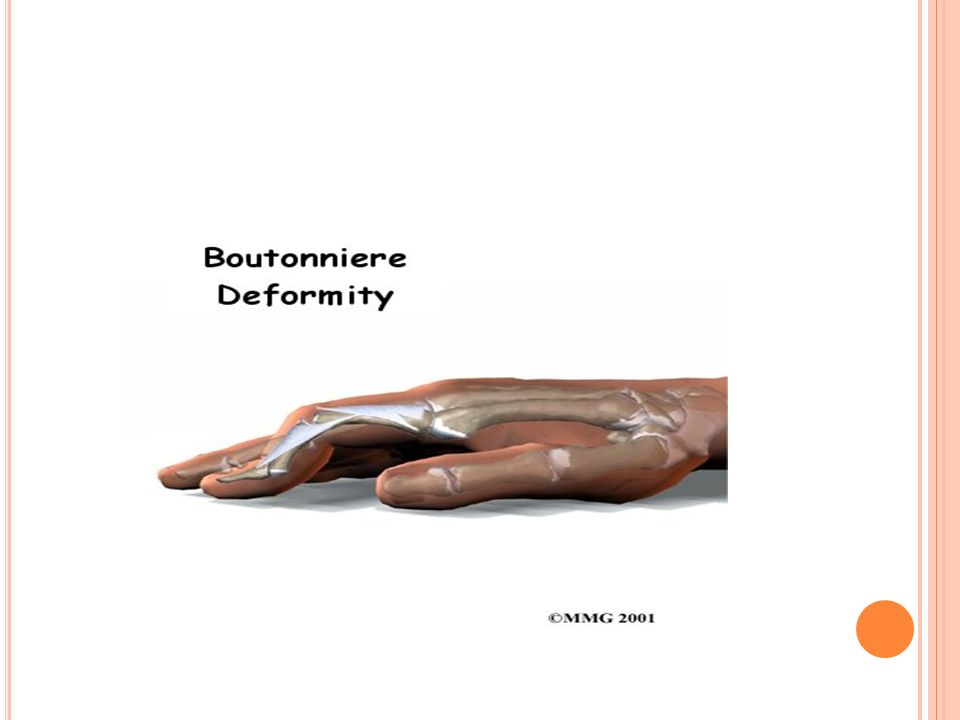
DEFORMITIES Z deformity
Swan neck deformity Boutonniere deformity Acute hand deformity: fusiform swelling of fingers due to synovitis of PIPs

35
Deformity- ra Swan neck deformity

37
Z - deformity Subcutaneous nodules

39
RF may be negative at onset and may remain negative in 15-20%!
RA is a clinical diagnosis, no laboratory test is diagnostic, just supportive!

40
Systemic erythematosus Lupus
Immune complex deposition disease, involving many organs Female:Male 10:1 ANA and other criterion will make the diagnosis

41
sle- non erosive arthritis
Musculoskeletal manifestation 90%. Most have arthralgia. May have acute inflammatory synovitis RA-like. Hand ,wrist & knee. Soft tissue inv- myositis, tendonitis. Do not develop erosions. Other clinical features help with DD: malar rash, photosensitivity, rashes, alopecia, oral ulceration.

42
Butterfly Rash – SLE

43
Photosensitivity

44
Alopecia - SLE

45
Criterion For Diagnosis of SLE Need 4 out of 11 to make the diagnosis
MalarRash :Rash spares nasolabialfolds Discoid Rash Photosensitivity Oral Ulcers: Painless observed by physician Arthritis: Nonserosive 2 or > joints Serositis: Pleuritis, Pericarditis Renal Disorder: Proteinuria> o.5g/day or casts Neurologic Disorder: seizures/ psychosis HematologicDisorder: Hemolysis, Leukopenia<4000, Lymphopenia <1500,Thrombocytopenia <100000 ANA Immunologic disorder: Anti-DNA, Anti-Sm, APS

47
Arthritis of Rheumatic Fever
Etiology: Streptococcus pyogenes (group A); immune response to antecedent infection Migratory polyarthritis, large joints: knees, ankles, elbows, wrists. Major manifestations: carditis, polyarthritis, chorea, erythema marginatum, subcutaneous nodules.
; immune response to antecedent infection. Migratory polyarthritis, large joints: knees, ankles, elbows, wrists. Major manifestations: carditis, polyarthritis, chorea, erythema marginatum, subcutaneous nodules.")
48
Seronegative spondoarthropathies
Psoriatic arthritis Reactive arthritis Enteropathic arthritis Ankylosing sponylosis

49
features of spondoarthropathies
Absence of RA Factor,subcut nodules Sacroiliatis/spondylitis + Assymetric peripheral joints Extra articular- ocular,oral,skin,enthesitis Familial aggregation HLA-B27 +

50
Distribution of spondoarthropathies
Asymmetric arthritis Axial spine & lower limb joints Soft tissues involvment Bursitis,achilles tendonitis,epichondyli tis,plantar fascitis

51
Psoriatic arthritis Psoriasis precedes in 60-70%
Wright & Molls 5 patterns of arthropathy Nail changes in 90% INVOLVEMENT OF DIP joints Dactylitis,enthesitis,tenosynovitis Arthritis mutilans

52
Psoriatic arthritis

53
Reactive arthritis Acute ,painful,assymetric
Knee,ankle ,ST,MT ,IP joints Dactylitis Constitutional symptoms Tendonitis,enthesitis,fascitis Ocular,muco-cutaneous lesions

54
Ankylosing spondylitis
Sacroiliatis Syndesmophytes Bamboo spine Inflamm. Backache Age<50 Improves with exercise not with rest

55
Enteropathic arthritis
Ankylosing spondylitis Peripheral arthritis-acute oligo & chronic polyarthritis Joint invl same in UC &CD Erosion and deformity rare

56
Soft tissue rheumatism
Most common cause of MSK pain Enthesopathy,bursitis,tedonitis,tenosynovitis Mostly associated with fibromyalgia Improves with local steriod inj.

57
SOFT TISSUE RHEUMATISM

58
Lab investigations Routine blood tests ESR,CRP Rheumatoid factor,CCP
ANA Autoimmune antibodies Complement levels

59
SYNOVIAL FLUID EXAMINATION

60
INTERPRETATION OF SYNOVIAL FLUID EXAMINATION
Strongly consider synovial fluid examination if Monoarthritis Trauma with joint effusion Mono arthritis in a pt. with chronic arthritis Suspicion of joint infection,crystal induced arthritis,heamarthrosi Appearance Viscocity WBC count Crystal identification Gram stain,culture if neded Is the effusion is hemorrhagic? Inflammatory or non inflammatory articular condition Is wbc / μl ? Consider noninflamm. Condition Osteoarthritis Trauma Other Consider inflamm. Or septic arthritis is the % PMNs.75% ? Consider noninflamm articular conditions Osteoarthrutis other Are crystals present? Consider other inflamm. Or septic arthritides.gram stain ,culture Is WBC /μl ? Probable inflamm arthritis Possible septic arthritis Crystal identification for specific diagnosis Gout or pseudogout Consider Trauma or mechanical derangement Coagulopathy Neuropathic arthropathy

62
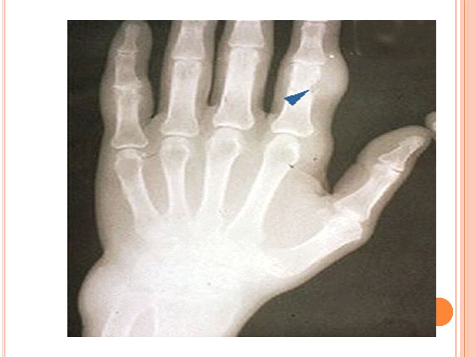
Diagnostic imaging Plain X-ray- show soft tissue swelling,erosions
they are indicated in patients with a history of trauma or patients who have had symptoms for several weeks. Occasionally, unsuspected bony lesions, such as osteomyelitis or malignancy, may be detected.

64
Ultrasonography CT Scan

65
5-MRI: Magnetic resonance imaging is superior in detecting ischemic necrosis, occult fractures, and meniscal and ligamentous injuries.

66
6-RADIONUCLIDE SCANS: Radionuclide scanning can detect infection in deep- seated joints. 7- OTHERS: Other diagnostic procedures, such as synovial biopsy or arthroscopy, may be useful to rule out deposition diseases (e.g., hemochromatosis, atypical infections) and intra-articular tumors.
 and intra-articular tumors.")
67
A 67 year old male presents with his first episode of knee pain and swelling together with the following x-ray. Which of the following investigations is the next investigation indicated diagnostically? (a) Thyroid function tests (b) Serum urate (c) Knee aspiration (d) Serum iron (e) Skeletal survey
 Thyroid function tests. (b) Serum urate. (c) Knee aspiration. (d) Serum iron. (e) Skeletal survey.")
68
Which of the following types of joint involvement is not seen in psoriatic arthritis?
(a) Symmetrical small joint arthropathy (b) Jaccoud’s arthropathy (c) Sacroiliitis (d) Monoarthritis (e) DIP joint arthropathy
 Symmetrical small joint arthropathy. (b) Jaccoud’s arthropathy. (c) Sacroiliitis. (d) Monoarthritis. (e) DIP joint arthropathy.")
69
ACUTE MONOARTHRITIS IS SEPTIC UNTIL PROVEN OTHERWISE !!

70
THANK YOU

Similar presentations


 Rogelio A Balagat MD ASMPH.>")

>")


 – Rheumatoid arthritis Inflammatory dz affecting synovial joints predominately Hyperplasia of synovial fibroblasts Severity is varied.>")

>")