Download presentation
Presentation is loading. Please wait.

1
Shark Lab Spiny Dog Fish

2
Scientific Name: Squalus acanthias
Habitat: Coastal Waters Diet: squid, fishes, crabs, shrimp and other invertebrates Size: 3 to 4 feet ( m) Range: Depths from the surface to 3,000 feet Life span : 25 years
 Range: Depths from the surface to 3,000 feet. Life span : 25 years.")
4
Spawning •Females mature at about age 12 and are around 30".
•In the North Atlantic spawning occurs in the winter. •Females are ovoviviparous (produce eggs that hatch within the body) and have a gestation period of 24 months, the longest of any vertebrate. •Eggs for the next litter begin developing for fertilization concurrent with the litter. •They generally have from 2 to 15 pups with an average of 6 pups. •Pups are about 14" when they are born.
 and have a gestation period of 24 months, the longest of any vertebrate. •Eggs for the next litter begin developing for fertilization concurrent with the litter. •They generally have from 2 to 15 pups with an average of 6 pups. •Pups are about 14 when they are born.")
5
External Anatomy of the Dogfish Shark

6
The shark has a graceful and streamlined body shape built for fast, long distance swimming. The body is divided into the head, trunk, and tail. The shark's body is dark gray above and almost white below (counter shading). Along the sides of the body is a light-colored horizontal stripe called the lateral line. The line is made up of a series of tiny pores that lead to receptors that are sensitive to the vibration movement of water and sudden changes of pressure. The spiny dogfish has a double dorsal fin. The anterior dorsal fin is larger than the posterior dorsal fin. The spiny dogfish has the presence two spines, one immediately in front of each dorsal fin. The spines carry a venom secreted by glands at their base. The caudal fin is divided into two lobes: a larger dorsal lobe and a smaller ventral lobe. This type of tail is known as a heterocercal tail.

7
Placoid Scales Cartilage

8
The rostrum is the pointed snout at the anterior end
The rostrum is the pointed snout at the anterior end. This tapered tip at the anterior end helps overcome water resistance in swimming. The eyes are prominent in sharks and are very similar to the eyes of man. A transparent cornea covers and protects the eye. A darkly pigmented iris can be seen below the cornea with the pupil at its center. Upper and lower eyelids protect the eye. Just inside the lower lid is a membrane that extends over the surface of the eye to cover the cornea. Large spiracle openings are located posterior and dorsal to the eyes.. The spiracle is an incurrent water passageway leading into the mouth for respiration. Most sharks have five external gill slits located on there sides behind the mouth and in front of the pectoral fins. Water taken in by the mouth and spiracles is passed over the internal gills and forced out by way of the gill slits

9
The opening to the mouth of sharks is always on the underside
The opening to the mouth of sharks is always on the underside. The teeth are sharp and point inward.There are several rows of flattened teeth lying behind the upright set ready to replace them when worn out or lost. The nares or external nostrils are located on the underside (ventral surface) of the rostrum anterior to the jaws. A nasal flap separates the incurrent from the excurrent opening. Water passes into and out of the olfactory sac, permitting the shark to detect the odors of the water. The patches of pores on the head in the areas of the eyes, snout, and nostrils are the openings of the ampullae of Lorenzini. These sense organs are sensitive to changes in temperature, water pressure, electrical fields, and salinity.
 of the rostrum anterior to the jaws. A nasal flap separates the incurrent from the excurrent opening. Water passes into and out of the olfactory sac, permitting the shark to detect the odors of the water. The patches of pores on the head in the areas of the eyes, snout, and nostrils are the openings of the ampullae of Lorenzini. These sense organs are sensitive to changes in temperature, water pressure, electrical fields, and salinity.")
10
The paired pectoral fins act like an airplane's wings to provide the lift needed to keep the shark from sinking. The paired pelvic fins are located on either side of the cloacal aperture. They are different in males and females.

11
Males have stout, grooved copulatory organs called claspers on the inner side of their pelvic fins. Fertilization in the dogfish shark is internal. The cloacal opening located on the ventral surface between the pelvic fins. It receives the products of the intestine, the urinary and the genital ducts. The name cloaca, meaning sewer, seems quite appropriate.

13
Somatic (body) Muscles
3 Skin Patches Somatic (body) Muscles Brachial muscle Fin muscle
 Muscles. Brachial muscle. Fin muscle.")
14
Somatic or Body muscles
W-shaped bundles called myotomes. The myotomes are separated from one another by connective tissue. Contractions of the myotomes produce the side to side motion of the body that propels the shark forward.

15
Fin muscle Tie the fins to the body wall. Used in locomotion.

16
Brachial muscles Muscles that surround the gills.
Control the respiratory process. Two types: Constrictors (close) Levitors (open)
 Levitors (open)")
17
Day one Pages 1-4 Questions 6 Drawings

18
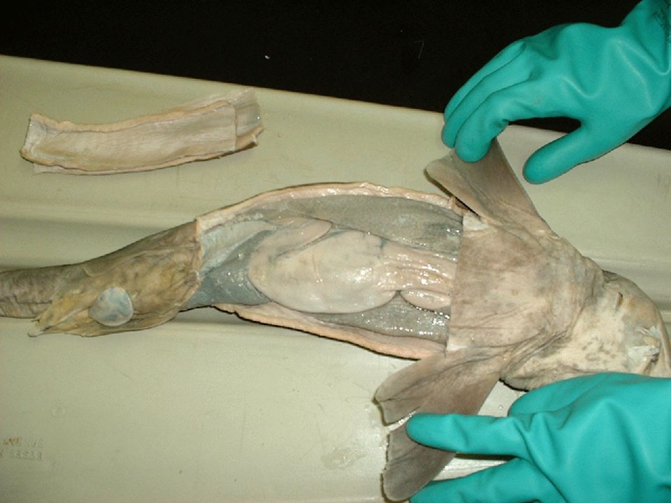
Internal Anatomy of the Dogfish Shark
Make an I cut starting at the pectoral fins down to the pelvic fins

20
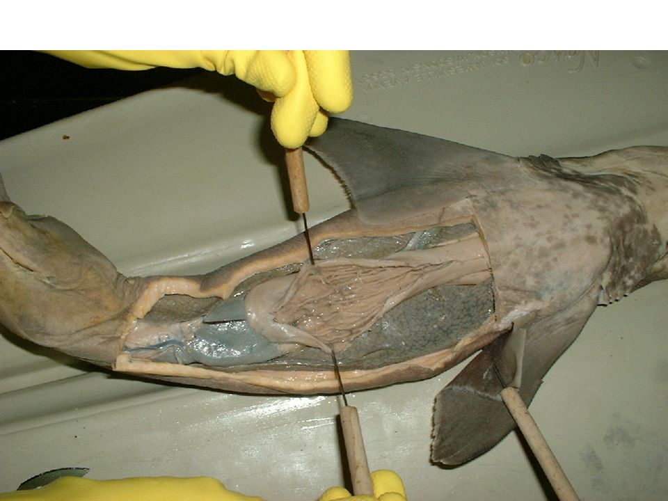
A smooth, shiny membrane called peritoneum can be seen lining the inside of the body wall. The visceral organs are suspended dorsally by a double membrane of peritoneum know as mesentery.

21
The liver is the largest organ lying within the body cavity
The liver is the largest organ lying within the body cavity. Its two main lobes, the right and left lobes, extend from the pectoral girdle posteriorly most of the length of the cavity. A third lobe much shorter lobe is located medially and contains the green gall bladder along its right edge. Bile is transported through the bile duct to the intestine.

23
The esophagus is the thick muscular tube extending from the top of the cavity connecting the oral cavity and pharynx with the stomach. The esophagus leads into the "J"-shaped stomach. The upper portion, the cardiac region, continues as the main body, and ends at the duodenal end. The stomach contains bumps and folds called rugae.

25
The duodenum is a short "U"-shaped portion of the small intestine that connects the stomach to the intestine. The bile duct from the gall bladder enters the duodenum. The pancreas is located on the duodenum and the lower stomach. The secretions of the pancreas enter the duodenum by way of the pancreatic duct.

26
The dark, triangular-shaped spleen is located near the posterior end of the stomach. Although a part the lymphatic system, the spleen is closely associated with the digestive organs in all vertebrates.

27
The spiral valve is the screw-like, symmetrical shape within the valvular intestine. It adds surface area for digestion and absorption to an otherwise relatively short intestine.

28
The colon is the narrowed continuation of the valvular intestine
The colon is the narrowed continuation of the valvular intestine. It is located at the posterior end of the body cavity. The rectal gland is a slender, blind-ended, finger-like structure that leads into the colon by means of a duct. It has been shown to excrete salt (NaCI) in concentrations higher than that of the shark's body fluids or sea water. It is thus an organ involved in regulating the shark's salt balance.
 in concentrations higher than that of the shark s body fluids or sea water. It is thus an organ involved in regulating the shark s salt balance.")
30
The cloaca is the last portion of the alimentary canal
The cloaca is the last portion of the alimentary canal. It collects the products of the colon as well as the urogenital ducts. It is a catch-all basin leading to the outside by means of the cloacal opening.

31
Urogenital System The Urogenital system is a combination of the two closely related systems - the excretory and the reproductive. The urinary and genital systems have distinct and unique functions. First, the removal of nitrogenous wastes and the maintenance of water balance. The reproduction of species.

32
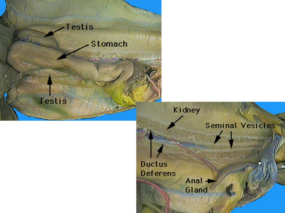
The kidneys are flattened, ribbon-like, darkly colored structures Iying dorsally on either side of the midline, along the entire length of the body cavity. A tough white glistening strip of connective tissue is found between the kidneys in the midline.

33
Male Paired testes lie near the anterior end of the body cavity, dorsal to the liver, adjacent to the anterior ends of the kidneys.The sperm pass from the testes to the kidneys within narrow tubules called efferent ductules. After passing through the anterior end of the kidney the sperm enter the ductus deferens and pass posteriorly toward the cloaca. In mature male specimens the ductus deferens may be seen on the ventral surface of the kidneys as a pair of highly coiled tubules.

35
Female The ovaries are two cream-colored elongated organs in the anterior part of the body cavity dorsal to the liver on either side of the mid-dorsal line. The shape of the ovaries will vary depending upon the maturity of the specimen. In immature females they will be undifferentiated and glandular in appearance. In mature specimens you may find two to three large eggs, about three centimeters in diameter, in each ovary.

37
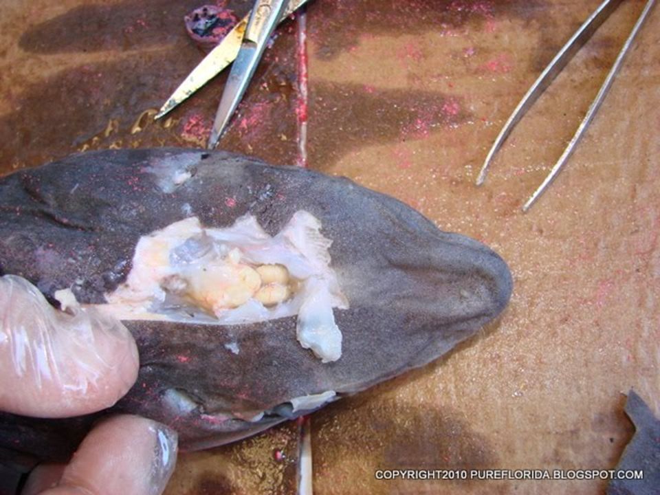
Fertilization in the dogfish shark is internal, usually taking place within the shell gland of the oviduct. The fertilized eggs continue to move posteriorly to the uterus. As they grow the pups are attached to the egg, now known as the yolk sac, by means of a stalk. During its period of gestation, which is nearly two years, the yolk is slowly absorbed by the shark "pup.“ At birth the young are about 14 inches long. This type of development, where the young are born as miniature adults but have not received any nutrition directly from the mother's uterus, is known as ovoviviparous.

39
Day 2 Pages 5, 6, 7, 8, 9 Matching 3 Drawings

40
Circulatory System

41
The pericardial cavity, lined by the pericardium, contains the heart and major blood vessels.
The ventricle is the thick muscular walled cavity that pumps blood to the gills and the body. The atrium is thin-walled with two lateral bulging lobes, that receives the blood.

43
Blood enters the heart through the sinus venosus which drains into the atrium.
The front of the conus arteriosus branches into five pairs of arteries which carry deoxygenated blood from the heart to the gills. Another set of vessels return oxygenated blood from the gills to the heart to be distributed to all parts of the body.

44
Nervous System There are two main parts of the nervous system:
Central nervous system - the brain and spinal cord. Peripheral nervous system - the sense organs, cranial and spinal nerves, and their branches.

46
Parts of the shark brain
Forebrain: The two cerebrum hemispheres are rounded lobes at the front of the brain. The first portion of the cerebrum is known as the olfactory lobes, responsible for the sense of smell. The second portion of the forebrain consists of the epithalamus, pineal body, thalamus, hypothalamus and pituitary body. Midbrain: The optic lobes are prominent bulges of the brain responsible for sight. Hindbrain: The cerebellum is an oval-shaped portion that partly overlaps the optic lobes. The medulla oblongata is the elongated region at the back of the brain that narrows into the spinal cord.

47
Cerebellum Olfactory lobe Medulla oblongata Optic lobe Cerebrum

48
NO LATE!!!!!!!!!!! Finish the lab Day 3 Pages 10, 11, 12
Due day of the TEST !!!!!!!!!!!! NO LATE!!!!!!!!!!!

Similar presentations


>")


 Dissection: Anatomy and Physiology>")


. This body shape reduces.>")


