Download presentation
Presentation is loading. Please wait.

1
CARDIAC CT IN SCREENING FOR CAD Hossein Nademi MD CARDIOLOGIST JAVADOL-A-EME HEART HOSPITAL OCT. 2011

2
Coronary CT Angio graphy

4
Computed Tomography (CT) was introduced into clinical practice in 1972 and revolutionized X ray imaging by providing high quality images which reproduced transverse cross sections of the body. Tissues are therefore not superimposed on the image as they are in conventional projections The technique offered in particular improved low contrast resolution for better visualization of soft tissue, but with relatively high absorbed radiation dose 4 Introduction

5
CORONARY Calcium scoring CORONARY CT angiography CURRENT CLINICAL USES OF CARDIAC CT

7
coronary calcium SCORING Cardiac CT, can detect and quantify coronary calcium, a marker of atherosclerosis The radiation dose is low, with a typical effective dose of 1.5 mSv CT calcium scoring produces the same amount of radiation as 1 to 2 mammograms performed on each breast

8
Because of radiation exposure and the general low prevalence of calcification in men 40 years of age and women 50 years of age, CT scanning should generally not be done in these younger- age patients.

9
Coronary calcium progresses at typically 10% to 20% of the baseline value per year among persons 45 years of age, approximately 7% per year of those without calcium develop detectable coronary calcium.

10
Score > 0 is indicative of significant CAD. -Sensitivity 85-100% -Specificity 31-62% -Negative predictive value 84-100% Nikolaou., Poon M., Sirol M., Becker C., Fayad Z., Complementary Results of Computed Tomography and Magnetic Resonance Imaging of the Heart and Coronary Arteries: A Review and Future Outlook. Cardiology Clinics. November 2003. Vol. 21, Nr. 4. Clinical application

11
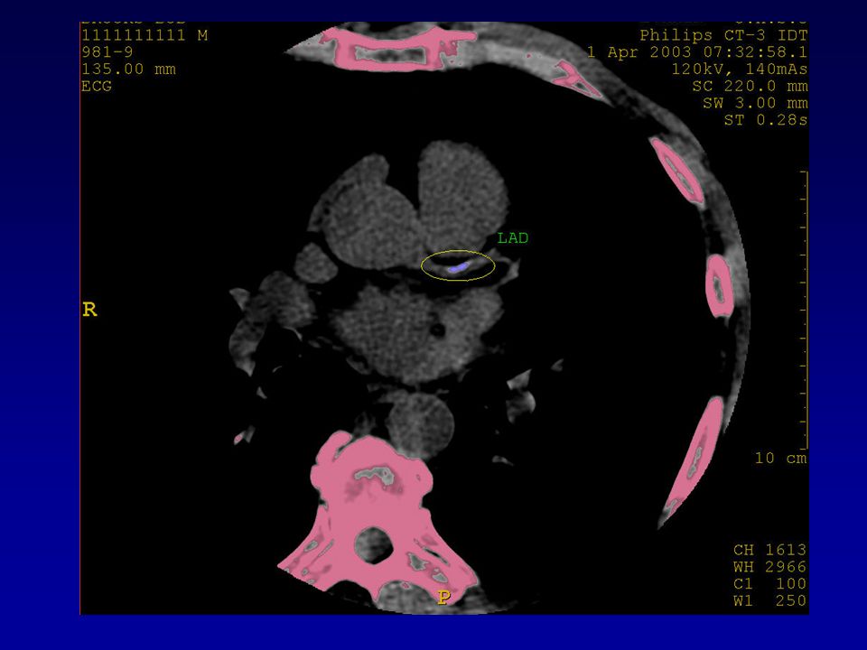
Soft plaque with stenosis of LAD

12
CHALLENGES OF MDCT SMALL VESSELSCARDIAC MOTION ARTIFACTSCORONARY CALCIFICATION/METAL OBJECTS(STENTS)IRREGULAR RHYTHMRAPID HR(>65/min)
IRREGULAR RHYTHMRAPID HR(>65/min)")
13
coronary CT angiography for detection of atherosclerosis (typically defined as a 50% diameter stenosis), Sensitivities and specificities from 40 studies are consistently in the range of 85% to 95%,
, Sensitivities and specificities from 40 studies are consistently in the range of 85% to 95%,")
14
64-slice coronary CT angiography is highly accurate for the exclusion of significant coronary artery stenosis (>50% luminal narrowing) with negative predictive values of 97%– 100%, in comparison with invasive selective coronary angiography. coronary CT angiography

15
Estimated more than 40% of invasive coronary angiograms are not followed up by subsequent interventional or surgical therapy, but are done to rule out coronary artery disease. multidetector CT Can serve as a non- invasive quick study - Especially in atypical chest pain without a significant CAD history ANOTHER WAY?

16
Plaque composition rather than the degree of lumen stenosis determines the risk of plaque rupture. AHA has categorized plaques: - Vulnerable or “high-risk” plaques have thin fibrous cap with extracellular lipid core. - Severely stenotic plaques usually have increased smoothe muscle cells and collagen at the core with little lipid component. These are less likely to rupture. Not visible by catheterization, but is being explored with CT angio. PLAQUE COMPONENTS

17
Classes of Recommendations and Levels of Evidence

18
CORONARY ARTERY CALCIUM SCORING CLASS IIa 1. Measurement of CAC is reasonable for cardiovascular risk assessment in asymptomatic adults at intermediate risk (10% to 20% 10-year risk)(Level of Evidence: B) CLASS IIb 1. Measurement of CAC may be reasonable for cardiovascular risk assessment in persons at low to intermediate risk (6% to 10% 10-year risk) (Level of Evidence: B) CLASS III: NO BENEFIT 1. Persons at low risk (6% 10-year risk) should not undergo CAC measurement for cardiovascular risk assessment(Level of Evidence: B)
(Level of Evidence: B) CLASS IIb 1. Measurement of CAC may be reasonable for cardiovascular risk assessment in persons at low to intermediate risk (6% to 10% 10-year risk) (Level of Evidence: B) CLASS III: NO BENEFIT 1. Persons at low risk (6% 10-year risk) should not undergo CAC measurement for cardiovascular risk assessment(Level of Evidence: B).")
19
Intermediate-risk patients with an elevated CAC score (intermediate FRS and CAC 300) had a 2.8% annual rate of cardiac death or MI (roughly equivalent to a 10-year rate of 28%) that would be considered high risk subjects who had CAC scores of 0 had a low rate of events over the subsequent 3 to 5 years (0.4%, a CAC score between 100 and 400 indicated a RR of 4.3 a score of 400 to 1000 indicated a RR of 7.2 and a score 1000 indicated a RR of 10.8 The corresponding pooled rates of 3- to 5-year CHD death or MI rates were 4.6% (for scores from 400 to 1000) and 7.1% (for scores 1000), resulting in a RR ratio of 7.2 (95% CI 5.2 to 9.9; p0.001) and 10.8
 had a 2.8% annual rate of cardiac death or MI (roughly equivalent to a 10-year rate of 28%) that would be considered high risk subjects who had CAC scores of 0 had a low rate of events over the subsequent 3 to 5 years (0.4%, a CAC score between 100 and 400 indicated a RR of 4.3 a score of 400 to 1000 indicated a RR of 7.2 and a score 1000 indicated a RR of 10.8 The corresponding pooled rates of 3- to 5-year CHD death or MI rates were 4.6% (for scores from 400 to 1000) and 7.1% (for scores 1000), resulting in a RR ratio of 7.2 (95% CI 5.2 to 9.9; p0.001) and 10.8")
20
the relationships between CAC outcomes are similar in men and women and different ethnic groups Each of these studies demonstrated that the AUC to predict coronary artery events is significantly higher with CAC than either Framingham or PROCAM (Münster Heart Study) risk stratification alone.
 risk stratification alone.")
21
USE AS A REPEAT MEASURE TO MONITOR EFFECTS OF THERAPY IN ASYMPTOMATIC PERSONS Although preliminary data suggest that a calcium scan progression rate of 15% per year is associated with a 17-fold increased risk for incident CHD events there are no data demonstrating that serial CAC testing leads to improved outcomes or changes in therapeutic decision making

22
USEFULNESS OF CORONARY CALCIUM SCORING IN GUIDING THERAPY Calcium scores 100 to 300 are associated with a high rate of incident CHD events over the ensuing 3 to 5 years, so that persons with calcium scores in this range are a suitable target group for stringent lifestyle recommendations

23
RECOMMENDATION FOR CORONARY COMPUTED TOMOGRAPHY ANGIOGRAPHY CLASS III: NO BENEFIT 1. Coronary computed tomography angiography is not recommended for cardiovascular risk assessment in asymptomatic adults (Level of Evidence: C)
.")
24
Effective doses for Cardiac Imaging ProceduresModality Effective Dose (mSv) Ca Scoring EBCT1.0 - 1.3 MDCT1.5 - 6.2* CTA EBCT1.5 - 2.0* MDCT6.7* - 25.0 Cardiac SPECT w Tc-99m or Tl-2016.0 - 15.0 CA (diagnostic only w fluoroscopy)2.1* - 6.0 Chest x-ray0.1 24 Average U.S. background radiation per year 3.6 mSv

Similar presentations


 Detection and Treatment of Asymptomatic Atherosclerosis for Primary Prevention.>")


 Dynamic scanning implies 15 or more scans in rapid sequence within one.>")


>")

 Electrons –Atomic particles –Have mass Wouldn’t a beam of particulate.>")