Lack of autophagy induces steroid-resistant airway inflammation Yuzo Suzuki, MD, PhD, Hadi Maazi, DVM, PhD, Ishwarya Sankaranarayanan, MS, Jonathan Lam, PhD, Bryant Khoo, BSc, Pejman Soroosh, PhD, Richard G. Barbers, MD, J.-H. James Ou, PhD, Jae U. Jung, PhD, Omid Akbari, PhD Journal of Allergy and Clinical Immunology Volume 137, Issue 5, Pages 1382-1389.e9 (May 2016) DOI: 10.1016/j.jaci.2015.09.033 Copyright © 2015 American Academy of Allergy, Asthma & Immunology Terms and Conditions
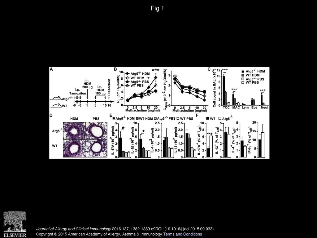
Fig 1 Lack of autophagy augments neutrophilic airway inflammation. A, Experimental timeline. i.n., Intranasal; i.p., intraperitoneal. B, Lung resistance (RL; left panel) and dynamic compliance (Cdyn; right panel) measured on day 16. C, Differential cell counts in BALF. Eos, Eosinophils; Lym, lymphocytes; MAC, macrophages; Neut, neutrophils; TCC, total cell number. D, Hematoxylin and eosin–stained lung sections (magnification ×200). E, Cytokine levels in whole-lung lysates. F, Frequency of IL-17A–, IL-4–, IL-5–, IL-13–, and IFN-γ–producing CD3+CD4+CD44+ effector T (Teff) cells. Data are representative of 2 to 3 experiments and shown as means ± SEMs (n = 5-10 per group). *P < .05 and ***P < .001. Journal of Allergy and Clinical Immunology 2016 137, 1382-1389.e9DOI: (10.1016/j.jaci.2015.09.033) Copyright © 2015 American Academy of Allergy, Asthma & Immunology Terms and Conditions
Fig 2 Lack of autophagy in immune cells contributes to induction of lung inflammation. A, WT or Atg5−/− BM cells were adoptively transferred into irradiated CD45.1+ WT mice and then immunized and challenged with HDM, as indicated. B, Reconstitution rate of transferred cells, as assessed by using the frequency of CD45.1+ and CD45.2+ neutrophils in BALF. C, Lung resistance (RL). D, Dynamic compliance (Cdyn). E, Differential cell counts in BALF. F, Hematoxylin and eosin–stained lung sections (magnification ×200). G, Atg5−/− mice were irradiated and received either WT or Atg5−/− BM, followed by HDM sensitization, as indicated. H, Reconstitution rate of transferred cells. I and J, Lung resistance (RL; Fig 2, I) and dynamic compliance (Cdyn; Fig 2, J) after sensitization, as shown in Fig 2, G. K, Differential cell counts in BALF. L, Hematoxylin and eosin–stained lung sections (magnification ×200). Eos, Eosinophils; i.n., intranasal; Lym, lymphocytes; MAC, macrophages; Neut, neutrophils; TCC, total cell number (n = 5 per group). Data are shown as means ± SEMs. *P < .05 and ***P < .001. Journal of Allergy and Clinical Immunology 2016 137, 1382-1389.e9DOI: (10.1016/j.jaci.2015.09.033) Copyright © 2015 American Academy of Allergy, Asthma & Immunology Terms and Conditions
Fig 3 Induction of asthma impairs autophagy in lung CD11c+ cells. A, Timeline for sensitization and challenge. WT or LC3-GFP mice were immunized with HDM, as indicated. i.n., Intranasal; i.p., intraperitoneal. B, Western blot analysis of expression levels of p62, LC3-I, and LC3-II in purified pulmonary CD11c+ cells. C, Levels of the indicated proteins are expressed relative to the β-actin gene (Actb). D, Confocal microscopy of purified lung CD11c+ cells from LC3-GFP (magnification ×600). DAPI, 4′-6-Diamidino-2-phenylindole dihydrochloride. E, Mean fluorescence intensity (MFI) of LC3. Data are expressed as means ± SEMs (n = 3-9 per group). *P < .05. Journal of Allergy and Clinical Immunology 2016 137, 1382-1389.e9DOI: (10.1016/j.jaci.2015.09.033) Copyright © 2015 American Academy of Allergy, Asthma & Immunology Terms and Conditions
Fig 4 Lack of autophagy in DCs induces IL-1 and IL-23 and TH17 polarization. A, BMDCs from WT and Atg5−/− mice were stimulated with or without HDM (50 μg/mL) for 36 hours. Cytokine levels in supernatants were measured by means of ELISA. B, Quantification of mRNA expression by means of RT-PCR. C, BMDCs from WT or Atg5−/− mice were cocultured with CD4+ T cells isolated from lungs of HDM-sensitized mice in the presence of HDM (10 μg/mL) for 3 days. HDM sensitization was performed according to Fig 1, A. Cytokine levels were measured by means of ELISA. Data are representative of 2 experiments and expressed as means ± SEMs (n = 4-8 per group). *P < .05 and **P < .01. Journal of Allergy and Clinical Immunology 2016 137, 1382-1389.e9DOI: (10.1016/j.jaci.2015.09.033) Copyright © 2015 American Academy of Allergy, Asthma & Immunology Terms and Conditions
Fig 5 Lack of Atg5 in CD11c+ cells causes unprovoked asthma and induces severe neutrophilic lung inflammation. A, Experimental timeline. Cultured BMDCs from WT or Atg5-/- mice were stimulated with HDM for 6 hours and then intravenously injected to WT mice. Then mice were intratracheally challenged with HDM-stimulated BMDCs on days 8 and 15. i.n., Intranasal; i.t., intratracheal. B and C, Lung resistance (RL; Fig 5, B) and dynamic compliance (Cdyn; Fig 5, C). D, Differential cell counts in BALF. E, Cytokine levels in whole-lung lysate. Pooled data from 2 experiments are shown (n = 7-8 per group). F, Lung resistance in CD11c-specific Atg5−/− and WT mice was measured at steady state. G, Dynamic compliance. H, Differential cell counts in BALF. Values are expressed as means ± SEMs. *P < .05, **P < .01, ***P < .001; n.s., Not significant. Eos, Eosinophils; Lym, lymphocytes; MAC, macrophages; Neut, neutrophils; TCC, total cell number. Journal of Allergy and Clinical Immunology 2016 137, 1382-1389.e9DOI: (10.1016/j.jaci.2015.09.033) Copyright © 2015 American Academy of Allergy, Asthma & Immunology Terms and Conditions
Fig 6 Atg5−/− mice have IL-17A–dependent steroid-resistant airway inflammation. A and D, WT and Atg5−/− mice were immunized and challenged with HDM, as described in Fig 1, A, and treated with dexamethasone (1 mg/kg) or anti–IL-17A antibody (300 μg) 1 day before HDM challenge. Mice were subsequently assessed for AHR by measuring lung resistance (RL). B and E, BAL cells from the mice in Fig 6, A and D, were analyzed after AHR measurements. C and F, Lung tissue sections from WT and Atg5−/− mice after dexamethasone or anti–IL-17A antibody treatment were stained with hematoxylin and eosin (magnification ×200). Data are expressed as means ± SEMs and representative of 2 independent experiments (n = 5-6 per group). *P < .05 and ***P < .001. Journal of Allergy and Clinical Immunology 2016 137, 1382-1389.e9DOI: (10.1016/j.jaci.2015.09.033) Copyright © 2015 American Academy of Allergy, Asthma & Immunology Terms and Conditions
Fig E1 Confirmation of depletion of Atg5 in the lungs and spleens of Atg5−/− mice. ROSA CRE ERT-Atg5 and WT mice received tamoxifen (800 μg per mouse per day) for 5 consecutive days. Six days after the last tamoxifen injection, mice were killed, and relative gene expression of Atg5 in lungs and spleens of ROSA CRE ERT-Atg5−/− and WT mice was assessed by using RT-PCR. Values are expressed as means ± SEMs. Journal of Allergy and Clinical Immunology 2016 137, 1382-1389.e9DOI: (10.1016/j.jaci.2015.09.033) Copyright © 2015 American Academy of Allergy, Asthma & Immunology Terms and Conditions
Fig E2 Representative dot plot presentation of cytokine production by T cells in the lung. Atg5−/− and WT mice were sensitized to HDM, as described in Fig 1, A. One day after the last sensitization, lung single cells were cultured in the presence of phorbol 12-myriststae 13-acetate/ionomycin, as described in the Methods section, followed by analysis of cytokine production by means of flow cytometry. Journal of Allergy and Clinical Immunology 2016 137, 1382-1389.e9DOI: (10.1016/j.jaci.2015.09.033) Copyright © 2015 American Academy of Allergy, Asthma & Immunology Terms and Conditions
Fig E3 T cells are the major source of IL-17 production in lungs of Atg5−/− mice. Atg5−/− and WT mice were sensitized, as mentioned in Fig 1, A. One day after the last sensitization, lungs were harvested and cultured in the presence of phorbol 12-myriststae 13-acetate/ionomycin for 5 hours, followed by evaluation of IL-17 production with flow cytometry. Live IL-17+CD45+ single cells were further gated to identify the source of IL-17A production. Bar graphs show the percentage of each subset within IL-17+ cells, as indicated. ILCs, Innate lymphoid cells. Journal of Allergy and Clinical Immunology 2016 137, 1382-1389.e9DOI: (10.1016/j.jaci.2015.09.033) Copyright © 2015 American Academy of Allergy, Asthma & Immunology Terms and Conditions
Fig E4 BAL neutrophils show similar viability in Atg5−/− and WT mice. BALF of HDM-sensitized or control Atg5−/− and WT mice were analyzed for neutrophil viability by using flow cytometry. Journal of Allergy and Clinical Immunology 2016 137, 1382-1389.e9DOI: (10.1016/j.jaci.2015.09.033) Copyright © 2015 American Academy of Allergy, Asthma & Immunology Terms and Conditions
Fig E5 Proportions of different subsets of antigen-presenting cells in lungs of Atg5−/− and WT mice are similar. Deletion of the Atg5 gene was induced in ROSA-Atg5−/− mice by using tamoxifen. Subsequently, Atg5−/− and WT mice were sensitized, as mentioned in Fig 1, A. One day after the last HDM sensitization, lungs of HDM-sensitized or control mice were analyzed for different subsets of antigen-presenting cells. A, Bar graph showing numbers of alveolar macrophages, plasmacytoid DCs, and CD103+ and CD11b+ DCs in the lungs. B, Dot plots show the gating strategy. Red rectangles and arrows show the gating hierarchy. Data are expressed as means ± SEMs. Alv. MAC, Alveolar macrophages; pDCs, plasmacytoid DCs. Journal of Allergy and Clinical Immunology 2016 137, 1382-1389.e9DOI: (10.1016/j.jaci.2015.09.033) Copyright © 2015 American Academy of Allergy, Asthma & Immunology Terms and Conditions
Fig E6 Lack of autophagy in lung epithelial cells does not contribute to AHR induction. A, Relative expression levels of the SPC-Cre gene were assessed by using RT-PCR. B, WT and SPC-Atg5−/− mice were immunized intranasally with HDM, as described in Fig 1, A. The mice were subsequently assessed for AHR. Pooled data from 2 experiments are shown (n = 8 mice per group). C, Lung tissues from WT and Atg5−/− mice were stained with hematoxylin and eosin (original magnification ×200). D, Cytokine levels in whole-lung lysates of asthmatic WT and SPC- Atg5−/− mice immunized as in Fig 1, A (n = 5 per group). Values are expressed as means ± SEMs. Cdyn, Dynamic compliance; N.S., not significant; RL, lung resistance. Journal of Allergy and Clinical Immunology 2016 137, 1382-1389.e9DOI: (10.1016/j.jaci.2015.09.033) Copyright © 2015 American Academy of Allergy, Asthma & Immunology Terms and Conditions
Fig E7 Lack of autophagy in DCs induces IL-1 and IL-23 and TH17 polarization. A, BMDCs from WT and Atg5−/− mice were stimulated with or without LPS (1 μg/mL) for 1 or 2 days. Cytokine levels in supernatants were measured by using ELISA (n = 6-8 per group). B, Quantification of mRNA expression by using RT-PCR (n = 6 per group). C, BMDCs from WT or Atg5−/− mice were cocultured with DO11.10 CD4+ T cells in the presence of the ovalbumin peptide OVA323-339 for 3 days. Cytokine levels in supernatants were measured by using ELISA. D, Caspase-1 level, as measured by using flow cytometry in Atg5−/− and WT BMDCs, in the presence or absence of LPS (1 μg/mL) for 24 hours. One of 2 representative experiments is shown (n = 4 per group). Data are expressed as means ± SEMs. P values were calculated with the Student t test. *P < .05. Journal of Allergy and Clinical Immunology 2016 137, 1382-1389.e9DOI: (10.1016/j.jaci.2015.09.033) Copyright © 2015 American Academy of Allergy, Asthma & Immunology Terms and Conditions