Download presentation
Presentation is loading. Please wait.

1
Diseases of the Pancreas Victor Politi, M.D., Medical Director SVCMC, School of Allied Health Professions, Physician Assistant Program

2
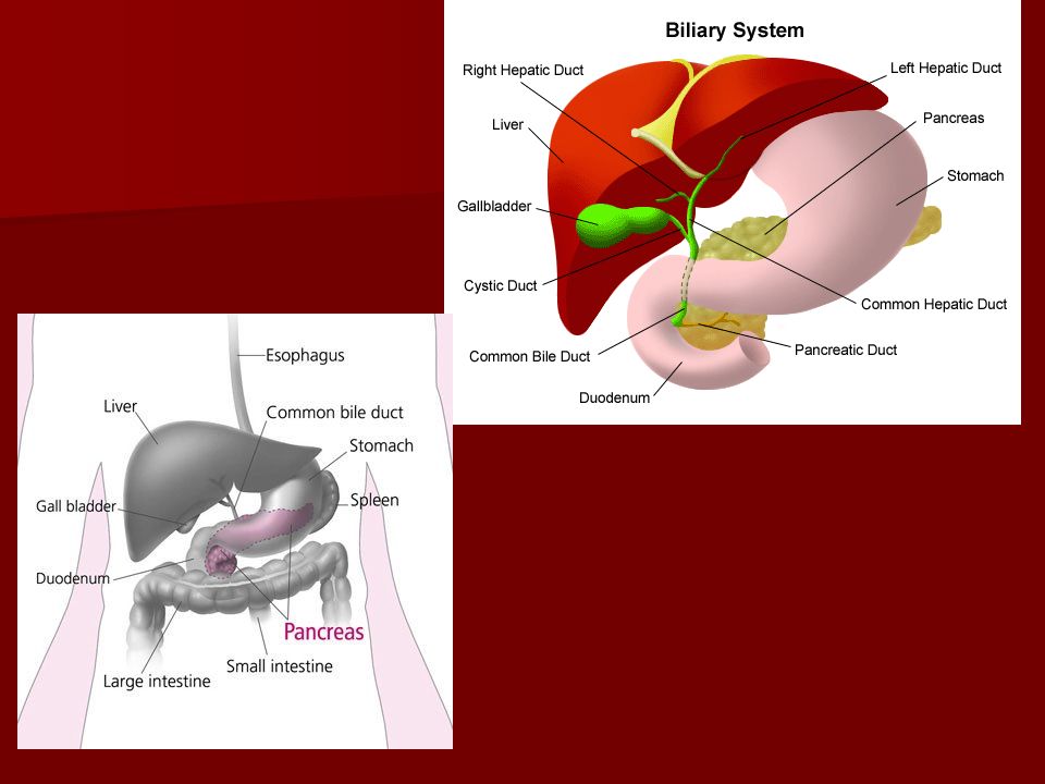
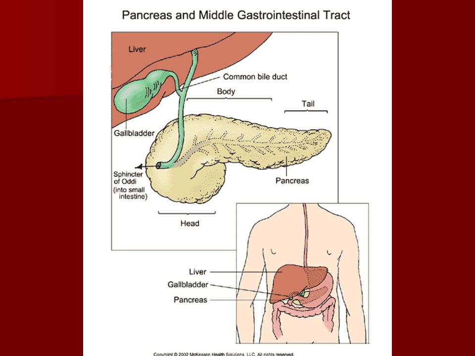
Pancreas anatomy The pancreas is an elongated, tapered organ located across the back of the abdomen, behind the stomach. The pancreas is an elongated, tapered organ located across the back of the abdomen, behind the stomach. The right side of the organ (called the head) is the widest part of the organ and lies in the curve of the duodenum (the first section of the small intestine). The right side of the organ (called the head) is the widest part of the organ and lies in the curve of the duodenum (the first section of the small intestine). The tapered left side extends slightly upward (called the body of the pancreas) and ends near the spleen (called the tail). The tapered left side extends slightly upward (called the body of the pancreas) and ends near the spleen (called the tail).
 is the widest part of the organ and lies in the curve of the duodenum (the first section of the small intestine). The right side of the organ (called the head) is the widest part of the organ and lies in the curve of the duodenum (the first section of the small intestine). The tapered left side extends slightly upward (called the body of the pancreas) and ends near the spleen (called the tail). The tapered left side extends slightly upward (called the body of the pancreas) and ends near the spleen (called the tail)..")
4
The pancreas is made up of two types of tissue: The pancreas is made up of two types of tissue: exocrine tissue exocrine tissue –The exocrine tissue secretes digestive enzymes. These are secreted into a network of ducts that join the main pancreatic duct, which runs the length of the pancreas. endocrine tissue endocrine tissue –The endocrine tissue, which consists of the islets of Langerhans, secretes hormones into the bloodstream. Pancreas anatomy

6
Accessory duct of Santorini Accessory duct of Santorini Duct of Wirsung Duct of Wirsung

7
The pancreas has digestive and hormonal functions: The pancreas has digestive and hormonal functions: –The enzymes secreted by the exocrine tissue in the pancreas help break down carbohydrates, fats, and proteins in the duodenum. –These enzymes travel down the pancreatic duct into the bile duct in an inactive form. –When they enter the duodenum, they are activated. –The exocrine tissue also secretes bicarbonate to neutralize stomach acid in the duodenum. Pancreas anatomy

8
The hormones secreted by the endocrine tissue in the pancreas are insulin, glucagon (which regulate the level of glucose in the blood), somatostatin (which prevents the release of the other two hormones), and many others. The hormones secreted by the endocrine tissue in the pancreas are insulin, glucagon (which regulate the level of glucose in the blood), somatostatin (which prevents the release of the other two hormones), and many others. Pancreas anatomy
, somatostatin (which prevents the release of the other two hormones), and many others. Pancreas anatomy.")
9
What is Pancreatitis? Pancreatitis is an inflammatory process in which pancreatic enzymes autodigest the gland Pancreatitis is an inflammatory process in which pancreatic enzymes autodigest the gland

10
Normally, digestive enzymes do not become active until they reach the small intestine, where they begin digesting food. Normally, digestive enzymes do not become active until they reach the small intestine, where they begin digesting food. But if these enzymes become active inside the pancreas, they start "digesting" the pancreas itself But if these enzymes become active inside the pancreas, they start "digesting" the pancreas itself

12
The gland can sometimes heal without any impairment of function or any morphologic changes. The gland can sometimes heal without any impairment of function or any morphologic changes. –This process is known as acute pancreatitis. It can recur intermittently, contributing to the functional and morphologic loss of the gland. It can recur intermittently, contributing to the functional and morphologic loss of the gland. –Recurrent attacks are referred to as chronic pancreatitis.

13
Acute pancreatitis occurs suddenly and lasts for a short period of time and usually resolves. Acute pancreatitis occurs suddenly and lasts for a short period of time and usually resolves. Chronic pancreatitis does not resolve itself and results in a slow destruction of the pancreas. Chronic pancreatitis does not resolve itself and results in a slow destruction of the pancreas.

14
Either form can cause serious complications. Either form can cause serious complications. In severe cases, bleeding, tissue damage, and infection may occur. In severe cases, bleeding, tissue damage, and infection may occur. Pseudocysts, accumulations of fluid and tissue debris, may also develop. Pseudocysts, accumulations of fluid and tissue debris, may also develop. Enzymes and toxins may enter the bloodstream, injuring the heart, lungs, and kidneys, or other organs. Enzymes and toxins may enter the bloodstream, injuring the heart, lungs, and kidneys, or other organs.

15
Acute edematous pancreatitis Since the pancreas is located in the retroperitoneal space with no capsule - inflammation can spread easily. Since the pancreas is located in the retroperitoneal space with no capsule - inflammation can spread easily. In acute pancreatitis, parenchymal edema and peripancreatic fat necrosis occur first. In acute pancreatitis, parenchymal edema and peripancreatic fat necrosis occur first. This process is known as acute edematous pancreatitis This process is known as acute edematous pancreatitis

16
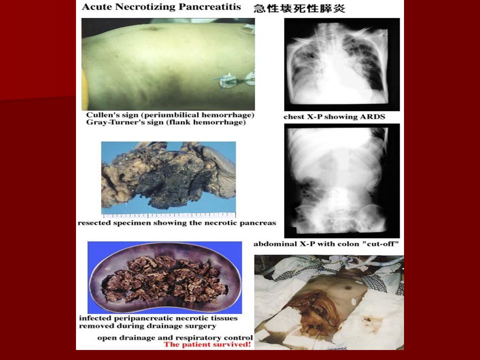
Necrotizing pancreatitis When necrosis involves the parenchyma, accompanied by hemorrhage and dysfunction of the gland, the inflammation evolves into hemorrhagic or necrotizing pancreatitis When necrosis involves the parenchyma, accompanied by hemorrhage and dysfunction of the gland, the inflammation evolves into hemorrhagic or necrotizing pancreatitis

18
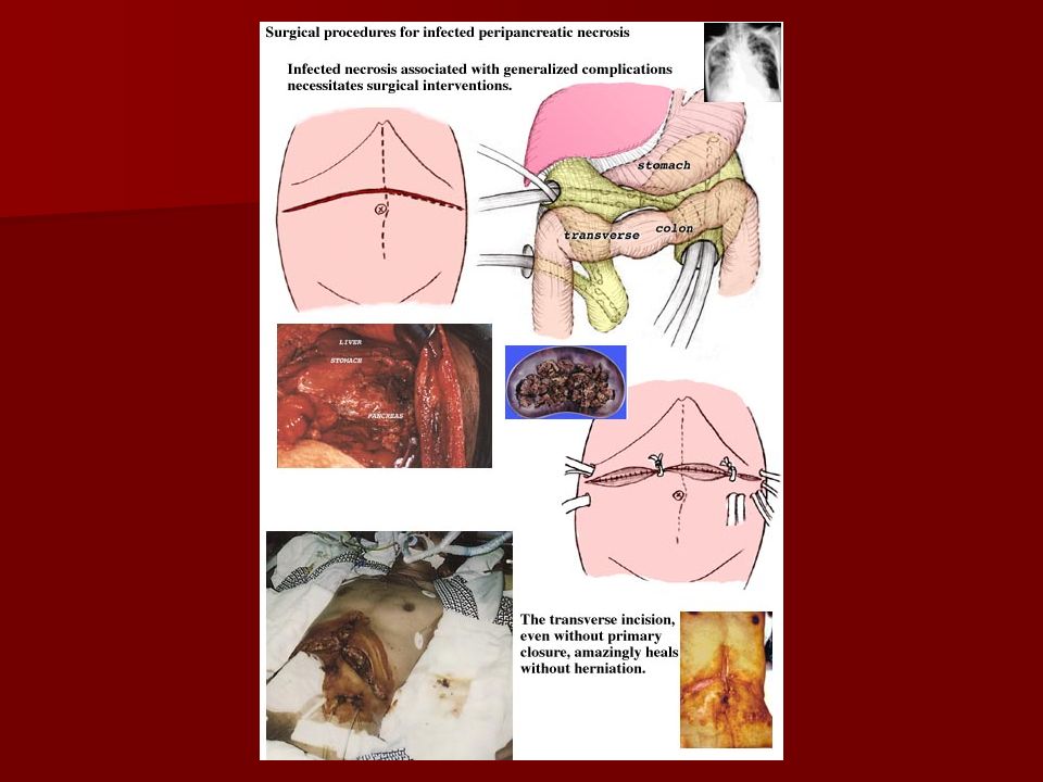
Necrotizing pancreatitis Pseudocysts and pancreatic abscesses can result from necrotizing pancreatitis because of enzymes being walled off by granulation tissue (ie, pseudocyst formation) or bacterial seeding of pancreatic or peripancreatic tissue (ie, pancreatic abscess formation). Pseudocysts and pancreatic abscesses can result from necrotizing pancreatitis because of enzymes being walled off by granulation tissue (ie, pseudocyst formation) or bacterial seeding of pancreatic or peripancreatic tissue (ie, pancreatic abscess formation). An ultrasound or, preferably, a CT scan can be used detect both. An ultrasound or, preferably, a CT scan can be used detect both.
 or bacterial seeding of pancreatic or peripancreatic tissue (ie, pancreatic abscess formation). An ultrasound or, preferably, a CT scan can be used detect both. An ultrasound or, preferably, a CT scan can be used detect both..")
20
The inflammatory process can cause systemic effects because of the presence of cytokines, such as bradykinins and phospholipase A. The inflammatory process can cause systemic effects because of the presence of cytokines, such as bradykinins and phospholipase A. These cytokines may cause vasodilation, increase in vascular permeability, pain, and leukocyte accumulation in the vessel walls. These cytokines may cause vasodilation, increase in vascular permeability, pain, and leukocyte accumulation in the vessel walls. Fat necrosis may cause hypocalcemia. Fat necrosis may cause hypocalcemia. Pancreatic B cell injury may lead to hyperglycemia. Pancreatic B cell injury may lead to hyperglycemia.

21
Mortality/Morbidity Although acute pancreatitis should be noted, chronic pancreatitis has a more severe presentation as episodes recur. Although acute pancreatitis should be noted, chronic pancreatitis has a more severe presentation as episodes recur. Acute respiratory distress syndrome (ARDS), acute renal failure, cardiac depression, hemorrhage, and hypotensive shock all may be systemic manifestations of acute pancreatitis in its most severe form. Acute respiratory distress syndrome (ARDS), acute renal failure, cardiac depression, hemorrhage, and hypotensive shock all may be systemic manifestations of acute pancreatitis in its most severe form.
, acute renal failure, cardiac depression, hemorrhage, and hypotensive shock all may be systemic manifestations of acute pancreatitis in its most severe form. Acute respiratory distress syndrome (ARDS), acute renal failure, cardiac depression, hemorrhage, and hypotensive shock all may be systemic manifestations of acute pancreatitis in its most severe form..")
22
Acute Pancreatitis Some people have more than one attack and recover completely after each, but acute pancreatitis can be a severe, life- threatening illness with many complications. Some people have more than one attack and recover completely after each, but acute pancreatitis can be a severe, life- threatening illness with many complications.

23
Acute Pancreatitis About 80,000 cases occur in the United States each year; some 20 percent of them are severe. About 80,000 cases occur in the United States each year; some 20 percent of them are severe. Acute pancreatitis occurs more often in men than women. Acute pancreatitis occurs more often in men than women.

24
The risk for African American persons aged 35-64 years is 10 times higher than for any other group. The risk for African American persons aged 35-64 years is 10 times higher than for any other group. African American persons are at higher risk than white persons in that same age group African American persons are at higher risk than white persons in that same age group

25
History The main presentation of acute pancreatitis is epigastric pain or right upper quadrant pain radiating to the back The main presentation of acute pancreatitis is epigastric pain or right upper quadrant pain radiating to the back –The pain may be severe and may become constant--just in the abdomen-or it may reach to the back and other areas. –It may be sudden and intense or begin as a mild pain that gets worse when food is eaten.

26
History Nausea and/or vomiting Nausea and/or vomiting Fever Fever Query the patient about recent surgeries and invasive procedures (ie, endoscopic retrograde cholangiopancreatography) or family history of hypertriglyceridemia. Query the patient about recent surgeries and invasive procedures (ie, endoscopic retrograde cholangiopancreatography) or family history of hypertriglyceridemia. Patients frequently have a history of previous biliary colic and binge alcohol consumption, the major causes of acute pancreatitis. Patients frequently have a history of previous biliary colic and binge alcohol consumption, the major causes of acute pancreatitis.
 or family history of hypertriglyceridemia. Patients frequently have a history of previous biliary colic and binge alcohol consumption, the major causes of acute pancreatitis. Patients frequently have a history of previous biliary colic and binge alcohol consumption, the major causes of acute pancreatitis..")
27
Tachycardia Tachycardia Tachypnea Tachypnea Hypotension Hypotension Fever Fever Abdominal tenderness, distension, guarding, and rigidity Abdominal tenderness, distension, guarding, and rigidity Physical

28
Mild jaundice Mild jaundice Diminished or absent bowel sounds Diminished or absent bowel sounds Because of contiguous spread of inflammation (effusion) from the pancreas, lung auscultation may reveal basilar rales, especially in the left lung. Because of contiguous spread of inflammation (effusion) from the pancreas, lung auscultation may reveal basilar rales, especially in the left lung. Occasionally, in the extremities, muscular spasm may be noted secondary to hypocalcemia. Occasionally, in the extremities, muscular spasm may be noted secondary to hypocalcemia. Physical
 from the pancreas, lung auscultation may reveal basilar rales, especially in the left lung. Occasionally, in the extremities, muscular spasm may be noted secondary to hypocalcemia. Occasionally, in the extremities, muscular spasm may be noted secondary to hypocalcemia. Physical.")
29
Physical Severe cases may have a Grey Turner sign (ie, bluish discoloration of the flanks) and Cullen sign (ie, bluish discoloration of the periumbilical area) caused by the retroperitoneal leak of blood from the pancreas in hemorrhagic pancreatitis. Severe cases may have a Grey Turner sign (ie, bluish discoloration of the flanks) and Cullen sign (ie, bluish discoloration of the periumbilical area) caused by the retroperitoneal leak of blood from the pancreas in hemorrhagic pancreatitis.
 and Cullen sign (ie, bluish discoloration of the periumbilical area) caused by the retroperitoneal leak of blood from the pancreas in hemorrhagic pancreatitis..")
30
This is Grey-Turner's sign with haemorrhage appearing in both flanks. It is due to extensive retro-peritoneal bleeding and typically occurs in haemorrhagic pancreatitis This is Grey-Turner's sign with haemorrhage appearing in both flanks. It is due to extensive retro-peritoneal bleeding and typically occurs in haemorrhagic pancreatitis

31
Causes The major causes are long-standing alcohol consumption and biliary stone disease. The major causes are long-standing alcohol consumption and biliary stone disease.

32
Causes In developed countries, the most common cause of acute pancreatitis is alcohol abuse In developed countries, the most common cause of acute pancreatitis is alcohol abuse –On the cellular level, ethanol leads to intracellular accumulation of digestive enzymes and their premature activation and release. –On the ductal level, ethanol increases the permeability of ductules, which allow enzymes to reach the parenchyma, resulting in pancreatic damage

33
Causes –Ethanol increases the protein content of the pancreatic juice and decreases bicarbonate levels and trypsin inhibitor concentrations. This leads to the formation of protein plugs that block the pancreatic outflow and obstruction

34
Causes Another major cause of acute pancreatitis is biliary stone disease (eg, cholelithiasis, choledocholithiasis). Another major cause of acute pancreatitis is biliary stone disease (eg, cholelithiasis, choledocholithiasis). A biliary stone may lodge in the pancreatic duct or ampulla of Vater and obstruct the pancreatic duct, leading to extravasation of enzymes into the parenchyma. A biliary stone may lodge in the pancreatic duct or ampulla of Vater and obstruct the pancreatic duct, leading to extravasation of enzymes into the parenchyma.
. A biliary stone may lodge in the pancreatic duct or ampulla of Vater and obstruct the pancreatic duct, leading to extravasation of enzymes into the parenchyma. A biliary stone may lodge in the pancreatic duct or ampulla of Vater and obstruct the pancreatic duct, leading to extravasation of enzymes into the parenchyma..")
35
Minor causes of acute pancreatitis –Medications, including azathioprine, corticosteroids, sulfonamides, thiazides, furosemides, NSAIDs, mercaptopurine, methyldopa, and tetracyclines –Endoscopic retrograde cholangiopancreatography (ERCP) –Hypertriglyceridemia (When the triglyceride (TG) level exceeds 1000 mg/U, an episode of pancreatitis is more likely.) –Peptic ulcer disease
 –Hypertriglyceridemia (When the triglyceride (TG) level exceeds 1000 mg/U, an episode of pancreatitis is more likely.) –Peptic ulcer disease")
36
–Abdominal or cardiopulmonary bypass surgery may insult the gland by ischemia –Trauma to the abdomen or back resulting in sudden compression of the gland against the spine posteriorly –Carcinoma of the pancreas which may lead to pancreatic outflow obstruction –Viral infections, including mumps, Coxsackievirus, cytomegalovirus (CMV), hepatitis virus, Epstein-Barr virus (EBV), and rubella –Bacterial infections such as mycoplasma Minor causes of acute pancreatitis
, hepatitis virus, Epstein-Barr virus (EBV), and rubella –Bacterial infections such as mycoplasma Minor causes of acute pancreatitis")
37
–Intestinal parasites, such as ascaris, which can block the pancreatic outflow –Pancreas divisum –Scorpion and snake bites Vascular factors, such as ischemia or vasculitis Vascular factors, such as ischemia or vasculitis Minor causes of acute pancreatitis

38
Other problems to be considered –Perforated viscus –Acute peritonitis –Choledocholithiasis –Macroamylasemia –Macrolipasemia –Intestinal obstruction –Pancreatic cancer –Malabsorption syndromes/processes

39
Acute Pancreatitis - Diagnosis History History Physical exam Physical exam Lab Studies Lab Studies –During acute attacks, the blood contains at least three times more amylase and lipase than usual. Amylase and lipase are digestive enzymes formed in the pancreas. –Changes may also occur in blood levels of glucose, calcium, magnesium, sodium, potassium, and bicarbonate. –After the pancreas improves, these levels usually return to normal.

40
Imaging Studies Imaging Studies –X-ray –ultrasound –CT Acute Pancreatitis - Diagnosis

41
Lab Studies A complete blood count (CBC) demonstrates leukocytosis (WBC >12000) with the differential being shifted towards the segmented polymorphs. A complete blood count (CBC) demonstrates leukocytosis (WBC >12000) with the differential being shifted towards the segmented polymorphs. If blood transfusion is necessary, as in cases of hemorrhagic pancreatitis, obtain type and crossmatch. If blood transfusion is necessary, as in cases of hemorrhagic pancreatitis, obtain type and crossmatch. Measure blood glucose level because it may be elevated from B cell injury in the pancreas. Measure blood glucose level because it may be elevated from B cell injury in the pancreas. Obtain measurements for BUN, creatine (Cr), and electrolytes (Na, K, Cl, CO2, P, Mg); a great disturbance in the electrolyte balance is usually found, secondary to third spacing of fluids Obtain measurements for BUN, creatine (Cr), and electrolytes (Na, K, Cl, CO2, P, Mg); a great disturbance in the electrolyte balance is usually found, secondary to third spacing of fluids
 demonstrates leukocytosis (WBC >12000) with the differential being shifted towards the segmented polymorphs. If blood transfusion is necessary, as in cases of hemorrhagic pancreatitis, obtain type and crossmatch. If blood transfusion is necessary, as in cases of hemorrhagic pancreatitis, obtain type and crossmatch. Measure blood glucose level because it may be elevated from B cell injury in the pancreas. Measure blood glucose level because it may be elevated from B cell injury in the pancreas. Obtain measurements for BUN, creatine (Cr), and electrolytes (Na, K, Cl, CO2, P, Mg); a great disturbance in the electrolyte balance is usually found, secondary to third spacing of fluids Obtain measurements for BUN, creatine (Cr), and electrolytes (Na, K, Cl, CO2, P, Mg); a great disturbance in the electrolyte balance is usually found, secondary to third spacing of fluids.")
42
Lab Studies Measure amylase levels, preferably the Amylase P, which is more specific to pancreatic pathology. Levels more than 3 times higher than normal strongly suggest the diagnosis of acute pancreatitis Measure amylase levels, preferably the Amylase P, which is more specific to pancreatic pathology. Levels more than 3 times higher than normal strongly suggest the diagnosis of acute pancreatitis Lipase levels also are elevated and remain high for 12 days. In patients with chronic pancreatitis (usually caused by alcohol abuse), lipase may be elevated in the presence of a normal serum amylase level Lipase levels also are elevated and remain high for 12 days. In patients with chronic pancreatitis (usually caused by alcohol abuse), lipase may be elevated in the presence of a normal serum amylase level
, lipase may be elevated in the presence of a normal serum amylase level Lipase levels also are elevated and remain high for 12 days. In patients with chronic pancreatitis (usually caused by alcohol abuse), lipase may be elevated in the presence of a normal serum amylase level.")
43
Lab Studies Perform liver function tests (eg, alkaline phosphatase, serum glutamic-pyruvic transaminase [SGPT], serum glutamic- oxaloacetic transaminase [SGOT], G-GT) and bilirubin, particularly with biliary origin pancreatitis. Perform liver function tests (eg, alkaline phosphatase, serum glutamic-pyruvic transaminase [SGPT], serum glutamic- oxaloacetic transaminase [SGOT], G-GT) and bilirubin, particularly with biliary origin pancreatitis. In chronic pancreatitis the enzymes may be normal or low due to pancreas burn out In chronic pancreatitis the enzymes may be normal or low due to pancreas burn out
![Lab Studies Perform liver function tests (eg, alkaline phosphatase, serum glutamic-pyruvic transaminase [SGPT], serum glutamic- oxaloacetic transaminase [SGOT], G-GT) and bilirubin, particularly with biliary origin pancreatitis.](http://images.slideplayer.com/32/9973326/slides/slide_43.jpg "Perform liver function tests (eg, alkaline phosphatase, serum glutamic-pyruvic transaminase [SGPT], serum glutamic- oxaloacetic transaminase [SGOT], G-GT) and bilirubin, particularly with biliary origin pancreatitis. In chronic pancreatitis the enzymes may be normal or low due to pancreas burn out In chronic pancreatitis the enzymes may be normal or low due to pancreas burn out.")
44
Imaging Studies Perform a plain KUB (Kidneys, ureters, bladder) with the patient in the upright position to exclude viscus perforation (ie, air under the diaphragm). Perform a plain KUB (Kidneys, ureters, bladder) with the patient in the upright position to exclude viscus perforation (ie, air under the diaphragm). In cases with a recurrent episode of chronic pancreatitis, peripancreatic calcifications may be noted. In cases with a recurrent episode of chronic pancreatitis, peripancreatic calcifications may be noted.
 with the patient in the upright position to exclude viscus perforation (ie, air under the diaphragm). In cases with a recurrent episode of chronic pancreatitis, peripancreatic calcifications may be noted. In cases with a recurrent episode of chronic pancreatitis, peripancreatic calcifications may be noted..")
45
Imaging Studies Ultrasound Ultrasound –can be used as a screening test. –If overlying gas shadows secondary to bowel distention are present, it may not be specific.

46
Imaging Studies CT scan is the most reliable imaging modality in the diagnosis of acute pancreatitis. CT scan is the most reliable imaging modality in the diagnosis of acute pancreatitis.

47
Pancreatitis, Acute - CT Scan

48
Pancreatitis, Chronic - CT Scan

49
Treatment depends on the severity of the attack. Treatment depends on the severity of the attack. If no kidney or lung complications occur, acute pancreatitis usually improves on its own. If no kidney or lung complications occur, acute pancreatitis usually improves on its own. Treatment, in general, is designed to support vital bodily functions and prevent complications. Treatment, in general, is designed to support vital bodily functions and prevent complications. Treatment

50
Most of the cases presenting to the ED are treated conservatively, and approximately 80% respond to such treatment Most of the cases presenting to the ED are treated conservatively, and approximately 80% respond to such treatment Treatment

51
Fluid resuscitation Fluid resuscitation –Monitor accurate intake/output and electrolyte balance of the patient. –Crystalloids are used, but other infusions, such as packed red blood cells (PRBCs), are occasionally administered, particularly in the case of hemorrhagic pancreatitis. –Central lines and Swan-Ganz catheters are used in patients with severe fluid loss and very low blood pressure. Treatment
, are occasionally administered, particularly in the case of hemorrhagic pancreatitis. –Central lines and Swan-Ganz catheters are used in patients with severe fluid loss and very low blood pressure. Treatment.")
52
Patients should have nothing by mouth, and a nasogastric tube should be inserted to assure an empty stomach and to keep the GI system at rest. Patients should have nothing by mouth, and a nasogastric tube should be inserted to assure an empty stomach and to keep the GI system at rest. Begin parenteral nutrition if the prognosis is poor and if the patient is going to be kept in the hospital for more than 4 days. Begin parenteral nutrition if the prognosis is poor and if the patient is going to be kept in the hospital for more than 4 days. Treatment

53
Analgesics are used to relieve pain. Meperidine is preferred over morphine because of the greater spastic effect of the latter on the sphincter of Oddi. Analgesics are used to relieve pain. Meperidine is preferred over morphine because of the greater spastic effect of the latter on the sphincter of Oddi. Antibiotics are used in severe cases associated with septic shock or when the CT scan indicates that a phlegmon of the pancreas has evolved. Antibiotics are used in severe cases associated with septic shock or when the CT scan indicates that a phlegmon of the pancreas has evolved. Treatment

54
Other conditions, such as biliary pancreatitis associated with cholangitis, also need antibiotic coverage. Other conditions, such as biliary pancreatitis associated with cholangitis, also need antibiotic coverage. The preferred antibiotics are the ones secreted by the biliary system, such as ampicillin and third generation cephalosporins. The preferred antibiotics are the ones secreted by the biliary system, such as ampicillin and third generation cephalosporins. Treatment

55
Continuous oxygen saturation should be monitored by pulse oxymetry and acidosis should be corrected. When tachypnea and pending respiratory failure develops, intubation should be performed. Continuous oxygen saturation should be monitored by pulse oxymetry and acidosis should be corrected. When tachypnea and pending respiratory failure develops, intubation should be performed. Perform CT-guided aspiration of necrotic areas, if necessary. Perform CT-guided aspiration of necrotic areas, if necessary. An ERCP may be indicated for common duct stone removal An ERCP may be indicated for common duct stone removal Treatment

56
Treatment Surgical Consult Surgical Consult –For phlegmon of the pancreas –Hemorrhagic pancreatitis –Patients who fail to improve despite optimal medical treatment –Patients who push the Ranson score even further –Biliary pancreatitis

57
Medications Antibiotics Antibiotics –Used to cover the microorganisms that may grow in biliary pancreatitis and acute necrotizing pancreatitis. –The empiric antibiotic regimen usually is based on the premise that enteric anaerobic and aerobic gram- bacilli microorganisms are often the cause of pancreatic infections. –Once culture sensitivities are made, adjustments in the antibiotic regimen can be done.

58
Antibiotics Antibiotics –Ceftriaxone (Rocephin), Unasyn, Mefoxitin –Ampicillin (Marcillin, Omnipen),Gent, Flagyl Analgesics Analgesics –Meperidine (Demerol)
, Unasyn, Mefoxitin –Ampicillin (Marcillin, Omnipen),Gent, Flagyl Analgesics Analgesics –Meperidine (Demerol)")
59
Ranson Scale Ranson developed a series of different criteria for the severity of acute pancreatitis Ranson developed a series of different criteria for the severity of acute pancreatitis For the following catagories- For the following catagories- –answer each question regarding the patient then add up total score for prognosis If answer is no (o point) If answer is yes (1 point)
 If answer is yes (1 point)")
60
Ranson Scale Present on admission Present on admission –Older than 55 years –WBC higher than 16,000 per mcL –Blood glucose higher than 200 mg/dL –Serum lactate dehydrogenase (LDH) more than 350 IU/L –SGOT (ie, aspartate aminotransferase [AST]) greater than 250 IU/L
![Ranson Scale Present on admission Present on admission –Older than 55 years –WBC higher than 16,000 per mcL –Blood glucose higher than 200 mg/dL –Serum lactate dehydrogenase (LDH) more than 350 IU/L –SGOT (ie, aspartate aminotransferase [AST]) greater than 250 IU/L](http://images.slideplayer.com/32/9973326/slides/slide_60.jpg "Ranson Scale Present on admission Present on admission –Older than 55 years –WBC higher than 16,000 per mcL –Blood glucose higher than 200 mg/dL –Serum lactate dehydrogenase (LDH) more than 350 IU/L –SGOT (ie, aspartate aminotransferase [AST]) greater than 250 IU/L")
61
Ranson Scale Developing during the first 48 hours Developing during the first 48 hours –Hematocrit fall more than 10% –BUN increase more than 8 mg/dL –Serum calcium less than 8 mg/dL –Arterial oxygen saturation less than 60 mm Hg –Base deficit higher than 4 mEq/L –Estimated fluid sequestration higher than 600 mL

62
Ranson Score A Ranson score of 0-2 has a minimal mortality rate. A Ranson score of 0-2 has a minimal mortality rate. A Ranson score of 3-5 has a 10%-20% mortality rate. A Ranson score of 3-5 has a 10%-20% mortality rate. A Ranson score higher than 5 has a mortality rate of more than 50% and is associated with more systemic complications A Ranson score higher than 5 has a mortality rate of more than 50% and is associated with more systemic complications

63
Complications Infected pancreatic necrosis may result from seeding of bacteria into the inflammation. Infected pancreatic necrosis may result from seeding of bacteria into the inflammation. An acute pseudocyst is an effusion of pancreatic juice that is walled off by granulation tissue after an episode of acute pancreatitis. An acute pseudocyst is an effusion of pancreatic juice that is walled off by granulation tissue after an episode of acute pancreatitis. Hemorrhage into the GI tract retroperitoneum or the peritoneal cavity is possible because of erosion of large vessels. Hemorrhage into the GI tract retroperitoneum or the peritoneal cavity is possible because of erosion of large vessels. Intestinal obstruction or necrosis may occur. Intestinal obstruction or necrosis may occur.

64
Other Disorders of the Pancreas

65
Pancreatic Cancer Pancreatic cancer is the fourth most common cancer in men and women in the US, according to the American Cancer Society. Pancreatic cancer is the fourth most common cancer in men and women in the US, according to the American Cancer Society. The majority of pancreatic cancer occurs in people 50 years of age or older The majority of pancreatic cancer occurs in people 50 years of age or older

66
In the United States, approximately 30,000 people die of pancreatic cancer each year. In the United States, approximately 30,000 people die of pancreatic cancer each year. Among cancers of the gastrointestinal tract, it is the third most common malignancy and the fifth leading cause of cancer-related mortality. Among cancers of the gastrointestinal tract, it is the third most common malignancy and the fifth leading cause of cancer-related mortality.

67
About 95% of cancerous tumors of the pancreas are adenocarcinomas. About 95% of cancerous tumors of the pancreas are adenocarcinomas. Adenocarcinomas usually originate in the glandular cells lining the pancreatic duct. Adenocarcinomas usually originate in the glandular cells lining the pancreatic duct. Most adenocarcinomas occur in the head of the pancreas, the part nearest the first segment of the small intestine (duodenum). Most adenocarcinomas occur in the head of the pancreas, the part nearest the first segment of the small intestine (duodenum).
. Most adenocarcinomas occur in the head of the pancreas, the part nearest the first segment of the small intestine (duodenum)..")
68
Adenocarcinoma usually does not develop before age 50; the average age at diagnosis is 55. Adenocarcinoma usually does not develop before age 50; the average age at diagnosis is 55. These tumors are nearly twice as common in men as in women and are slightly more common in blacks than in whites. These tumors are nearly twice as common in men as in women and are slightly more common in blacks than in whites. Adenocarcinoma of the pancreas is 2 to 3 times more common in heavy smokers than in nonsmokers. Adenocarcinoma of the pancreas is 2 to 3 times more common in heavy smokers than in nonsmokers. People with chronic pancreatitis are at greater risk as well People with chronic pancreatitis are at greater risk as well

69
The disease is difficult to diagnose in its early stages, and most patients have incurable disease by the time they present with symptoms. The disease is difficult to diagnose in its early stages, and most patients have incurable disease by the time they present with symptoms. The overall 5-year survival rate for this disease is less than 5%. The overall 5-year survival rate for this disease is less than 5%.

70
Pancreatic cancers can arise from both the exocrine and endocrine portions of the pancreas. Pancreatic cancers can arise from both the exocrine and endocrine portions of the pancreas. Of pancreatic tumors, 95% develop from the exocrine portion of the pancreas, including the ductal epithelium, acinar cells, connective tissue, and lymphatic tissue. Of pancreatic tumors, 95% develop from the exocrine portion of the pancreas, including the ductal epithelium, acinar cells, connective tissue, and lymphatic tissue. Approximately 75% of all pancreatic carcinomas occur within the head or neck of the pancreas Approximately 75% of all pancreatic carcinomas occur within the head or neck of the pancreas

71
Typically, pancreatic cancer first metastasizes to regional lymph nodes, then to the liver, and less commonly, to the lungs. It can also directly invade surrounding visceral organs such as the duodenum, stomach, and colon. Typically, pancreatic cancer first metastasizes to regional lymph nodes, then to the liver, and less commonly, to the lungs. It can also directly invade surrounding visceral organs such as the duodenum, stomach, and colon.

72
As in other organs, chronic inflammation is a predisposing factor in the development of pancreatic cancer. As in other organs, chronic inflammation is a predisposing factor in the development of pancreatic cancer. Patients with chronic pancreatitis from alcohol, especially those with familial forms, have much higher incidence and an earlier age of onset of pancreatic carcinoma. Patients with chronic pancreatitis from alcohol, especially those with familial forms, have much higher incidence and an earlier age of onset of pancreatic carcinoma.

73
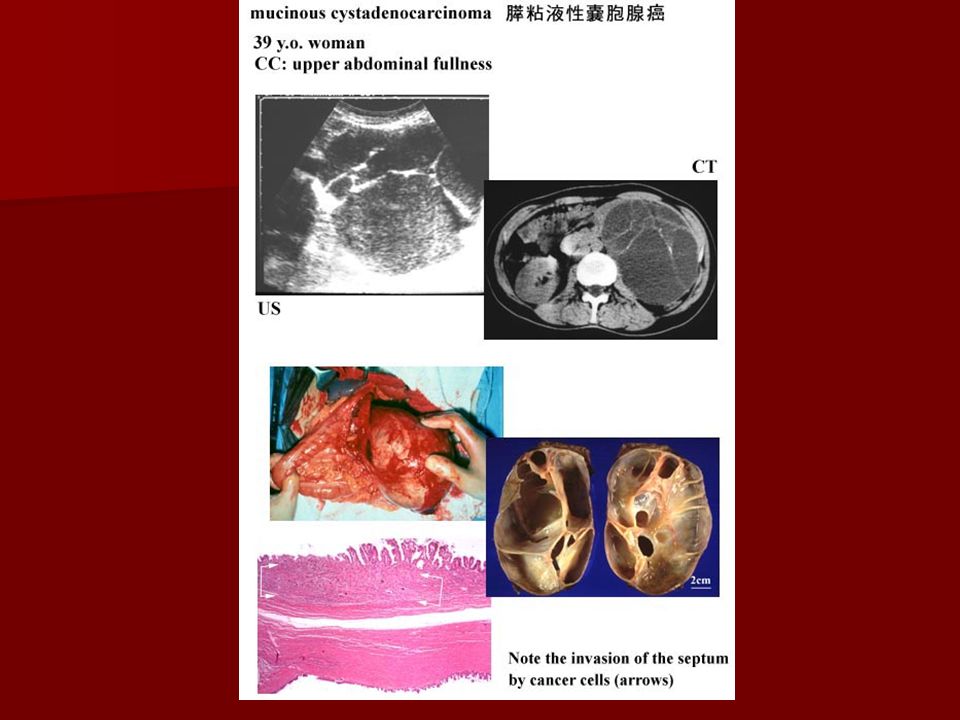
Cystadenocarcinoma Cystadenocarcinoma of the pancreas is a rare type of pancreatic cancer that develops from a fluid-filled noncancerous tumor called a cystadenoma. Cystadenocarcinoma of the pancreas is a rare type of pancreatic cancer that develops from a fluid-filled noncancerous tumor called a cystadenoma. It often causes upper abdominal pain and may grow large enough for a doctor to feel it through the abdominal wall. It often causes upper abdominal pain and may grow large enough for a doctor to feel it through the abdominal wall.

75
Mortality/Morbidity Pancreatic carcinoma is unfortunately usually a fatal disease. Pancreatic carcinoma is unfortunately usually a fatal disease. Most patients eventually succumb to the consequences of local invasion and metastatic cancer, and true long-term cures are rare. Most patients eventually succumb to the consequences of local invasion and metastatic cancer, and true long-term cures are rare. Endocrine and cystic neoplasms of the pancreas have much better survival rates than pancreatic adenocarcinoma. Endocrine and cystic neoplasms of the pancreas have much better survival rates than pancreatic adenocarcinoma.

76
History Unfortunately, the initial symptoms are often quite nonspecific and subtle in onset. Unfortunately, the initial symptoms are often quite nonspecific and subtle in onset. Patients typically report the gradual onset of nonspecific symptoms such as anorexia, malaise, nausea, fatigue, and midepigastric or back pain. Patients typically report the gradual onset of nonspecific symptoms such as anorexia, malaise, nausea, fatigue, and midepigastric or back pain. Significant weight loss is a characteristic feature of pancreatic cancer. Significant weight loss is a characteristic feature of pancreatic cancer.

77
History Pain is the most common presenting symptom in patients with pancreatic cancer. Pain is the most common presenting symptom in patients with pancreatic cancer. Typically, it is midepigastric in location, with radiation of the pain sometimes occurring to the mid- or lower-back region. Typically, it is midepigastric in location, with radiation of the pain sometimes occurring to the mid- or lower-back region.

78
History The most characteristic sign of pancreatic carcinoma of the head of the pancreas is painless obstructive jaundice. The most characteristic sign of pancreatic carcinoma of the head of the pancreas is painless obstructive jaundice. –Patients with this sign may come to medical attention before their tumor grows large enough to cause abdominal pain. Pruritus may accompany obstructive jaundice. Pruritus may accompany obstructive jaundice.

79
History Migratory thrombophlebitis (ie, Trousseau sign) and venous thrombosis also occur with higher frequency in patients with pancreatic cancer. Migratory thrombophlebitis (ie, Trousseau sign) and venous thrombosis also occur with higher frequency in patients with pancreatic cancer. Depression is reported to be more common in patients with pancreatic cancer than in patients with other abdominal tumors. Depression is reported to be more common in patients with pancreatic cancer than in patients with other abdominal tumors. –In some patients, depression may be the most prominent presenting symptom.
 and venous thrombosis also occur with higher frequency in patients with pancreatic cancer. Depression is reported to be more common in patients with pancreatic cancer than in patients with other abdominal tumors. Depression is reported to be more common in patients with pancreatic cancer than in patients with other abdominal tumors. –In some patients, depression may be the most prominent presenting symptom..")
81
Physical The physical examination findings in a patient with pancreatic cancer are usually limited to evidence of significant weight loss and some mild-to-moderate midepigastric tenderness. The physical examination findings in a patient with pancreatic cancer are usually limited to evidence of significant weight loss and some mild-to-moderate midepigastric tenderness. Patients with jaundice may have a palpable gallbladder (ie, Courvoisier sign) and may have evidence of skin excoriations from pruritus. Patients with jaundice may have a palpable gallbladder (ie, Courvoisier sign) and may have evidence of skin excoriations from pruritus.
 and may have evidence of skin excoriations from pruritus. Patients with jaundice may have a palpable gallbladder (ie, Courvoisier sign) and may have evidence of skin excoriations from pruritus..")
82
Physical Patients presenting with end-stage disease may have ascites, a palpable abdominal mass, hepatomegaly from liver metastases, or splenomegaly from portal vein obstruction. Patients presenting with end-stage disease may have ascites, a palpable abdominal mass, hepatomegaly from liver metastases, or splenomegaly from portal vein obstruction.

83
Labs –The laboratory findings in patients with pancreatic cancer are usually nonspecific. As with many chronic diseases, a mild normochromic anemia may be present. –Thrombocytosis is also sometimes observed in patients with cancer. –The major useful tumor marker for pancreatic carcinoma is carbohydrate antigen 19-9 (CA 19-9).
..")
84
Gross section of an adenocarcinoma of the pancreas measuring 5 X 6 cm resected from the pancreatic body and tail. Although the tumor was considered to have been fully resected and had not spread to any nodes, the patient died of recurrent cancer within 1 year Gross section of an adenocarcinoma of the pancreas measuring 5 X 6 cm resected from the pancreatic body and tail. Although the tumor was considered to have been fully resected and had not spread to any nodes, the patient died of recurrent cancer within 1 year

85
CTshowing a pancreatic adenocarcinoma of the pancreatic head. The gallbladder (gb) is distended because of biliary obstruction. The superior mesenteric artery (sma) is surrounded by tumor, making this an unresectable T4 lesion. CTshowing a pancreatic adenocarcinoma of the pancreatic head. The gallbladder (gb) is distended because of biliary obstruction. The superior mesenteric artery (sma) is surrounded by tumor, making this an unresectable T4 lesion.
 is distended because of biliary obstruction. The superior mesenteric artery (sma) is surrounded by tumor, making this an unresectable T4 lesion. CTshowing a pancreatic adenocarcinoma of the pancreatic head. The gallbladder (gb) is distended because of biliary obstruction. The superior mesenteric artery (sma) is surrounded by tumor, making this an unresectable T4 lesion..")
86
Treatment The only therapy that has definitively been shown to increase the survival of patients with pancreatic cancer is surgical resection. The only therapy that has definitively been shown to increase the survival of patients with pancreatic cancer is surgical resection. For patients with disease not amenable to curative resection, little has been shown to significantly impact survival. For patients with disease not amenable to curative resection, little has been shown to significantly impact survival. –The mean survival for patients with unresectable disease remains 4-6 months.

87
Other therapies for pancreatic cancer should include palliation of the major symptoms of disease. Other therapies for pancreatic cancer should include palliation of the major symptoms of disease. Chemotherapy Chemotherapy Radiation therapy Radiation therapy Treatment

88
Pancreaticoduodenectomy (Whipple operation) The standard operation for carcinoma of the head of the pancreas is a pancreaticoduodenectomy (Whipple procedure). The standard operation for carcinoma of the head of the pancreas is a pancreaticoduodenectomy (Whipple procedure). This operation involves resection of the pancreatic head; the first, second, and third portions of the duodenum; the distal antrum; and the distal common bile duct This operation involves resection of the pancreatic head; the first, second, and third portions of the duodenum; the distal antrum; and the distal common bile duct
. This operation involves resection of the pancreatic head; the first, second, and third portions of the duodenum; the distal antrum; and the distal common bile duct This operation involves resection of the pancreatic head; the first, second, and third portions of the duodenum; the distal antrum; and the distal common bile duct.")
89
Deterrence/Prevention Smoking is the most significant reversible risk factor for pancreatic cancer. Estimates indicate that smoking accounts for up to 30% of cases of pancreatic cancer. Smoking is the most significant reversible risk factor for pancreatic cancer. Estimates indicate that smoking accounts for up to 30% of cases of pancreatic cancer. A diet high in energy intake and low in fresh fruits and vegetables increases the risk of pancreatic cancer. A diet high in energy intake and low in fresh fruits and vegetables increases the risk of pancreatic cancer. Alcohol consumption does not increase the risk of pancreatic cancer unless it leads to chronic pancreatitis. A multicenter study of more than 2000 patients with chronic pancreatitis showed a 26-fold increase in the risk of developing pancreatic cancer. Alcohol consumption does not increase the risk of pancreatic cancer unless it leads to chronic pancreatitis. A multicenter study of more than 2000 patients with chronic pancreatitis showed a 26-fold increase in the risk of developing pancreatic cancer.

90
Prognosis The mean survival for patients with unresectable disease remains 4-6 months, with a 5-year survival rate of less than 3%. The mean survival for patients with unresectable disease remains 4-6 months, with a 5-year survival rate of less than 3%. The median survival for patients who undergo successful resection (only 20% of patients) is approximately 12-19 months, with a 5-year survival rate of 15-20%. The median survival for patients who undergo successful resection (only 20% of patients) is approximately 12-19 months, with a 5-year survival rate of 15-20%. Although discouraging, these results are still markedly better than those for patients with unresectable pancreatic carcinoma. Although discouraging, these results are still markedly better than those for patients with unresectable pancreatic carcinoma.
 is approximately months, with a 5-year survival rate of 15-20%. The median survival for patients who undergo successful resection (only 20% of patients) is approximately months, with a 5-year survival rate of 15-20%. Although discouraging, these results are still markedly better than those for patients with unresectable pancreatic carcinoma. Although discouraging, these results are still markedly better than those for patients with unresectable pancreatic carcinoma..")
91
Management The management of pancreatic carcinoma is a multidisciplinary process. The management of pancreatic carcinoma is a multidisciplinary process. Most patients initially present to their primary care practitioner with general symptoms such as abdominal pain, weight loss, or fatigue. Most patients initially present to their primary care practitioner with general symptoms such as abdominal pain, weight loss, or fatigue. Patients may also be seen initially by a gastroenterologist if they present with obstructive jaundice. Patients may also be seen initially by a gastroenterologist if they present with obstructive jaundice. Typically, the management of pancreatic cancer would entail consultations with a gastroenterologist, medical oncologist, general surgeon or surgical oncologist, and possibly a radiation oncologist. Typically, the management of pancreatic cancer would entail consultations with a gastroenterologist, medical oncologist, general surgeon or surgical oncologist, and possibly a radiation oncologist.

92
Benign Tumors of the Pancreas –Insulinoma – rare pancreatic tumor secretes insulin 10% cancerous –Gastrinoma – secretes above average levels of gastrin can cause peptic ulcers 50% cancerous –Glucagonoma – secretes glucagon Causes rash 80% cancerous

93
Insulinoma An insulinoma is a rare type of pancreatic tumor that secretes insulin, a hormone that lowers the levels of sugar (glucose) in the blood. An insulinoma is a rare type of pancreatic tumor that secretes insulin, a hormone that lowers the levels of sugar (glucose) in the blood. Only 10% of insulinomas are cancerous. Only 10% of insulinomas are cancerous.
 in the blood. Only 10% of insulinomas are cancerous. Only 10% of insulinomas are cancerous..")
94
Symptoms result from low levels of sugar in the blood. Symptoms result from low levels of sugar in the blood. The symptoms include faintness, weakness, trembling, awareness of the heartbeat (palpitations), sweating, nervousness, and profound hunger. The symptoms include faintness, weakness, trembling, awareness of the heartbeat (palpitations), sweating, nervousness, and profound hunger. Other symptoms include headache, confusion, vision abnormalities, unsteadiness, and marked changes in personality. Other symptoms include headache, confusion, vision abnormalities, unsteadiness, and marked changes in personality. The low levels of sugar in the blood may even lead to a loss of consciousness, seizures, and coma. The low levels of sugar in the blood may even lead to a loss of consciousness, seizures, and coma. Insulinoma
, sweating, nervousness, and profound hunger. The symptoms include faintness, weakness, trembling, awareness of the heartbeat (palpitations), sweating, nervousness, and profound hunger. Other symptoms include headache, confusion, vision abnormalities, unsteadiness, and marked changes in personality. Other symptoms include headache, confusion, vision abnormalities, unsteadiness, and marked changes in personality. The low levels of sugar in the blood may even lead to a loss of consciousness, seizures, and coma. The low levels of sugar in the blood may even lead to a loss of consciousness, seizures, and coma. Insulinoma.")
95
Very low levels of sugar and high levels of insulin in the blood can indicate the presence of an insulinoma. Very low levels of sugar and high levels of insulin in the blood can indicate the presence of an insulinoma. The location must be pinpointed. Imaging tests—such as CT, ultrasound, and arteriography of the intestinal arteries— can be used to locate the tumor, but sometimes exploratory surgery is needed The location must be pinpointed. Imaging tests—such as CT, ultrasound, and arteriography of the intestinal arteries— can be used to locate the tumor, but sometimes exploratory surgery is needed Insulinoma

96
The primary treatment for an insulinoma is surgical removal - cure rate of about 90%. The primary treatment for an insulinoma is surgical removal - cure rate of about 90%. When the insulinoma cannot be completely removed and symptoms continue, several drugs (for example, streptozocin (Zanosar) and octreotide (Sandostatin)can be helpful When the insulinoma cannot be completely removed and symptoms continue, several drugs (for example, streptozocin (Zanosar) and octreotide (Sandostatin)can be helpful Insulinoma
 and octreotide (Sandostatin)can be helpful When the insulinoma cannot be completely removed and symptoms continue, several drugs (for example, streptozocin (Zanosar) and octreotide (Sandostatin)can be helpful Insulinoma.")
98
GASTRINOMA A gastrinoma is a tumor usually in the pancreas or duodenum (the first segment of the small intestine) that produces excessive levels of the hormone gastrin, which stimulates the stomach to secrete acid and enzymes, causing peptic ulcers. A gastrinoma is a tumor usually in the pancreas or duodenum (the first segment of the small intestine) that produces excessive levels of the hormone gastrin, which stimulates the stomach to secrete acid and enzymes, causing peptic ulcers.
 that produces excessive levels of the hormone gastrin, which stimulates the stomach to secrete acid and enzymes, causing peptic ulcers..")
99
Most people with gastrinomas have several tumors clustered in or near the pancreas. Most people with gastrinomas have several tumors clustered in or near the pancreas. About half of the tumors are cancerous. About half of the tumors are cancerous. Sometimes a gastrinoma occurs as part of multiple endocrine neoplasia, a hereditary disorder in which tumors arise from the cells of various endocrine glands, such as the insulin producing cells of the pancreas. Sometimes a gastrinoma occurs as part of multiple endocrine neoplasia, a hereditary disorder in which tumors arise from the cells of various endocrine glands, such as the insulin producing cells of the pancreas. GASTRINOMA

100
The excess gastrin secreted by the gastrinoma causes Zollinger-Ellison syndrome (ZES) The excess gastrin secreted by the gastrinoma causes Zollinger-Ellison syndrome (ZES) –a rare disorder that causes tumors in the pancreas and duodenum and aggressive peptic ulcers in the stomach and duodenum GASTRINOMA
 The excess gastrin secreted by the gastrinoma causes Zollinger-Ellison syndrome (ZES) –a rare disorder that causes tumors in the pancreas and duodenum and aggressive peptic ulcers in the stomach and duodenum GASTRINOMA")
101
Synonyms of Zollinger Ellison Syndrome Gastrinoma Gastrinoma Pancreatic Ulcerogenic Tumor Syndrome Pancreatic Ulcerogenic Tumor Syndrome Z-E Syndrome Z-E Syndrome ZES ZES

102
However, as many as 25% of people with (ZES) Zollinger-Ellison syndrome may not have an ulcer when the diagnosis is made. However, as many as 25% of people with (ZES) Zollinger-Ellison syndrome may not have an ulcer when the diagnosis is made. Rupture, bleeding, and obstruction of the intestine can occur and are life threatening. Rupture, bleeding, and obstruction of the intestine can occur and are life threatening. GASTRINOMA
 Zollinger-Ellison syndrome may not have an ulcer when the diagnosis is made. Rupture, bleeding, and obstruction of the intestine can occur and are life threatening. Rupture, bleeding, and obstruction of the intestine can occur and are life threatening. GASTRINOMA.")
103
For more than half of the people with a gastrinoma, symptoms are no worse than those experienced by people with ordinary peptic ulcer disease. For more than half of the people with a gastrinoma, symptoms are no worse than those experienced by people with ordinary peptic ulcer disease. In 25 to 40% of people, diarrhea is the first symptom. In 25 to 40% of people, diarrhea is the first symptom. GASTRINOMA

104
a gastrinoma is suspected when a person has frequent peptic ulcers or several peptic ulcers that do not respond to the usual ulcer treatments. a gastrinoma is suspected when a person has frequent peptic ulcers or several peptic ulcers that do not respond to the usual ulcer treatments. Blood tests to detect abnormally high levels of gastrin are the most reliable diagnostic tests. Blood tests to detect abnormally high levels of gastrin are the most reliable diagnostic tests. GASTRINOMA

105
High doses of proton pump inhibitors may be effective for reducing acid levels and relieving symptoms temporarily. High doses of proton pump inhibitors may be effective for reducing acid levels and relieving symptoms temporarily. About 20% of people who do not have multiple endocrine neoplasia can be cured with surgical removal of the gastrinoma. About 20% of people who do not have multiple endocrine neoplasia can be cured with surgical removal of the gastrinoma. If these treatments fail, a total gastrectomy may be necessary. If these treatments fail, a total gastrectomy may be necessary. GASTRINOMA

106
This operation does not remove the tumor, but the gastrin can no longer create ulcers after the acid-producing stomach is removed. This operation does not remove the tumor, but the gastrin can no longer create ulcers after the acid-producing stomach is removed. If the stomach is removed, daily oral iron and calcium supplements and monthly injections of vitamin B12 are needed, because absorption of these nutrients is impaired when stomach juices that prepare these nutrients for absorption are no longer available. If the stomach is removed, daily oral iron and calcium supplements and monthly injections of vitamin B12 are needed, because absorption of these nutrients is impaired when stomach juices that prepare these nutrients for absorption are no longer available. GASTRINOMA

107
Gastrinoma

108
If cancerous tumors have spread to other parts of the body, chemotherapy may help reduce the number of tumor cells and the levels of gastrin in the blood. If cancerous tumors have spread to other parts of the body, chemotherapy may help reduce the number of tumor cells and the levels of gastrin in the blood. However, such therapy does not cure the cancer, which is ultimately fatal. However, such therapy does not cure the cancer, which is ultimately fatal. GASTRINOMA

109
Glucagonoma A glucagonoma is a tumor of the pancreas that produces the hormone glucagon, which raises the level of sugar (glucose) in the blood and produces a distinctive rash. A glucagonoma is a tumor of the pancreas that produces the hormone glucagon, which raises the level of sugar (glucose) in the blood and produces a distinctive rash.
 in the blood and produces a distinctive rash..")
110
About 80% of glucagonomas are cancerous. About 80% of glucagonomas are cancerous. However, they grow slowly, and many people survive for 15 years or more after the diagnosis. However, they grow slowly, and many people survive for 15 years or more after the diagnosis. The average age at which symptoms begin is 50. The average age at which symptoms begin is 50. About 80% of people with glucagonomas are women. About 80% of people with glucagonomas are women. Glucagonoma

111
High levels of glucagon in the blood cause the symptoms of diabetes mellitus. High levels of glucagon in the blood cause the symptoms of diabetes mellitus. Often, the person loses weight. Often, the person loses weight. In 90% of people, the most distinctive features are a chronic reddish brown skin rash (necrolytic migratory erythema) and a smooth, shiny, bright red-orange tongue. In 90% of people, the most distinctive features are a chronic reddish brown skin rash (necrolytic migratory erythema) and a smooth, shiny, bright red-orange tongue. Glucagonoma
 and a smooth, shiny, bright red-orange tongue. In 90% of people, the most distinctive features are a chronic reddish brown skin rash (necrolytic migratory erythema) and a smooth, shiny, bright red-orange tongue. Glucagonoma.")
112
The mouth also may have cracks at the corners. The mouth also may have cracks at the corners. The rash, which causes scaling, starts in the groin and moves to the buttocks, forearms, and legs. The rash, which causes scaling, starts in the groin and moves to the buttocks, forearms, and legs. Glucagonoma

113
Glucagonoma syndrome -- necrolytic migratory erythema

114
The diagnosis is made by identifying high levels of glucagon in the blood and then locating the tumor by arteriography The diagnosis is made by identifying high levels of glucagon in the blood and then locating the tumor by arteriography Glucagonoma

115
Ideally, the tumor is surgically removed, which eliminates all symptoms. Ideally, the tumor is surgically removed, which eliminates all symptoms. However, if removal is not possible or if the tumor has spread, chemotherapy may reduce the levels of glucagon and lessen the symptoms. However, if removal is not possible or if the tumor has spread, chemotherapy may reduce the levels of glucagon and lessen the symptoms. However, chemotherapy does not improve survival. However, chemotherapy does not improve survival. Glucagonoma

116
The drug octreotide also reduces glucagon levels, may clear up the rash, and may restore appetite, facilitating weight gain. The drug octreotide also reduces glucagon levels, may clear up the rash, and may restore appetite, facilitating weight gain. Octreotide may elevate the levels of sugar in the blood even more. Octreotide may elevate the levels of sugar in the blood even more. Glucagonoma

117
Zinc ointment may be used to treat the skin rash. Zinc ointment may be used to treat the skin rash. Sometimes the rash is treated with intravenous amino acids or fatty acids. Sometimes the rash is treated with intravenous amino acids or fatty acids. Glucagonoma

Similar presentations


 EXOCRINE PANCREASE Lecture Dr. Gamal Hamra Wednesday 01/12/1430 (18/11/2009)>")
>")
 The McGraw-Hill Companies, Inc. Permission required for reproduction or display. 23-1 Chapter 23 Abdominal and Gastrointestinal Disorders.>")


