Download presentation
Presentation is loading. Please wait.

1
FUNCTIONAL ORGANIZATION OF THE NERVOUS SYSTEM
1. Overview: The Central and Peripheral Nervous Systems The central nervous system Brain Spinal cord The peripheral nervous system Peripheral nerves somatic portion

2
The nervous system consists of the central nervous system (CNS) and the peripheral nervous system (PNS).
 and the peripheral nervous system (PNS).")
3
autonomic portion sympathetic nerves parasympathelic nerves Ganglia 2. Cells of the Nervous System A. Neurons Structures Dendrites Cell body - soma Axon Synaptic terminal

5
Classification By structure Multipolar Bipolar Unipolar By function Sensory neuron (afferent neuron) Motor neuron (efferent neuron) Interneuron
 Interneuron.")
6
Pseudounipolar sensory neurons appear to have a single process
Pseudounipolar sensory neurons appear to have a single process*However, during development, the axon and dendrite fused, creating a single process called an axon*Bipolar sensory neurons have two relatively equal fibers extending off central cell body

7
Axonal transport Antigrade transport Fast: 400mm/day (organelles: synaptic vesicles, mitochondria, etc.) Slow:1-2mm/day (structural proteins: actin, microtubules, etc.) Kinesin: a protein motor Retrograde transport Dynein
 Kinesin: a protein motor. Retrograde transport. Dynein.")
8
Slow axonal transport moves slowly by axoplasmic flow from body out axon*Fast axonal transport uses microtubules as tracks and quickly moves substances back and forth between body and axon Axonal transport of membranous organelles occurs in two forms: slow axonal transport, and fast axonal transport.

9
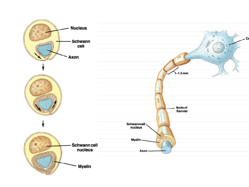
B. Glia (Glial cells) Oligodendroglia (CNS)/Schwann cells (PNS) Myelination Nodes of Ranvier astroglia Fibrous astrocyte Protoplasmic astrocyte Microglia

11
In the CNS, oligodendrocytes form myelin around portions of several interneuron axons.
IAstrocytes contact both neurons and blood vessels, but do not form myelin*They may transfer nutrients between blood vessels and neurons

12
Structural map of the location and function of glial cells.
Map indicates which glial cells are found in PNS, and which in CNS, and functions for each*Note which glial cells produce the myelin sheath in each region, which is important in repair. Structural map of the location and function of glial cells.

13
3. The Membrane Potential and the Action Potential
A. Resting membrane potential (RMP) RMP: -60 TO -90 mV predominately diffusion potential (largely K+) Ion channels of nervous membranes Passive ion channels found in all areas of the nerve cell channel specificity Chemical activated ion channels located predominantly on dendrites and the soma
 RMP: -60 TO -90 mV. predominately diffusion potential (largely K+) Ion channels of nervous membranes. Passive ion channels. found in all areas of the nerve cell. channel specificity. Chemical activated ion channels. located predominantly on dendrites and the soma.")
14
normally closed also known as receptors Voltage-activated ion channels found in axons and soma open at certain voltage responsible for generating and propagating action potential Electrochemical gradients across the cell membrane e.g. K+ RMP = Equilibrium potential (E), can be mathematically calculated
, can be mathematically calculated.")
15
Nernst equation: E[ion]= RT/zF x ln([ion]o/[ion]i) where z is the ion valance and F is the faraday constant or EK = 58 x log10([K+]o/[ K+]i) at 20° C e.g. EK = -75mV, ENa = +55 mV Goldman equation:
![Nernst equation: E[ion]= RT/zF x ln([ion]o/[ion]i) where z is the ion valance and F is the faraday constant.](http://slideplayer.com/slide/9924215/32/images/15/Nernst+equation%3A+E%5Bion%5D%3D+RT%2FzF+x+ln%28%5Bion%5Do%2F%5Bion%5Di%29+where+z+is+the+ion+valance+and+F+is+the+faraday+constant..jpg "or EK = 58 x log10([K+]o/[ K+]i) at 20° C. e.g. EK = -75mV, ENa = +55 mV. Goldman equation:")
16
B. The action potential A temporary change in the membrane potential Components of action potential Resting potential membrane depolarization threshold action potention repolariztion hyperpolarization Ion channels and the action potential Na+ channel: Resting state Activation state Inactivation state

17
K+ channel: Resting state Slow activation state Phases of the action potential and corresponding movements of ions Refractory periods Absolute refractory period Relative refractory period Propagation of the action potential

18
Membrane potential begins at resting potential, rises to threshold & rapidly depolarizes and then repolarizes (period of hyperpolarization) back to resting*Membrane becomes very permeable to Na+ during depolarization, and to K+ during repolarization Diagram indicates the membrane potential events and the changes in membrane ion permeability during an action potential.

19
Sodium cannot diffuse into neuron with channel closed
Sodium cannot diffuse into neuron with channel closed*Resting membrane potential is maintained Depolarizing stimulus arrival opens the activation gate in the channel. Entry of sodium ions depolarizes the membrane potential Inactivation gate finally closed*Membrane potential has reached +30 mvolts

20
Once the two gates have reset to their original positions, the absolute refractory period for the neuron is finished This is a positive feedback loop since depolarization is enhanced as more sodium ions enter cell, allowing more entry*Repolarization as potassium leaves breaks the loop

21
During the relative refractory period, a larger-than-normal stimulus can initiate a new action potential*During this period, the Na+ channel gates are resetting to their resting positions, and the K+ channels are open

22
There are many action potentials taking place along an axon at any point in time
Twelve electrodes have been placed along the axon; the recordings from them are shown below the axon In the distal parts of the axon, local current flow from the active region causes new sections of the membrane to depolarize Voltage-gated Na+ channels open and Na+ enters the axon

23
Velocity of the action potential Unmyelinated axon: diameter
Myelinated nerve fibers Diameter of the axon Distance between nodes of Ranvier - saltatory conduction Action potentials appear to jump from one node of Ranvier to the next when local current flow moves rapidly through the myelinated sections between the nodes*Voltage-gated Na+ channels are scarce between the nodes Since there are few Na+ channels in the demyelinated regions, the action potentials are not conducted as strongly between the nodes

24
Subthreshold graded potential does not fire action potential when blood K+ conc. is in the normal range (normokalemia). Above-threshold (suprathreshold) stimulus will fire an action potential when K+ concentration is normal. Increased blood K+ conc. brings membrane closer to threshold. Now a subthreshold stimulus can trigger action potential. Decreased blood K+ concentration hyperpolarizes membrane and makes the neuron less likely to fire an action potential.
 stimulus will fire an action potential when K+ concentration is normal. Increased blood K+ conc. brings membrane closer to threshold. Now a subthreshold stimulus can trigger action potential. Decreased blood K+ concentration hyperpolarizes membrane and makes the neuron less likely to fire an action potential.")
25
Communication between nerve cells: synaptic transmission
Electrical synapses Gap junctions Connexon Connexin Communication between adjacent cells By passing molecules between cells Found between axons/soma axons/dendrites dendrites/dendrites soma/soma

26
Provide a rapid communication between cells
Synchronize the activity of many adjoining cells Chemical synapses Mediate communication between distant cells by transmitter-receptor interaction Components of chemical synapses Presynaptic cell Postsynaptic cell Synaptic cleft

27
Action potential invading synaptic terminal activation of voltage-sensitive Ca2+ channels Ca2+ in the terminal release of neurotransmitters coupling of the transmitter with the receptor change in postsynaptic potential The axon terminal contains mitochondria and synaptic vesicles filled with neurotransmitter*The postsynaptic membrane has receptors for neurotransmitter that diffuses across the synaptic cleft

28
Postsynaptic potential
Single channel current Synaptic current Unitary postsynaptic potential Summation of postsynaptic potential Excitatory postsynaptic potentials (EPSPs) Inhibitory postsynaptic potentials (IPSPs) Termination of synaptic transmission Reuptake Degradation
 Inhibitory postsynaptic potentials (IPSPs) Termination of synaptic transmission. Reuptake. Degradation.")
29
Acetylcholine (ACh) is made from choline and acetyl CoA in the axon terminal*In the synaptic cleft ACh is rapidly broken down by the enzyme acetylcholinesterase*Choline is transported back into the axon terminal and is used to make more Ach
 is made from choline and acetyl CoA in the axon terminal*In the synaptic cleft ACh is rapidly broken down by the enzyme acetylcholinesterase*Choline is transported back into the axon terminal and is used to make more Ach")
30
Many neurotransmitters create rapid, short-acting responses by opening ion channels*Some neurotransmitters create slower, longer-lasting responses by activating second messenger systems

31
The enzymes stop neurotransmitter activity by breaking down the molecule

32
Divergent pathways, such as the one shown, can be responsible for the widespread effects of the sympathetic nervous system, whose neurons show wide divergence Convergent pathways, such as the one shown, allow input from many different source to influence the activity of the postsynaptic cell, by summation

33
A single neuron many have synapses with as many as 10,000 presynaptic neurons, creating an amazing capacity for summation

34
The 3 neurons are excitatory neurons
The 3 neurons are excitatory neurons*Their graded potentials separately are all below threshold*But if their graded potentials arrive at trigger zone together they sum to create a suprathreshold signal

35
The two excitatory potentials are diminished by summation with the inhibitory potential*No action potential is generated since the summed potentials are below threshold.

36
The same two subthreshold potentials must arrive at the trigger zone within a period of time shorter than the total destruction of their released neurotranmitters, so that their summation can create an action potential

37
Note that in this way, one target of the neuron can be selectively inhibited
In postsynaptic inhibition, all targets of the postsynaptic cell will be inhibited equally

Similar presentations


 - made.>")


