Download presentation
Presentation is loading. Please wait.

1
The Skeleton

2
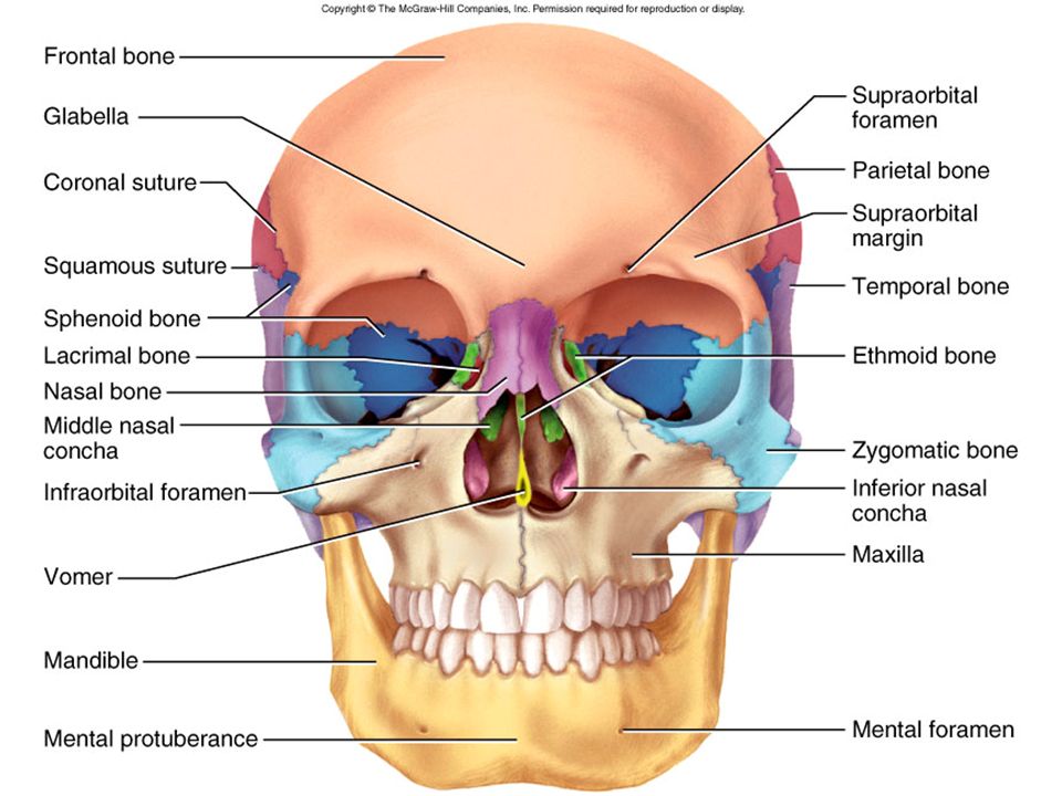
Figure 8.1a

3
Figure 8.1a

4
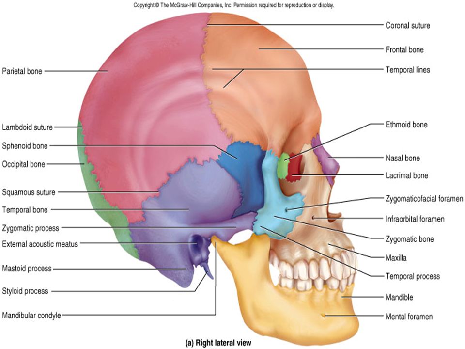
Figure 8.1b

5
Figure 8.1b

8
Figure 8.5a

9
Figure 8.5b

10
Figure 8.6

11
Head Trochanter Tubercle Head Epicondyle Sulcus Sinus Tuberosity
Copyright © The McGraw-Hill Companies, Inc. Permission required for reproduction or display. Head Trochanter Tubercle Head Epicondyle Sulcus Sinus Tuberosity Fissure Process Canal (meatus) Ramus Foramen Epicondyle Condyle Fossa Alveolus Epicondyle Condyle Ramus Trochlea Femur Skull, anterior view Skull, sagittal view Humerus Facet Crest Fossa Spine Line Foramen Ramus Pelvis
 Ramus. Foramen. Epicondyle. Condyle. Fossa. Alveolus. Epicondyle. Condyle. Ramus. Trochlea. Femur. Skull, anterior view. Skull, sagittal view. Humerus. Facet. Crest. Fossa. Spine. Line. Foramen. Ramus. Pelvis.")
12
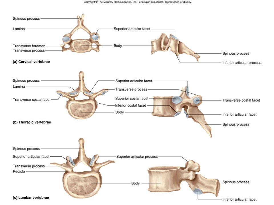
Transverse process (a) Superior view
Copyright © The McGraw-Hill Companies, Inc. Permission required for reproduction or display. Spinous process Transverse process Lamina Superior articular process and facet Vertebral arch Pedicle Vertebral foramen Body (a) Superior view
 Superior view.")
13
(a) 2nd lumbar vertebra (L2)
Copyright © The McGraw-Hill Companies, Inc. Permission required for reproduction or display. Posterior Spinous process Superior articular facet Lamina Vertebral arch Transverse process Pedicle Vertebral foramen Body Anterior (a) 2nd lumbar vertebra (L2) Nucleus pulposus Anulus fibrosus (b) Intervertebral disc
 2nd lumbar vertebra (L2) Nucleus pulposus. Anulus fibrosus. (b) Intervertebral disc.")
14
Slightly Movable Joints Freely Movable Joint Fibrous Joints
Copyright © The McGraw-Hill Companies, Inc. Permission required for reproduction or display. Immovable Joint Bone Suture Fibrous connective tissue a. Slightly Movable Joints Freely Movable Joint Fibrous Joints Fibrous capsule Synovial membrane Synovial fluid Articular cartilage Fibrous joints Cartilaginous Joints c. Body of vertebra Intervertebral disk Articular cartilage b.

15
Lateral (axillary) border
Copyright © The McGraw-Hill Companies, Inc. Permission required for reproduction or display. Acromion process Acromion process Coracoid process Glenoid cavity Glenoid cavity Spine Lateral (axillary) border Medial (vertebral) border Lateral (axillary) border Inferior angle (a) Anterior view (b) Posterior view Body of clavicle Sternal (medial) end Acromial (lateral) end (c) Superior view
 border. Medial (vertebral) border. Lateral (axillary) border. Inferior angle. (a) Anterior view. (b) Posterior view. Body of clavicle. Sternal (medial) end. Acromial (lateral) end. (c) Superior view.")
16
A A B C C D F F D E (a) Anterior view (b) Posterior view
Copyright © The McGraw-Hill Companies, Inc. Permission required for reproduction or display. A A B C C D F F D (a) Anterior view (b) Posterior view E (c) Superior view
 Anterior view. (b) Posterior view. E. (c) Superior view.")
17
Anatomical neck Head Greater tubercle Lesser tubercle Anatomical neck
Copyright © The McGraw-Hill Companies, Inc. Permission required for reproduction or display. Anatomical neck Head Greater tubercle Lesser tubercle Anatomical neck Intertubercular (bicipital) groove Deltoid tuberosity Olecranon fossa Lateral epicondyle Lateral epicondyle Medial epicondyle Capitulum Trochlea Trochlea (a) Anterior view (b) Posterior view
 groove. Deltoid. tuberosity. Olecranon. fossa. Lateral. epicondyle. Lateral. epicondyle. Medial. epicondyle. Capitulum. Trochlea. Trochlea. (a) Anterior view. (b) Posterior view.")
18
Anatomical neck Head Greater tubercle Lesser tubercle Anatomical neck
Copyright © The McGraw-Hill Companies, Inc. Permission required for reproduction or display. Anatomical neck Head Greater tubercle Lesser tubercle Anatomical neck Intertubercular (bicipital) groove Deltoid tuberosity Olecranon fossa Lateral epicondyle Lateral epicondyle Medial epicondyle Capitulum Trochlea Trochlea (a) Anterior view (b) Posterior view
 groove. Deltoid. tuberosity. Olecranon. fossa. Lateral. epicondyle. Lateral. epicondyle. Medial. epicondyle. Capitulum. Trochlea. Trochlea. (a) Anterior view. (b) Posterior view.")
19
Olecranon Olecranon Trochlear notch Head Coronoid process Neck
Copyright © The McGraw-Hill Companies, Inc. Permission required for reproduction or display. Olecranon Olecranon Trochlear notch Head Coronoid process Neck Proximal Radioular joint Neck Radial tuberosity Radius Ulna Radius Ulna Interosseous membrane Distal Radioulnar joint Head Styloid process Styloid process Styloid process

20
Middle phalanx of finger
Copyright © The McGraw-Hill Companies, Inc. Permission required for reproduction or display. Radius Ulna Carpals (distal row) Scaphoid bone Scaphoid bone Carpals (proximal row) Hamate bone Carpals (proximal row) Lunate bone Lunate bone Triquetrum bone Capitate bone Triquetrum bone Pisiform bone Trapezoid bone Pisiform bone Trapezium bone 1 1 Metacarpals 5 2 4 3 2 5 3 4 Proximal phalanx of thumb Distal phalanx of thumb Digits Proximal phalanx of finger Middle phalanx of finger Distal phalanx of finger (a) Posterior view (b) Anterior view
 Scaphoid bone. Scaphoid bone. Carpals. (proximal. row) Hamate bone. Carpals. (proximal. row) Lunate bone. Lunate bone. Triquetrum bone. Capitate bone. Triquetrum bone. Pisiform bone. Trapezoid bone. Pisiform bone. Trapezium bone Metacarpals Proximal. phalanx. of thumb. Distal. phalanx. of thumb. Digits. Proximal. phalanx. of finger. Middle phalanx of finger. Distal phalanx of finger. (a) Posterior view. (b) Anterior view.")
21
Anterior superior iliac spine
Copyright © The McGraw-Hill Companies, Inc. Permission required for reproduction or display. Iliac crest Scrum Sacroiliac joint Ilium Anterior superior iliac spine Os coxae Coccyx Pubis Acetabulum Obturator foramen Ischium Pubic symphysis

22
Head Head Fovea capitis Greater trochanter Greater trochanter Neck
Copyright © The McGraw-Hill Companies, Inc. Permission required for reproduction or display. Head Head Fovea capitis Greater trochanter Greater trochanter Neck Neck Lesser trochanter Gluteal tuberosity Linea aspera Body (shaft) of femur Adductor tubercle Medial epicondyle Lateral epicondyle Lateral epicondyle Intercondylar fossa Lateral condyle Medial condyle Patellar groove (a) Anterior view (b) Posterior view
 of femur. Adductor tubercle. Medial. epicondyle. Lateral epicondyle. Lateral epicondyle. Intercondylar fossa. Lateral condyle. Medial. condyle. Patellar groove. (a) Anterior view. (b) Posterior view.")
23
Articular surface for talus
Copyright © The McGraw-Hill Companies, Inc. Permission required for reproduction or display. Medial condyle Lateral condyle Lateral condyle Superior tibiofibular joint Medial condyle Articular surface of fibular head Head Head Neck of fibula Neck Tibial tuberosity Tibia Fibula Fibula Tibia Inferior Tibiofibular joint Medial malleolus Lateral malleolus Lateral malleolus Medial malleolus (a) Right tibia and fibula, anterior view Articular surface for talus Articular surface for talus
 Right tibia and fibula, anterior view. Articular surface for talus. Articular surface for talus.")
24
Medial longitudinal arch
Copyright © The McGraw-Hill Companies, Inc. Permission required for reproduction or display. Calcaneus Talus Cuboid Tarsals Navicular Medial cuneiform Intermediate cuneiform Metatarsals 5 Lateral cuneiform 4 3 2 1 Proximal phalanx Digits Middle phalanx Distal phalanx Proximal phalanx of great toe Distal phalanx of great toe (a) Superior view Talus Fibula Navicular Tibia Intermediate cuneiform Medial cuneiform Medial longitudinal arch Talus Lateral longitudinal arch Transverse arch Calcaneus Cuboid Phalanges Metatarsals Tarsals (b) Medial inferior view
 Superior view. Talus. Fibula. Navicular. Tibia. Intermediate cuneiform. Medial cuneiform. Medial longitudinal arch. Talus. Lateral. longitudinal. arch. Transverse. arch. Calcaneus. Cuboid. Phalanges. Metatarsals. Tarsals. (b) Medial inferior view.")
25
A B C D E

26
Transverse process (a) Superior view
Copyright © The McGraw-Hill Companies, Inc. Permission required for reproduction or display. Spinous process Transverse process Lamina Superior articular process and facet Vertebral arch Pedicle Vertebral foramen Body (a) Superior view
 Superior view.")
27
(a) 2nd lumbar vertebra (L2)
Copyright © The McGraw-Hill Companies, Inc. Permission required for reproduction or display. Posterior Spinous process Superior articular facet Lamina Vertebral arch Transverse process Pedicle Vertebral foramen Body Anterior (a) 2nd lumbar vertebra (L2) Nucleus pulposus Anulus fibrosus (b) Intervertebral disc
 2nd lumbar vertebra (L2) Nucleus pulposus. Anulus fibrosus. (b) Intervertebral disc.")
28
Olecranon Olecranon Trochlear notch Head Coronoid process Neck
Copyright © The McGraw-Hill Companies, Inc. Permission required for reproduction or display. Olecranon Olecranon Trochlear notch Head Coronoid process Neck Proximal Radioular joint Neck Radial tuberosity Radius Ulna Radius Ulna Interosseous membrane Interosseous borders Distal Radioulnar joint Head Styloid process Styloid process Styloid process

29
Figure 8.22

30
Figure 8.22a

31
Figure 8.23

32
Figure 8.23b

33
Figure 8.24

34
Figure 8.24c

36
Figure 8.26

37
Figure 8.27

38
Figure 8.29

39
Figure 8.31

40
Figure 8.31a

41
Figure 8.31b

42
Figure 8.32

43
Figure 8.33

44
Figure 8.34a

45
Figure 8.35a

46
Figure 8.35a

47
Figure 8.38

48
Figure 8.39

49
Figure 8.40a

50
Figure 8.40b

51
Figure 8.1

52
Figure 8.1b

53
Figure 8.2c

54
Figure 8.2d

55
Figure 8.19

56
Figure 8.22

57
Figure 8.22c

58
Figure 8.23

59
Figure 8.23a

60
Figure 8.23b

61
Figure 8.24

62
Figure 8.24c

63
Figure 8.25

64
Figure 8.33a

65
Figure 8.33b

66
Figure 8.34a

67
Figure 8.35a

68
Figure 8.38

69
Figure 8.38a

70
Figure 8.38b

71
Figure 8.39

72
Figure 8.39a

73
Figure 8.39b

74
Figure 8.40

Similar presentations