Download presentation
Presentation is loading. Please wait.

1
ألأستاذ الدكتورعبدالسلام المختار Lec.(3). ENTAMOEBA HISTOLYTICA OBJECTIVES: 1.Morphology and Life cycle. 2.Amoebiasis.

2
Morphology ( Trophozoite ): 1- Clear ectoplasm. 2- Large finger – like pseudopdia 3- The endoplasm is granular and may contain RBCs. 4- It has one nucleous, contain small central keryosome and fine chromatin granules arrenged regularly beneath nuclear membrane.

3
Trohozoites of E.histolytica,one show very clear pseudopodia and the other show very clear central kariosome and periferal chromatin granules.

4
Morphology ( mature cyst) : 1- Small, spherical in shape, containing 4 nuclei is usually found in feces. Each nucleous contain similar nuclear morphology like the trophozoite.

5
Life cycle of E. histolytica: Cysts are passed in feces. Infection by E. histolytica occurs by ingestion of mature cysts in fecally contaminated food, water, or hands. Excystation occurs in the small intestine and trophozoites are released which migrate to the large intestine. The trophozoites multiply by binary fission and produce cysts, which are passed in the feces.

6
cyst trophozoite

7
Flagellates: Giardia lamblia Dientamoeba fragilis Chilomastix mesnili Trichomonas hominis Enteromonas hominis Retortamonas intestinalis Ameba: Entamoeba histolytica Entamoeba dispar Entamoeba coli Entamoeba hartmanni Endolimax nana Iodamoeba bütschlii Apicomplexa: Cryptosporidium hominis Cryptosporidium parvum Cyclospora cayetanensis Isospora belli Other: Blastocystis hominis Balantidium coli INTESTINAL PROTOZOA Life cycle of E.histolytica.

8
Extraintestinal Amebiasis metastasis via blood stream primarily liver (portal vein) other sites less frequent ameba-free stools common high antibody titers
 other sites less frequent ameba-free stools common high antibody titers")
9
Because of the protection conferred by their walls, the cysts can survive days to-weeks in the external environment and are responsible for ttransmission. Trophozoites can also be passed in diarrheal stools, but are rapidly destroyed once outside the body, and if ingested rapidly destroyed by gastric juice. In many cases, the trophozoites remain in intestinal lumen as noninvasive infection of individuals who

10
are asymptomatic carriers, passing cysts in their stool only. In some patients the trophozoites invade the intestinal mucosa and cause intestinal disease or developed perforated ulcer and the trophozoites migrate through the blood stream to invade the extraintestinal organs such as the liver, brain, and lungs and it will cause amoebic infection in these organs.

11
Epidemiology :. * The incidence of Amebiasis is common & high in tropical & subtropical especially in areas of lower socioeconomic status due to: (1) poor sanitation(2) overcrowding & (3)malntrition. It is estimated that up to 10% of the world`s population may infected with E.hist. Transmision of amoebiasis occure through:
 poor sanitation(2) overcrowding & (3)malntrition. It is estimated that up to 10% of the world`s population may infected with E.hist. Transmision of amoebiasis occure through:.")
12
1. Mature cyst is the main sourse of the infection which passing with the feces of chronic patients or asymptomatic carrier. 2. Human being acquire the infection via contamination of food, drings, vegetables or hands with infective cysts especially in restorants. 3. Flise (House fly) play an important roles in trasnmission of these cysts to the food of human.
 play an important roles in trasnmission of these cysts to the food of human..")
13
Pathogenesis of E.histolytica: The Pathogenic activity of E. histolytica depend upon :

14
1- The resistant of the host. 2- The number of the amebas. 3- Presence of pathogenic bacteria. 4.Presence of physical & chemical injury of the mucosa.

15
The lesions produced by E. histolytica are primarily in large intestine andseconderily extraintestinal especially the liver or may be the brain or every organ of the body may be affected.

16
Pathogenisis of Intestinal lesion : 1.The lesion vary from small ulcer to a large typical flask shape ulcer. 2.The ulcer charecterised with large area of tissue necrosis, cell infiltration & rapid lysis of inflamatory cells. 3.The ulcer has a wide base and narrow opening with irregular elevated edges. 4.The amoebas usually found on the floor of the base of ulcer.

17
E. histolytica in the large intestine ( Flask shape ulser )
")
18
Clinical features of intestinal lesion : 1- The incubation period range from 2 – 4 weeks. 2- The majority of infections with E.histolytica show no symptoms (Asymptomatic cyst) or show symptoms which varies from mild to intense and long lasting.
 or show symptoms which varies from mild to intense and long lasting..")
19
The typical symptoms include : 1- Diarrohea, The diarrohea frequently alternates with constipation or soft stools may contain mucous but no visible blood. 2-Abdominal cramps. 3-Nausia. 4-Anoroxia.

20
5- Dysentery : Which is usually starts slowly with abdominal cramps and associated with loose stools and diarrohea with blood و mucus and necrotic tissues. 6- Few patients especially children may show fever, vomiting, abdominal tender ness.

21
The complications of intestinal amoebiasis: 1- Appendicitis. 2- Intestinal perforation. 3- Hemorrhage. 4- Liver abscess. 5- Ameboma (Granulomas) : a. Are a painful abdominal mass which occur most frequently in the caecum and assending colon.
 : a. Are a painful abdominal mass which occur most frequently in the caecum and assending colon..")
22
b. This lesion may be confused with carcinomas or tumour. c.Obstructive symptoms or dysentery may also be associated with ameboma.

23
Extraintestinal Amoebiasis : 1.The metastasis of amoeba usually via blood streem or by direct extension after intestinal perforation to the peritonium the amoeba may cause local abcsess or peritonitis or migrate to the liver is the most commonly affected than other organs e.g, lungs, perianal skin or brain.

24
Extraintestinal Amebiasi S

25
2.Amoebic liver abscesses: Are the most common extraintestinal amebiasis and characterised By: a.Hepatomegaly, Liver tenderness, fever and anorexia. b. Liver function tests are usually normal or slightly abnormal. c. Liver abscesses will occasionally rupture into the peritoneum causing peritonits

26
3.Pulmonery amoebiasis : a. This infection due to the direct extension of the liver abscess through the diaphragm, or via blood. b.The clinical symptoms are: cough, chest pain, dysnea and fever. c.The sputum may be purulent or contain blood and trophozoites of E. histolytica.

27
4. Cutaneous amoebiasis : It is caused by contact of the skin with amoebic abscess which lead to fistula in the skin.

28
Diagnosis of Amoebiasis : 1- Stool of patient should be examined by : a- Direct method with saline for motile trophozoite. b- Stool specimens should be stained usually with ioden and microscopically examined for cysts opf E.histolytica.

29
2- Culture of stool. 3- Sigmoidoscopy may reveal the charecteristic flask-shaped ulcers especially in sever cases.

30
4- Biopsy & fluid from large intestine aspirates also be examined microscopically for trophozoites. 5- Serology, is very important for the diagnosis of extraintestinal amoebiasis e,g, Indirect haemagglutination (IHA) & Polymirase Chain Reaction (PCR test).
 & Polymirase Chain Reaction (PCR test)..")
31
6- Ultrasound, CTscan, MRI can be used to detect hepatic abscesses. 7- The typical amoebic stool is contain blood, mucous, few WBC & Bacteria.

32
Treatment : 1. A symptomatic patients can be treated with Diiodohydroxyquine with tetracycline.

33
Treatment : 2. Symptomatic patients with diarrhoea or dysentary or extraintesinal amebiasis should be treated as follows : a- Patients should remain in bed and receive a high protein and high vitamin with adequate fluids.

34
b. Chemotherapy for sever amoebiasis: 1.Metronidazol (Flagel) is the drug of choice : 750 mg three times a day, orally for 5 – 10 day. 2.Tetracycline & diiodohydroxyquine are recommended to be given to the patient since metronidzal may not always cure the intestinal infection.
 is the drug of choice : 750 mg three times a day, orally for 5 – 10 day. 2.Tetracycline & diiodohydroxyquine are recommended to be given to the patient since metronidzal may not always cure the intestinal infection..")
35
The side effects of Mitronidazole are : Nausea,haedach, diarrohea and may be carcinogenic in experimental animal.

36
3. Alternative drugs of amoebic dysentery are Emetine hydrochloride, it is effective but it is toxic.Dehydroemtin Dihydrochloride is less toxic than emetine and is equally effective.

37
Prevention & Control : 1- All human infections should be treated because human being is the only chief source of infection. 2- A symptomatic carriers should be treated especially those working in restorants.

38
3- Effective enviromental sanitation is necessary to prevent water,food, and vegitable contamination, e.g. Sewage disposal should be treated with chemical before used as fertiliser in gardens. 4- Chlorination & filtered water supply are important to kill the cyst of E.histolytica.

39
5- Insects should be controlled by insecticides. 6- Uncooked vegetables should be washed with running water..

40
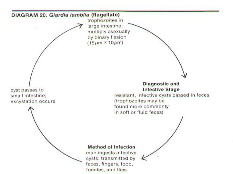
Lec.(5) الدكتور عبد السلام المختار Intestinal and Luminal Flagilates of Human Being. Giardia lambilia (intestinalis) : OBJECTIVES: 1.Medical importance of Giardia lambilia. 2.Morphology and Life cycle. 3.Epidemiology.
 : OBJECTIVES: 1.Medical importance of Giardia lambilia. 2.Morphology and Life cycle. 3.Epidemiology..")
41
Giardia lambilia (intestinalis) : Disease : Giardiasis or lambiliasis.
 : Disease : Giardiasis or lambiliasis.")
42
Morphology : of trophozoite : 1- Bilaterally symetrical,12–15 Mm.in length. 2- Pear shaped flagellate with rounded anterior end and tapering posterior end. 3- The dorsal surface is convex & it has a concave sucking disc ventraly.

43
4- It has two nuclei with large central karyosome 5- There are two axostyle & and two deeply staining parabasal bodies. 6-It has four pair of flagella.

44
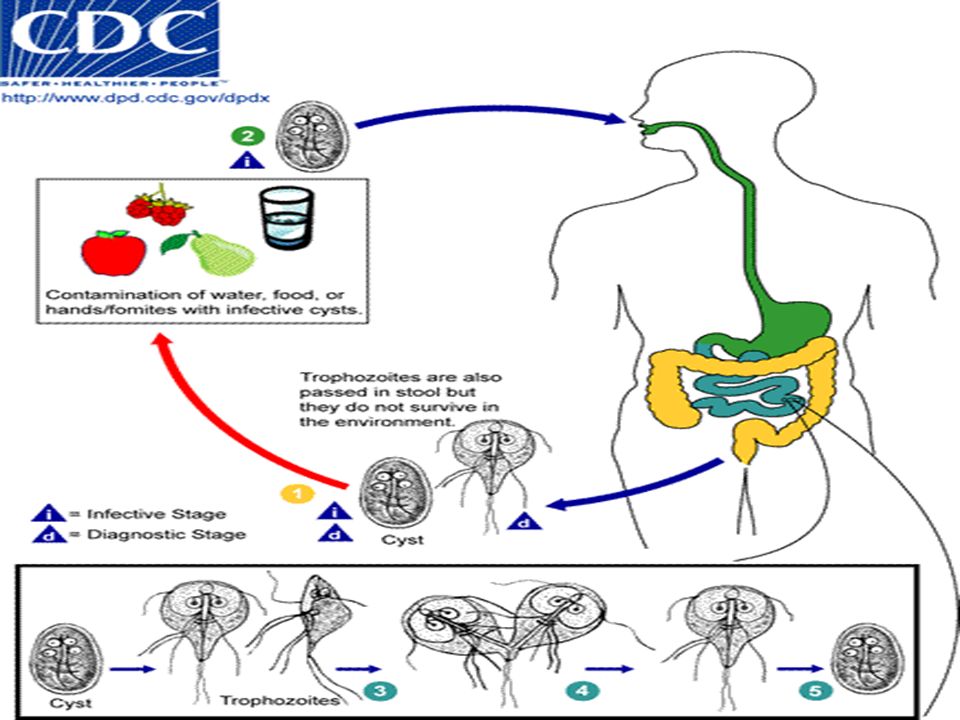
Giardia lamblia CYST infective stage passed in feces TROPHOZOITE replicative stage small intestine

46
Morphology of the cyst: 1- Small, 9 – 12 Mm. in size. 2-Rround or oval in shape & contain four nuclei. 3- It contain remaining parts of the trophozoites.

49
Life cycle : Human being is the natural host of Giardia lamblia inhabit the upper part of small intestine especially duodenum. Multiplication occurs by mitotic division during cyst formation, which pass with feces into soil outside the host and can remain viable for months under moist conditions.

50
Man especially children acquired the infection through the ingestion of cysts via contamineted food, these cysts passes through the stomach into duodenum where excystation takes place & within 30 minutes after emerging from the cyst resulting in two binucleated trophozoites.

51
Epidemiology: Worldwide in its distribution especially in chilidren.It is present in Iraq. The disease transmitted by contamination of food and drinks with infected cysts of G,lambilia. House flys and insects can transmit the infected cysts to the human being by mechanical way.

52
Lec.(6). ألدكتورعبدالسلام المختار OBJECTIVE: 1.Pathogenesis, 2.Clinicai picture of the disease. 3.Diagnosis andTreatment. 4.prevention and control.

53
Pathogenesis: 1.The specific mechanisum of Giardia leading to fatty diarrohea (steatorea). 2.Intestinal malabsorption due to attachment of trophozoites to cover large areas of the intestinal epithelium to produce a mechanical irritation & prevent absorption of food especially fat.

54
3.Giardia infection can also lead to Lactase deficiencies as well as other enyzme deficiencies and this may explain the malabsorption syndromes in giardiasis.

55
Giardia lamblia CYST infective stage passed in feces TROPHOZOITE replicative stage small intestine

56
Adhesive Disk and Attachment

57
Pathogenesis epithelial damage villus blunting crypt cell hypertrophy cellular infiltration malabsorbtion enzyme deficiencies lactase (lactose intolerance) Possible Mechanisms mechanical irritation obstruction of absorption
 Possible Mechanisms mechanical irritation obstruction of absorption")
58
Clinical picture of Giardiasis: The incubation periods is about 1 – 2 weeks. The clinical features associated with Giardia infection are range from Asymptomatic, to acute diarrhea,or may lead to chronic syndromes.

59
Acute Giardiasis : 1.The first signs include : Nausea, loss of appetite and an upper gastro – intestinal pain. 2.Watery or loose fatty diarrhea with absence blood or mucus which may help to distinguish giardiasis from other acute diarrheas.

60
3. Abdominal cramps, malabsorption, steatorrhea (excessive loss of fat in the feces), loss of weight, and bad smelling of the feces.
, loss of weight, and bad smelling of the feces..")
61
Acute infection may also lead to: Chronic infection : which characterised with : 1- Recurrent loose of stools. 2- Abdominal distention. 3- Normal stools or constipation ulternate with watery stools diarrhea.

62
4- Anorexia, nausea and epigastric pain. 5- Nutritional disorder. 6- Weight loss.

63
Diagnosis : 1- By direct examination with Saline & ioden : Is usually made by finding cysts of Giardia in soft stools and trophozoites & cysts in diarroheaic feces

64
2- Examination of duodenal content by aspiration given higher percentage of positive finding of trophozoites than that of the feces.

65
Treatment: Several drugs are available to treat giardiasis but metronidazole and tinidazole are the drugs of choice.

66
Prevention and control : Is just like that of E.histolytica. 1- All human infections should be treated because human being is the only chief source of infection. 2- A symptomatic carriers should be treated especially those working in restorants.

67
3- Effective enviromental sanitation is necessary to prevent water,food, and vegitable contamination, e.g. Sewage disposal should be treated with chemical before used as fertiliser in gardens.

68
4- Chlorination & filtered water supply are important to kill the cyst of G.lamblia. 5-Flies and Insects should be controlled by insecticides because they transmit the infective stage by mechanical methods. 6- Uncooked vegetables should be washed with running water..

Similar presentations


 Giardia lamblia ( 蓝氏贾第鞭毛虫 ) Intestinal flagellate Giardia lambilia lives in small intestine Giardiasis Diarrhea “traveler’s diarrhea”>")


