Download presentation
Presentation is loading. Please wait.

1
The Extracellular Space
Epithelial tissues closely packed cells lining spaces in the body (e.g. skin, intestine, etc) Connective tissues Largely composed of non-living extracellular material (e.g. cartilage, tendon, dermis)
 Connective tissues. Largely composed of non-living extracellular material (e.g. cartilage, tendon, dermis)")
2
The Extracellular Space
Proteins in plasma membrane have sugars attached Glycocalyx Mediate cell interactions Provide mechanical support Barrier to particles Binding sites for regulatory factors

3
The Extracellular Matrix (ECM)
An organized network of materials located beyond the plasma membrane

4
The Extracellular Matrix (ECM)
Basement membranes Thick regions of ECM Surround muscle/fat Underlie the basal surface of epithelial tissues

5
The Extracellular Matrix (ECM)
Basement membranes Separate different tissues Provide mechanical support Barrier to macromolecule and cellular movement Substrate for cell migration Generate signals that maintain cell survival

6
The Extracellular Matrix (ECM)
Collagens (27 different types) High tensile strength (resistant to pulling forces) Alpha-helical trimers bundle together into fibrils Types I, II, III (fibrillar) form rigid cables Adjacent collagens are strengthened by covalent cross-links Hydroxylysine - lysine Type IV (nonfibrillar) can form an interconnected lattice
 High tensile strength (resistant to pulling forces) Alpha-helical trimers bundle together into fibrils. Types I, II, III (fibrillar) form rigid cables. Adjacent collagens are strengthened by covalent cross-links. Hydroxylysine - lysine. Type IV (nonfibrillar) can form an interconnected lattice.")
7
The Extracellular Matrix (ECM)
Collagens (27 different types) Type IV (nonfibrillar) can form an interconnected lattice Composed of helical and non-helical segments (flexibility) Globular domains at each end (lattice contact points) Collagens bind: Fibronectins Integrins (cell surface)
 Type IV (nonfibrillar) can form an interconnected lattice. Composed of helical and non-helical segments (flexibility) Globular domains at each end (lattice contact points) Collagens bind: Fibronectins. Integrins (cell surface)")
8
The Extracellular Matrix (ECM)
Diseases caused by defects in collagen genes Osteogenesis imperfecta Fragile bones Ehlers-Danlos syndrome Hyperflexible joints, highly extensible skin

9
The Extracellular Matrix (ECM)
Proteoglycans Protein core + glycosaminoglycan (GAG) polysaccharide complex Chondroitin sulfate & keratin sulfate High amount of negative charge binds cations and H2O Hydrated gel resists compressive forces Hyaluronic acid links many proteoglycans to form extremely large molecules Fill the scaffold created by collagens
 polysaccharide complex. Chondroitin sulfate & keratin sulfate. High amount of negative charge binds cations and H2O. Hydrated gel resists compressive forces. Hyaluronic acid links many proteoglycans to form extremely large molecules. Fill the scaffold created by collagens.")
10
The Extracellular Matrix (ECM)
Fibronectins Modular domains for interactions Bind collagens, proteoglycans, integrins at cell surface Important for: linking ECM components together, cell attachment to matrix, cell migration NC cells

11
The Extracellular Matrix (ECM)
Laminins 3 polypeptides linked by disulfide bonds Form a second lattice interwoven with Collagen IV lattice Bind to proteoglycans, integrins at cell surface PGC on laminin

12
ECM Remodeling Matrix metalloproteinases (MMPs)
Enzymes that degrade ECM proteins Tissue remodeling Cell migration Wound healing

13
Steps leading to metastatic spread
MMP activity

14
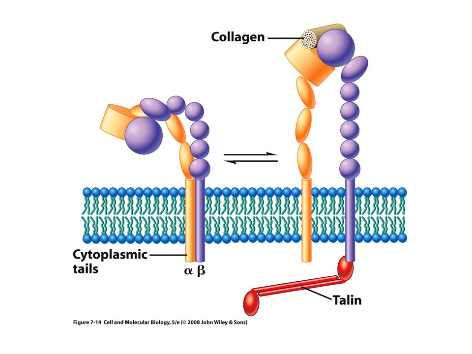
Cell - ECM Interactions
Integrins Only found in animals Heterodimer of alpha and beta subunits 18 alpha and 8 beta subunits known 12 different alpha/beta combinations known Transmembrane proteins Extracellular domain, transmembrane domain, intracellular domain Inside-out signaling Post-translational alterations to cytoplasmic tail regulate conformation changes in extracellular domain Talin separates beta from alpha to open receptor to active state Plasma membrane talin

15
Inactive

16
Active

18
Cell - ECM Interactions
Ligand binding RGD loop of Fibronectin binds to integrin receptor extracellular domain Isolated RGD Loop can be exploited to block platelet aggregation / blood clotting

19
Cell - ECM Interactions
Integrins Two major functions Adhesion to substrate Receptors cluster increasing overall strength Signal transmission Binding of ligand (collagen) can change cytoplasmic domain Cytoplasmic domain can activate kinases such as FAK and Src Activated kinases can transmit signals to nucleus and change gene expression
 can change cytoplasmic domain. Cytoplasmic domain can activate kinases such as FAK and Src. Activated kinases can transmit signals to nucleus and change gene expression.")
20
Cell - ECM Interactions
Structures important for adhesion to substrate Focal adhesions: Scattered, discrete, transient, dynamic, rapidly form and break Clusters of integrins bound to collagen / Fibronectin Cytoplasmic domains attach to cytoskeleton connecting exterior forces to internal signals Actin filaments Focal adhesion kinase (FAK)
")
21
Forces exerted by focal adhesions

22
Cell - ECM Interactions
Structures important for adhesion to substrate Hemidesmosome more permanent anchor to basement membrane Integrins bound to laminin to dense collection of intermediate filaments

23
Cell - ECM Interactions
Structures important for adhesion to substrate Hemidesmosome Disease: epidermolysis bullosa Epidermis poorly connected to basement membrane / dermis Fluid accumulates in between = blister (keratins)
")
24
Cell - ECM Interactions

25
Cell - Cell Interactions
Cadherins: Ca2+ dependent adhesion Homophilic interactions allow self-sorting of mixed cell populations Disease role: metastasis of cancer Lose adhesion by downregulating cadherin expression Penetrate / invade barriers by upregulating MMP expression

26
Cell - Cell Interactions
Structures important for cell-cell adhesion Adherens junctions (30nm gap between cells) Cadherin-cadherin interactions in belt-like strips holding two cells together Cytoplasmic domains link via beta-catenin and alpha-catenin to the cytoskeleton
 Cadherin-cadherin interactions in belt-like strips holding two cells together. Cytoplasmic domains link via beta-catenin and alpha-catenin to the cytoskeleton.")
27
Cell - Cell Interactions
Structures important for cell-cell adhesion Desmosomes (1 um diameter disc) Resist mechanical stress Cadherin-cadherin interactions linked to cytoskeleton (intermediate filaments)
 Resist mechanical stress. Cadherin-cadherin interactions linked to cytoskeleton (intermediate filaments)")
28
Cell - Cell Interactions
Tight junctions Seal two membranes together Block paracellular movement Occludin and claudins (24 genes) Different claudins have different permeabilities #1 doesn’t allow H2O to pass, #16 is permeable to Mg2+ Important for maintaining blood-brain barrier
 Different claudins have different permeabilities. #1 doesn’t allow H2O to pass, #16 is permeable to Mg2+ Important for maintaining blood-brain barrier.")
29
Cell - Cell Interactions
Gap junctions Join cytoplasmic spaces between adjacent cells via a narrow pore 1.5nm diameter 1kD cutoff, small molecules freely pass (ATP, cAMP, Ca2+, etc) Subunits are connexins Open / close regulated by phosphorylation Integrates cells of a tissue into a functional unit
 Subunits are connexins. Open / close regulated by phosphorylation. Integrates cells of a tissue into a functional unit.")
31
Cell - Cell Interactions

32
Plant cell-cell interactions
Plasmodesmata Join adjacent plant cytoplasmic spaces Capable of dilation, 1kD cutoff can open to a 50kD cutoff Exploited by some plant viruses

33
Roles of the plant cell wall
Cell wall functions Structural role supporting and protecting plant cells Cellulose microfibrils confer tensile strength Signaling roles Cell wall-associated transmembrane protein kinases Dynamic not static, undergoes significant remodeling

Similar presentations


 through specialized integral membrane.>")


? Something that is made by virtually all multi-cellular organisms. Elaborate.>")


>")


