Download presentation
Presentation is loading. Please wait.

1
Anatomy of spinal anesthesia Dr. S. Parthasarathy MD., DA., DNB, MD (Acu), Dip. Diab. DCA, Dip. Software statistics- PhD ( physiology)
.")
2
Vertebrae Thirty-three vertebrae in the body cervical, thoracic, lumbar, sacral and coccygeal. seven Presacral : cervical, - C 7 twelve thoracic – T12 five lumbar vertebrae L5 Fibro cartilaginous intervertebral discs separate them. The five sacral (S 5)and four coccygeal vertebrae (Co4) are composite in nature and they form sacral and coccygeal bone respectively. C7, T12 L5 S5 Co4
and four coccygeal vertebrae (Co4) are composite in nature and they form sacral and coccygeal bone respectively. C7, T12 L5 S5 Co4.")
3
Adult vertebral column has four curvatures the thoracic and sacrococcygeal curvatures are convex posteriorly and are called primary curvatures. the cervical and lumbar curvatures are convex anteriorly and they are secondary or compensatory curvatures that develop after birth,

4
Lordosis and kyphosis

5
Vertebra – individual differences – but Anterior body Posterior arch Pedicle(superior and inferior notch), lamina, (facets and transverse process) Two lamina – spine All are paired except spine !!
, lamina, (facets and transverse process) Two lamina – spine All are paired except spine !!")
6
Pedicle, lamina,spinous process junction of lamina and pedicle – articular facets

7
Between the body and the arch is the vertebral foramen Foramen continues with each vertebra down for the vertebral canal Spinal cord and meninges are safe inside the canal

8
Vertebral canal Width Cervical – 25 Thoracic – 17 Lumbar – 22 to 27 The AP dia is 15 – 17 mm

9
Superior and inferior notch – foramen for exit of the nerve

10
The intervertebral foramen contains (a) both the ends of the anterior and posterior nerve root with dorsal root ganglia, (b) the beginning of mixed spinal nerve, (c) the beginning of two rami of spinal nerve –anterior and posterior, (d) a spinal artery, (e) an intervertebral vein.
 both the ends of the anterior and posterior nerve root with dorsal root ganglia, (b) the beginning of mixed spinal nerve, (c) the beginning of two rami of spinal nerve –anterior and posterior, (d) a spinal artery, (e) an intervertebral vein.")
11
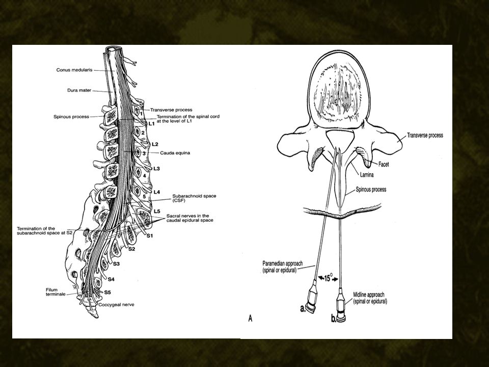
Cord anatomy Cord ends at mid or lower end of L 1 Dura continues to end at S2 – the CSF Bunch of nerves – cauda equina Expansion of subarachnoid space – lumbar cistern – injection of intrathecal local anesthetics Pial remnant – filum terminale

13
Spine is straight

14
Median approach Skin Subcutaneous tissue Supraspinous ligament Interspinous ligament Ligamentum flavum Epidural space Dura Arachnoid CSF

15
Paramedian approach Skin Subcutaneous tissue Lumbar aponeurosis Paravertebral muscles Ligamentum flavum Epidural space Dura Arachnoid CSF

16
Lig. flavum may be having gap in the midline – the “ gives”

17
The space between the laminae of two adjacent vertebrae and the interarticular joint is called the interlaminar foramen Narrow in extended position That’s why we flex !!

18
interlaminar and Interspinous

19
but below the cord level

20
Taylor’s approach

21
Surface anatomy C7 - vertebra prominens T3 - spine of scapula T7 – inferior angle of scapula Highest point of iliac crest – L4 – tuffier s line Posterior superior iliac spine – S2 Highly variable -- tuffier s line and L4L5 interspace

22
Possible ?? !! 14 % may have two nerves in single foramen When we plan segmental neurolysis Think of this anomaly

23
The most dependent points are T5 and S2

24
Spinal cord

25
The spinal cord as a part of CNS is a continuation of brain.- medulla – Ends as conus medullaris. The length of a spinal cord is 42 to 45 cm in an adult. Rarely goes to L 2-3. The spinal cord has two enlargements cervical and lumbar, corresponding to the increased nerve supply to the upper and lower limbs

26
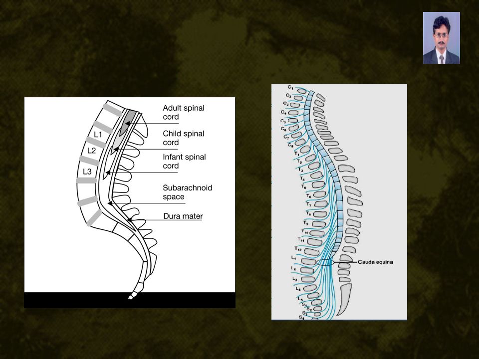
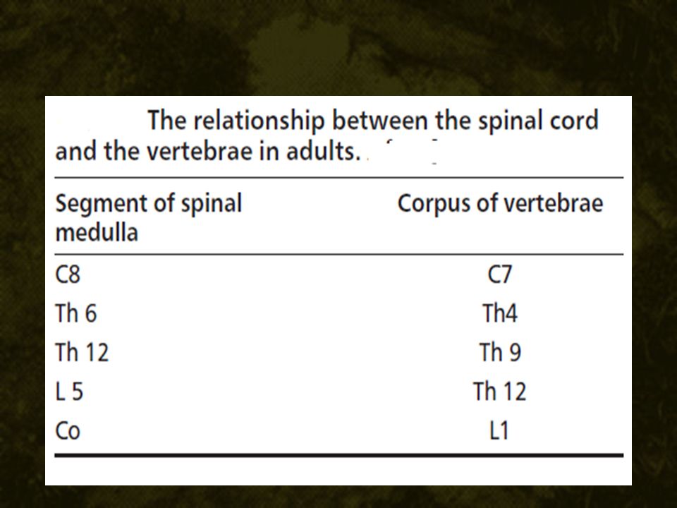
Spinal segment = vertebral level in fetus But at the time of birth, cord grows less fast – L3 Adult – L1 So what happens – After lumbar segments – bunch of nerves – cauda equina.. Segments don’t correspond !!

29
The spinal cord is mainly supplied by two posterior and one anterior spinal arteries. Posterior – Posterior inferior cerebellar arteries – reinforcement ++++ But anterior descending – a twig from both vertebral arteries – only at T11 – adamkiewieze

30
No connection between posterior and anterior

31
The normal intraspinal capillary pressure is 30 mm of Hg. Deprivation of blood supply for 2 minutes may result in infarction of cord Vertebral and azygous veins

32
31 pairs of spinal nerves --- 33 vertebrae

33
31 pairs 8 cervicals, 12 thoracic, 5 lumbar, 5 sacral 1 coccygeal The length of the spinal cord, giving origin to the rootlets for one spinal nerve constitutes one spinal segment. So, the spinal cord is made up of thirty one such segments:

34
Rootlets – dorsal and ventral roots Spinal nerve Anterior and posterior primary rami

35
DRGDRG PPRPPR APRAPR DRDR VR VR DuralcufDuralcuf

36
Rootlets, roots, nerve and rami

37
5000 neurons in anterior root The small diameter of the anterior sacral roots is a risk factor in their damage during technique 1 lakh in dorsal root dorsal root is packed in loose bundles So accessible more easy for local anesthetics See-- sensory root is more easily attacked by local anesthetics Cervical 1 and coccygeal 1 – can have no dorsal roots

38
CSF and the site of action of SA

39
Spinal CSF volume is decreased in pregnancy,obesity and increased intra abdominal pressure ?? Dose ?? During cough, there is cranial movement of spinal CSF ?? Level – beware

40
After radiological examination with contrast, the unequal distribution of CSF in 45–84% of the population has been observed. So don’t worry about segmental, patch blocks – we may not be the reason

41
How CSF ?? CSF is a clear fluid that fills the subarachnoid space. pH – 7.4 total volume of CSF in the adult varies between 100-150 ml. CSF within the spinal subarachnoid space is 50 – 75 ml approx Cerebral spinal fluid is continually produced at a rate of 450 ml per day by the choroid plexuses, which are located in the lateral. 3rd, and 4th ventricles. Cerebral spinal fluid is reabsorbed into the bloodstream through the arachnoid villi and granulations and to a small extent through epidural veins.

42
Normal pressure – 100-180 mm water Upright – 370 -500 mm water Further increase during epidural injection may cause dizziness

43
After Dural puncture – Albumin rises rate of production rises many times

44
Specific gravity CSF = 1.0001 to 1.000028 Density = mass/volume – alcohol = 1030 kg/m 3 Specific gravity Density / density of water Baricity Density / density of the other solution (CSF)
")
45
We are talking in terms of baricity because – CSF and bupivacaine

46
Duramater – large collagen fibres with a maximal interspace Thick but let fluids go easily Arachnoid – ( actually thin ) overlapping flattened epithelial cells with tight junctions – more resistance
 overlapping flattened epithelial cells with tight junctions – more resistance")
47
Subdural !! potential space between the dura mater and the arachnoid, the subdural space, that contains only small amounts of serous fluid. not intentionally given injection into it during spinal anesthesia may explain the occasional failed spinal anesthetic and the rare “total spinal” after pucca epidural anesthesia

48
Ankle ?? S1 root is usually very thick May be upto 7.7 mm Resistance and delayed action

49
Sono anatomy Nothing is complete without USG in anesthesia !! Obese, pregnant, kyphoscoliosis Previous laminectomy Depth of space Direction of needle Find out the interspinous and interlaminar space

50
Sono anatomy PD = posterior dura SC = spinal canal AD = anterior dura VB = vertebral body

51
USG The real time ultrasound guidance for needle visualization and performance of central neuraxial blockade is a three hand technique needing two individuals USG guided spinal In the next decades – this will be a normal phenomenon

52
Summary Vertebrae – number Body, arch and foraminae Spinal nerves – number Differential growth and positioning Roots, anterior posterior, thickness CSF Baricity Sonoanatomy

53
Thank you all

Similar presentations


 and Nerves. NERVOUS SYSTEM 1.Collect sensory input 2.Integrate sensory input 3.Motor output Functions of Nervous System.>")


: Brain, spinal cord 2.PERIPHERAL.>")
, Dip. Diab. DCA, Dip. Software statistics PhD (physio) Mahatma Gandhi medical college and research institute,>")
