Download presentation
Presentation is loading. Please wait.

1
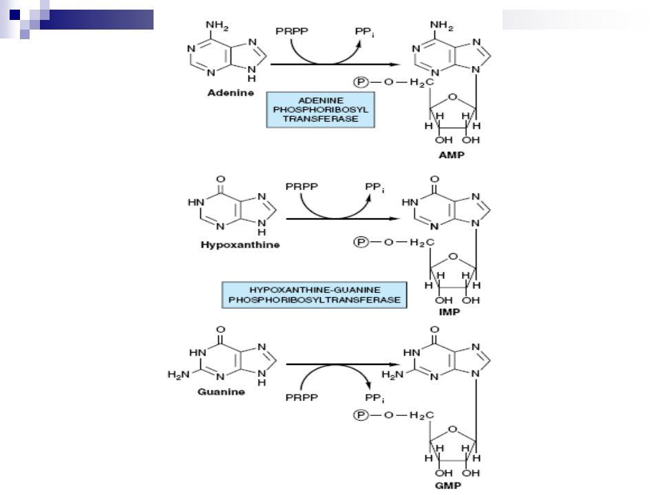
Salvage Pathway of Purines

2
Purines that result from the normal turnover of cellular nucleic acids, or that are obtained from the diet and not degraded, can be utilized again and converted to nucleoside triphosphates. This is referred to as the “salvage pathway” for purines.

3
Two enzymes are involved: 1. adenine phosphoribosyltransferase and 2. hypoxanthine-guanine phosphoribosyltransferase (HGPRT). Both enzymes use PRPP as the source of the ribose 5-phosphate group. The release of pyrophosphate (PPi) and its subsequent hydrolysis by pyrophosphatase makes these reactions irreversible.
. Both enzymes use PRPP as the source of the ribose 5-phosphate group. The release of pyrophosphate (PPi) and its subsequent hydrolysis by pyrophosphatase makes these reactions irreversible..")
5
There is an X-linked, recessive inherited disorder that is associated with a virtually complete deficiency of HGPRT called Lesch-Nyhan syndrome. This deficiency results in an inability to salvage hypoxanthine or guanine, from which excessive amounts of uric acid are produced. In addition, the lack of this salvage pathway causes increased PRPP levels and decreased IMP and GMP levels.

6
The combination of decreased purine reutilization and increased purine synthesis results in increased degradation of purines and the production of large amounts of uric acid, making Lesch-Nyhan a heritable cause of hyperuricemia. As a result, glutamine:phosphoribosylpyrophosphate amidotransferase (the committed step in purine synthesis) has excess substrate and decreased inhibitors available, and de novo purine synthesis is increased.
 has excess substrate and decreased inhibitors available, and de novo purine synthesis is increased..")
7
In patients with Lesch-Nyhan syndrome, the hyperuricemia frequently results in the formation of uric acid stones in the kidneys and the deposition of urate crystals in the joints (gouty arthritis) and soft tissues. In addition, the syndrome is characterized by motor dysfunction, cognitive deficits, and behavioral disturbances that include self-mutilation (biting of lips and fingers).
..")
9
Synthesis of deoxyribonucleotides In DNA, nucleotides are 2′ -deoxyribonucleotides which are produced by the activity of the enzyme “ribonucleotide reductase “. This enzyme is composed of two dimeric subunits R1 &R2. It is specific for the reduction of nucleoside (ADP, GDP, CDP, UDP) to their deoxy forms (dADP, dGDP, dCDP, dUDP).
 to their deoxy forms (dADP, dGDP, dCDP, dUDP)..")
10
The immediate donors of the hydrogen atoms needed for the reduction of the 2′- hydroxyl group are two sulfhydryl groups (SH) on the enzyme itself, which, during the reaction, form a disulfide bond (S-S).
 on the enzyme itself, which, during the reaction, form a disulfide bond (S-S).")
12
Degradation of Purine Nucleotides Degradation of dietary nucleic acids takes place in the small intestine by a family of pancreatic enzymes that hydrolyzes the nucleotides to nucleosides and free bases. Inside cells, purine nucleotides are sequentially degraded by specific enzymes, with uric acid as the end product of this pathway. [Note: Mammals other than primates oxidize uric acid further to allantoin, which, in some organisms other than mammals, may be further degraded to urea or ammonia.]

13
The Nucleases (Ribonucleases and deoxyribonucleases) secreted by the pancreas, hydrolyze RNA and DNA present in the diet primarily to oligonucleotides. Oligonucleotides are further hydrolyzed by pancreatic phosphodiesterases, producing a mixture of 3′- and 5′-mononucleotides. A family of nucleotidases removes the phosphate groups hydrolytically, releasing nucleosides that may be absorbed by the intestinal mucosal cells, or be further degraded to free bases before uptake.

14
Dietary purines and pyrimidines are not used to a large extent for the synthesis of tissue nucleic acids. Instead, the dietary purines are generally converted to uric acid by intestinal mucosal cells. Most of the uric acid enters the blood, and is eventually excreted in the urine.

16
Formation of uric acid

17
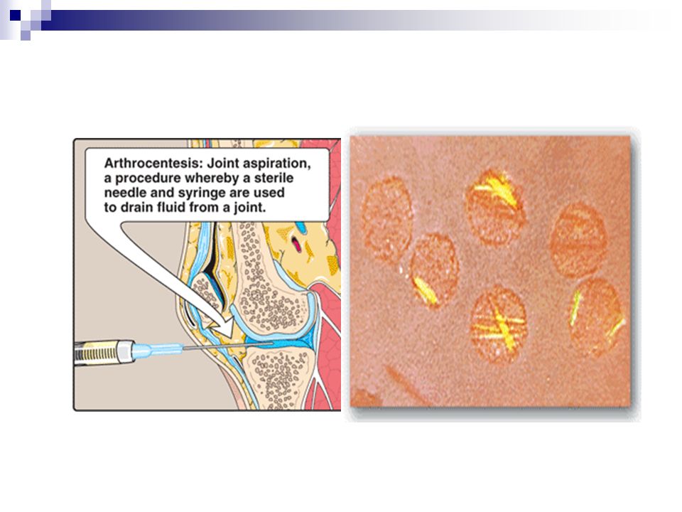
Diseases associated with purine degradation 1. Gout: It is a disorder characterized by high levels of uric in blood (hyperuricemia), either due to overproduction or underexcretion of uric acid. The precipitation of monosodium urate crystals in the joints, and the inflammatory response to the crystals, cause first acute and then chronic gouty arthritis. Nodular masses of monosodium urate crystals (tophi) may be deposited in the soft tissues, resulting in chronic tophaceous gout.
, either due to overproduction or underexcretion of uric acid. The precipitation of monosodium urate crystals in the joints, and the inflammatory response to the crystals, cause first acute and then chronic gouty arthritis. Nodular masses of monosodium urate crystals (tophi) may be deposited in the soft tissues, resulting in chronic tophaceous gout..")
19
Formation of uric acid stones in the kidney may also be seen (urate renal stones). The definitive diagnosis of gout requires aspiration and examination of synovial fluid from an affected joint (or material from a tophus) using polarized light microscopy to confirm the presence of needle-shaped monosodium urate crystals
 using polarized light microscopy to confirm the presence of needle-shaped monosodium urate crystals.")
21
In the vast majority of patients, the hyperuricemia leading to gout is caused by underexcretion of uric acid. Underexcretion can be primary, due to as-yet-unidentified inherent excretory defects, or secondary to known disease processes that affect how the kidney handles urate, for example lactic acidosis, and to environmental factors such as the use of drugs, for example, thiazide diuretics, or exposure to lead.

22
A less common cause of gout is hyperuricemia from the overproduction of uric acid. Primary hyperuricemia is, for the most part, idiopathic (having no known cause). However, there are several reasons for overproduction of uric acid including:
. However, there are several reasons for overproduction of uric acid including:.")
23
1. Several identified mutations in the X-linked PRPP synthetase gene increase availability of PRPP and increases purine production, resulting in elevated levels of plasma uric. 2. Lesch-Nyhan syndrome; is another cause of hyperuricaemia in which there will be an inherited decrease salvage of hypoxanthine and guanine with subsequent increased availability of PRPP and thus purines De Novo synthesis.

24
3. myeloproliferative disorders or who are undergoing chemotherapy and so have a high rate of cell turnover. 4. Some unrelated metabolic diseases; - Fructose intolerance - Von Gierke disease

25
Adenosine deaminase (ADA) deficiency This is an autosomal recessive disorder causes a type of severe combined immunodeficiency disease (SCID), involving a decrease in both T cells and B cells. Deficiency of ADA results in an accumulation of adenosine, which is converted to its ribonucleotide or deoxyribonucleotide forms by cellular kinases. As dATP levels rise, ribonucleotide reductase is inhibited, thus preventing the production of all deoxyribose-containing nucleotides and thus DNA leading to decrease cellular proliferation mainly the lymphocytes (which normally contain the highest ADA activity).
..")
26
Without appropriate treatment, children with this disorder usually die by the age of two due to severe uncontrollable infections. Treatment requires either bone marrow replacement or enzyme replacement therapy

Similar presentations


 Purine degradation pathway Fate of uric acid in humans Gout and hyperuricemia: Biochemistry Types Treatment.>")
 RNAs.>")


>")


 Mononucleotides.>")
.>")