Download presentation
Presentation is loading. Please wait.

1
Medical Virology Virological Tests Dr. Sameer Naji, MB, BCh, PhD (UK) Dean Assistant Head of Basic Medical Sciences Dept. Faculty of Medicine The Hashemite University
 Dean Assistant Head of Basic Medical Sciences Dept. Faculty of Medicine The Hashemite University.")
2
Virological Tests An Overview

3
Overview Clinical virology lab can provide significant benefit to patient care Traditionally epidemiologic and academic role Current rapid assays impact on therapeutic and public health decisions. Change largely due to molecular methods

4
Why Expanding Role for Diagnostic Virology Lab Increased pool of immunocompromised Increasing antiviral agents Results in increasing demand for rapid methods, viral load testing, antiviral susceptibility, genotyping.

5
Methods in use in virology Detecting Active Infection: Electron Microscopy Viral culture Detection of viral antigens Detection of viral nucleic acid. Histopathology Assessing virus-specific immune response Serologic testing

6
Methods

7
Specimen choice and collection Specimen quality limits test quality Pathogen detection depends on: Appropriate collection site. Proper timing of specimen collection. Effective and timely processing of sufficient specimen.

8
Specimens for Routine Tests

9
Specimen storage and transport Keep specimens other than blood at 4˚C If delay >24hrs, freeze at -70˚C or below. Avoid any storage at -20˚C: greater loss in infectivity Nonenveloped viruses (adenovirus, enteroviruses) more stable than enveloped (e.g. RSV, VZV, CMV).
 more stable than enveloped (e.g. RSV, VZV, CMV)..")
10
Diagnosis of viral diseases More difficult than other agents Consider overall clinical picture Take appropriate sample Infect cell culture- look for characteristic cytopathic effects Screen for parts of the virus Detect for antibodies using serological or molecular techniques

11
BASIC DIAGNOSTIC METHODS Diagnostic tests can be grouped into 3 categories: 1. Direct detection 2. Indirect detection ( virus isolation) 3. Serology
 3. Serology.")
12
Direct Examination 1. Electron Microscopy morphology of virus particles immune electron microscopy 2. Light Microscopy histological appearance inclusion bodies 3. Viral Genome Detection hybridization with specific nucleic acid probes polymerase chain reaction (PCR)
.")
13
Indirect Examination 1.Cell Culture cytopathic effect (CPE) haemadsorption immunofluorescence 2. Eggs pocks on CAM haemagglutination inclusion bodies 3. Animals disease or death

14
Serology Detection of rising titres of antibody between acute and convalescent stages of infection, or the detection of IgM in primary infection.

15
Cell Culture Viruses are obligate intracellular organisms – require living cells for virus isolation Advantages: Relatively sensitive and specific Can detect many different viruses Provides a viral isolate for further characterization (serotyping, genotyping, susceptibility)
")
16
Virus Isolation Cell Cultures are most widely used for virus isolation, there are 3 types of cell cultures: 1. Primary cells - 1-2 passages ( Monkey Kidney) 2. Semi-continuous cells - 20-50 passages ( Human embryonic kidney and skin fibroblasts) 3. Continuous cells - Indefinite passages ( HeLa, Vero, Hep2, LLC-MK2, MDCK) Primary cell culture are widely acknowledged as the best cell culture systems available since they support the widest range of viruses. However, they are very expensive and it is often difficult to obtain a reliable supply. Continuous cells are the most easy to handle but the range of viruses supported is often limited.
 2. Semi-continuous cells passages ( Human embryonic kidney and skin fibroblasts) 3. Continuous cells - Indefinite passages ( HeLa, Vero, Hep2, LLC-MK2, MDCK) Primary cell culture are widely acknowledged as the best cell culture systems available since they support the widest range of viruses. However, they are very expensive and it is often difficult to obtain a reliable supply. Continuous cells are the most easy to handle but the range of viruses supported is often limited..")
17
1. Cell Cultures Growing virus may produce 1. Cytopathic Effect (CPE) - such as the ballooning of cells or syncytia formation, may be specific or non-specific. 2. Haemadsorption - cells acquire the ability to stick to mammalian red blood cells. Confirmation of the identity of the virus may be carried out using neutralization, haemadsorption-inhibition or immunofluorescence tests.
 - such as the ballooning of cells or syncytia formation, may be specific or non-specific. 2. Haemadsorption - cells acquire the ability to stick to mammalian red blood cells. Confirmation of the identity of the virus may be carried out using neutralization, haemadsorption-inhibition or immunofluorescence tests..")
18
Cytopathic Effect (1) Cytopathic effect of enterovirus 71 and HSV in cell culture: note the ballooning of cells. (Virology Laboratory, Yale-New Haven Hospital, Linda Stannard, University of Cape Town)
.")
19
Cytopathic Effect (2) Syncytium formation in cell culture caused by RSV (top), and measles virus (bottom). (courtesy of Linda Stannard, University of Cape Town, S.A.)
.")
20
Haemadsorption Syncytial formation caused by mumps virus and haemadsorption of erythrocytes onto the surface of the cell sheet. (courtesy of Linda Stannard, University of Cape Town, S.A.) Orthomyxoviruses (influenza) and some paramyxoviruses (parainfluenza, measles, mumps) Insert viral glycoproteins (haemaglutinin) into host cell membrane. Promotes attachment of RBC of certain species (e.g guinea pig) to cell membrane.
 Orthomyxoviruses (influenza) and some paramyxoviruses (parainfluenza, measles, mumps) Insert viral glycoproteins (haemaglutinin) into host cell membrane. Promotes attachment of RBC of certain species (e.g guinea pig) to cell membrane..")
21
Problems with cell culture Long period (up to 4 weeks) required for result. Often very poor sensitivity, sensitivity depends on a large extent on the condition of the specimen. Susceptible to bacterial contamination. Susceptible to toxic substances which may be present in the specimen. Many viruses will not grow in cell culture e.g. Hepatitis B, Diarrhoeal viruses, parvovirus, papillomavirus.

22
Viruses Isolated by Cell Culture

23
Egg culture Eggs are used mainly for the isolation of influenza viruses. Ten to 12 day-old chick embryos are used. Routes of Inoculation Viruses can be cultivated in embryonated hen’s egg at different stages of development by the following routes: 1. Amniotic 2. Yolk sac 3. Allantoic 4. Chorioallantoic membrane

24
Direct Detection of Virus or Viral Antigen: Electron Microscopy Quick Looks for many viruses Useful if unknown pathogen Less prone to cross contamination vs molecular. Expensive equipment, need expertise to read Not well suited to screening large numbers of samples. Low sensitivity – need 10 5 -10 8 viral particles/ml to detect.

25
Electron Microscopy 10 6 virus particles per ml required for visualization, 50,000 - 60,000 magnification normally used. Viruses may be detected in the following specimens. FaecesRotavirus, Adenovirus Norwalk like viruses Astrovirus, Calicivirus Vesicle FluidHSV VZV Skin scrapingspapillomavirus, orf molluscum contagiosum

26
Electronmicrographs Adenovirus Rotavirus (courtesy of Linda Stannard, University of Cape Town, S.A.)
")
27
Paramyxovirus (Parainfluenza)
")
28
Problems with Electron Microscopy Expensive equipment Expensive maintenance Require experienced observer Sensitivity often low

29
Light Microscopy Replicating virus often produce histological changes in infected cells. These changes may be characteristic or non- specific. Viral inclusion bodies are basically collections of replicating virus particles either in the nucleus or cytoplasm. Examples of inclusion bodies include 1. the negri bodies found in rabies infection 2. cytomegalic inclusion bodies found in CMV infection Although not sensitive or specific, histology nevertheless serves as a useful adjunct in the diagnosis of certain viral infections.

30
Molecular Methods Methods based on the detection of viral genome are also commonly known as molecular methods. It is often said that molecular methods is the future direction of viral diagnosis. However in practice, although the use of these methods is indeed increasing, the role played by molecular methods in a routine diagnostic virus laboratory is still small compared to conventional methods. Classical molecular techniques include: 1. dot-blot and Southern-blot which depend on the use of specific DNA/RNA probes for hybridization. 2. the polymerase chain reaction (PCR) and RT-PCR which depend on the use of specific primers 3. ligase chain reaction (LCR), 4. nucleic acid based amplification (NASBA), and 5. branched DNA (bDNA)
 and RT-PCR which depend on the use of specific primers 3. ligase chain reaction (LCR), 4. nucleic acid based amplification (NASBA), and 5. branched DNA (bDNA).")
31
Nucleic Acid Detection Short length of viral genome makes them ideal candidate for nucleic-acid based diagnosis PCR conventional PCR – agarose gel detection of product Real-time PCR- products detected using probes or intercalating dyes within the reaction.

32
Polymerase Chain Reaction PCR allows the in vitro amplification of specific target DNA sequences by a factor of 10 6 and is thus an extremely sensitive technique. It is based on an enzymatic reaction involving the use of synthetic oligonucleotides flanking the target nucleic sequence of interest. These oligonucleotides act as primers for the thermostable Taq polymerase. Repeated cycles (usually 25 to 40) of denaturation of the template DNA (at 94 o C), annealing of primers to their complementary sequences (50 o C), and primer extension (72 o C) result in the exponential production of the specific target fragment. Further sensitivity and specificity may be obtained by the nested PCR. Detection and identification of the PCR product is usually carried out by agarose gel electrophoresis, hybridization with a specific oligonucleotide probe, restriction enzyme analysis, or DNA sequencing.
 of denaturation of the template DNA (at 94 o C), annealing of primers to their complementary sequences (50 o C), and primer extension (72 o C) result in the exponential production of the specific target fragment. Further sensitivity and specificity may be obtained by the nested PCR. Detection and identification of the PCR product is usually carried out by agarose gel electrophoresis, hybridization with a specific oligonucleotide probe, restriction enzyme analysis, or DNA sequencing..")
33
Polymerase Chain Reaction Advantages of PCR: Extremely high sensitivity, may detect down to one viral genome per sample volume Easy to set up Fast turnaround time Disadvantages of PCR Extremely liable to contamination High degree of operator skill required Not easy to set up a quantitative assay. A positive result may be difficult to interpret, especially with latent viruses such as CMV, where any seropositive person will have virus present in their blood irrespective whether they have disease or not. These problems are being addressed by the arrival of commercial closed systems such as the Roche Cobas Amplicor which requires minimum handling. The use of synthetic internal competitive targets in these commercial assays has facilitated the accurate quantification of results. However, these assays are very expensive.

34
Schematic of PCR Each cycle doubles the copy number of the target

36
Serology Criteria for diagnosing Primary Infection 4 fold or more increase in titre of IgG or total antibody between acute and convalescent sera Presence of IgM Seroconversion - is the development of detectable specific antibodies to microorganisms in the blood serum as a result of infection or immunization. A single high titre of IgG (or total antibody) - very unreliable Criteria for diagnosing Reinfection fold or more increase in titre of IgG or total antibody between acute and convalescent sera Absence or slight increase in IgM
 - very unreliable Criteria for diagnosing Reinfection fold or more increase in titre of IgG or total antibody between acute and convalescent sera Absence or slight increase in IgM.")
37
Typical Serological Profile After Acute Infection Note that during reinfection, IgM may be absent or present at a low level transiently

38
Complement fixation test The complement fixation test is an immunological medical test looking for evidence of infection. It tests for the presence of either specific antibody or specific antigen in a patient's serum. It uses sheep red blood cells (sRBC), anti-sRBC antibody and complement, plus specific antigen (if looking for antibody in serum) or specific antibody (if looking for antigen in serum). If either the antibody or antigen is present in the patient's serum, then the complement is completely utilized, so the sRBCs are not lysed. But if the antibody (or antigen) is not present, then the complement is not used up, so it binds anti-sRBC antibody, and the sRBCs are lysed. The Wassermann test is one form of complement fixation test.
, anti-sRBC antibody and complement, plus specific antigen (if looking for antibody in serum) or specific antibody (if looking for antigen in serum). If either the antibody or antigen is present in the patient s serum, then the complement is completely utilized, so the sRBCs are not lysed. But if the antibody (or antigen) is not present, then the complement is not used up, so it binds anti-sRBC antibody, and the sRBCs are lysed. The Wassermann test is one form of complement fixation test..")
39
Complement fixation test

40
ELISA Surface of solid phase (microtitre plate) coated with antibody Antigen of interest binds if present. Second enzyme-conjugated antibody added Substrate added and colour generated/read by spectrophotometer.

42
ELISA for HIV antibody Microplate ELISA for HIV antibody: coloured wells indicate reactivity

43
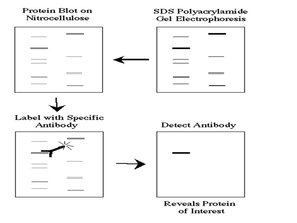
Western Blot Western blots allow investigators to determine the molecular weight of a protein and to measure relative amounts of the protein present in different samples.

44
Western Blot Proteins are separated by gel electrophoresis, usually SDS-PAGE. The proteins are transferred to a sheet of special blotting paper called nitrocellulose. The proteins retain the same pattern of separation they had on the gel.

45
Western Blot The blot is incubated with a generic protein (such as milk proteins) to bind to any remaining sticky places on the nitrocellulose. An antibody is then added to the solution which is able to bind to its specific protein. The antibody has an enzyme (e.g. alkaline phosphatase or horseradish peroxidase) or dye attached to it which cannot be seen at this time.
 or dye attached to it which cannot be seen at this time..")
46
Western Blot The location of the antibody is revealed by incubating it with a colorless substrate that the attached enzyme converts to a colored product that can be seen and photographed.

48
Western Blot HIV-1 Western Blot Lane1: Positive Control Lane 2: Negative Control Sample A: Negative Sample B: Indeterminate Sample C: Positive

49
Rapid Diagnosis Based on the Detection of Viral Antigens Nasopharyngeal AspirateRSV Influenza A and B Parainfluenza Adenovirus FaecesRotaviruses Adenoviruses Astrovirus SkinHSV VZV BloodCMV (pp65 antigenaemia test)
")
50
Direct immunofluorescence

51
Indirect immunofluorescence

52
Immunofluorescence Positive immunofluorescence test for rabies virus antigen. (Source: CDC) (Virology Laboratory, Yale-New Haven Hospital)
 (Virology Laboratory, Yale-New Haven Hospital).")
53
Advantages and Disadvantages Advantages Result available quickly, usually within a few hours. Potential Problems Often very much reduced sensitivity compared to cell culture, can be as low as 20%. Specificity often poor as well. Requires good specimens. The procedures involved are often tedious and time- consuming and thus expensive in terms of laboratory time.

54
Usefulness of Serological Results How useful a serological result is depends on the individual virus. For example, for viruses such as rubella, the onset of clinical symptoms coincide with the development of antibodies. The detection of IgM or rising titres of IgG in the serum of the patient would indicate active disease. However, many viruses often produce clinical disease before the appearance of antibodies such as respiratory and diarrhoeal viruses. So in this case, any serological diagnosis would be retrospective and therefore will not be that useful. There are also viruses which produce clinical disease months or years after seroconversion e.g. HIV and rabies. In the case of these viruses, the mere presence of antibody is sufficient to make a definitive diagnosis.

55
Problems with Serology Long period of time required for diagnosis for paired acute and convalescent sera. Mild local infections such as HSV genitalis may not produce a detectable humoral immune response. Extensive antigenic cross-reactivity between related viruses e.g. HSV and VZV, Japanese B encephalitis and Dengue, may lead to false positive results. immunocompromised patients often give a reduced or absent humoral immune response. Patients with infectious mononucleosis and those with connective tissue diseases such as SLE may react non-specifically giving a false positive result. Patients given blood or blood products may give a false positive result due to the transfer of antibody.

56
CSF antibodies Used mainly for the diagnosis of herpes simplex and VZV encephalitis CSF normally contain little or no antibodies presence of antibodies suggest meningitis or meningoencephalitis CSF antibody titre > _1_ is indicative of meningitis Serum antibody titre 100 Diagnosis depends on the presence of an intact blood-brain barrier

Similar presentations


 that polioviruses could be cultured tissue, cell culture has become a very.>")


