Download presentation
Presentation is loading. Please wait.

1
HODGKIN’S LYMPHOMA

4
Anemia,bleeding tendency. Hepatosplenomegaly.

5
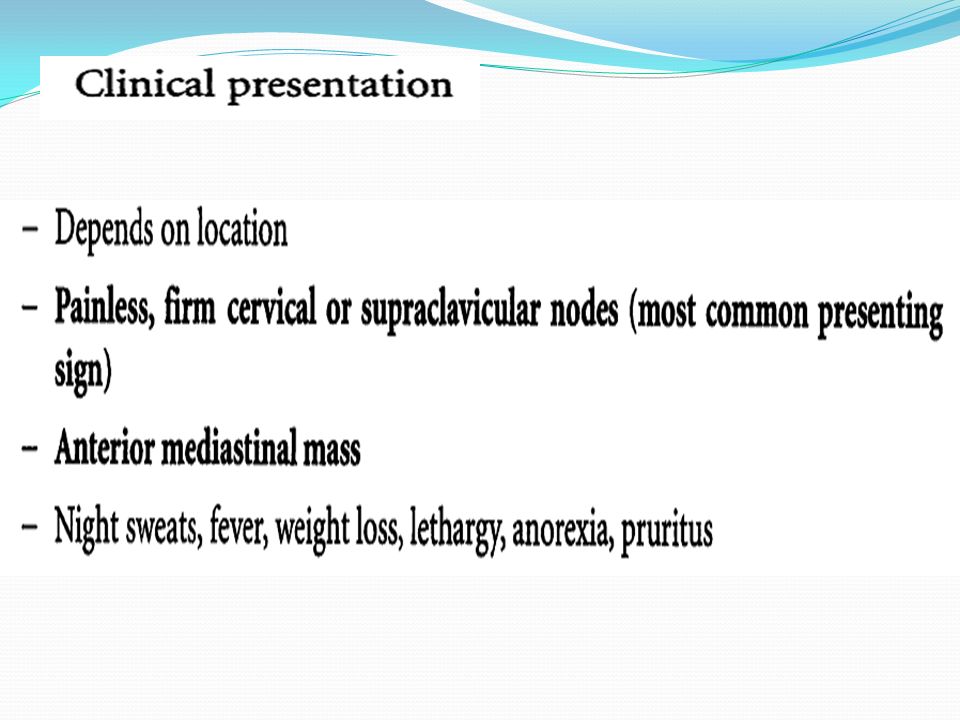
DIAGNOSIS

6
The investigation are a. Chest x ray b. USG of spleen,liver and abdominal lymphnode. c. CT scan d. Liver function test e. Bone marrow,spleen and liver biopsy. f. Staging laparotomy. Hb estimation low Total count of WBC may increase due to over spill of the lymphoma cells into peripheral blood. Serum uric acid increased Complete or partial destruction of the nodal architecture (reticulin stain) Presence of abnormal reticulum cells. Presence of Sternberg Reed giant cells. Character of Sternberg Reed : Large cell,paired nuclei (mirror image or owl eye),cytoplasm eosinophilic
 Presence of abnormal reticulum cells. Presence of Sternberg Reed giant cells. Character of Sternberg Reed : Large cell,paired nuclei (mirror image or owl eye),cytoplasm eosinophilic.")
7
Staging of Hodgkin’s lymphoma (Ann Arbor classification) Stage I : involvement of single lymphnode region or extra lymphatic site. Stage II : involvement of 2 or more lymphnodes region or an extralymphatic site and lymphnode region on the same side of (above or below )the diaphragm. Stage III : involvement of lymphnode regions on both sides of the diaphragm with or without localized extralymphatic involvement or involvement of the brain or spleen.
the diaphragm. Stage III : involvement of lymphnode regions on both sides of the diaphragm with or without localized extralymphatic involvement or involvement of the brain or spleen..")
8
Stage IV: diffuse involvement of one or more extralymphatic tissue. E.g –liver or bone marrow. Each stage is subdivided into A and B categories: A—no systemic symptoms B-- with B symptoms (unexplained fever above 38 C,heavy night sweats,unexplained weight loss of more than 10% of bd wt. in previous 6 months.

9
Treatment 1. Radiotherapy 2. Chemotherapy –ChlVPP regimen i. Chlorambucil 6 mg/m sq. Days 1 to 14 orally. ii. Vinblastine 6 mg/ m sq. Days 1 and 8 i.v iii. Procarbazine 100 mg/m sq. Days 1 to 14 orally. iv. Prednisolone 40 mg/m sq. Days 1 to 14 orally

10
3. Combined modality treatment : chemotherapy followed by radiotherapy to original sites of bulk disease which have been shrunk by chemotherapy

11
NON-HODGKIN’S LYMPHOMA

12
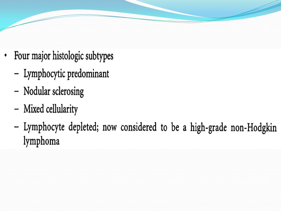
THREE HISTOLOGIC SUBTYPES:

13
St. Judes staging system for non Hodgkin’s lymphoma Stage I- Single nodal or extra nodal site excluding mediastinum and abdomen. Stage II- single extranodal site with regional node involvement. -two or more nodal areas on the same side of the diaphragm. -two single (extranodal) tumors with or without regional node involvement on the same side of the diaphragm. -primary git tumor usually in the ileocecal areas with or without involvement of associated mesenteric nodes (resectable ).
 tumors with or without regional node involvement on the same side of the diaphragm. -primary git tumor usually in the ileocecal areas with or without involvement of associated mesenteric nodes (resectable )..")
14
Stage III-two extra nodal sites on the opposite of the diaphragm. -two or more nodal areas above and below the diaphragm. -primary intra thoracic tumors(mediatinal,pleural,thymic) -extensive intrabdominal disease -paraspinal or epidural tumors. Stage IV –any of the above with bone marrow or CNS involvement.
 -extensive intrabdominal disease -paraspinal or epidural tumors. Stage IV –any of the above with bone marrow or CNS involvement..")
16
DIAGNOSIS Biopsy

17
Treatment 1.Low grade lymphoma: T cell type Stage I and II A –involved field radiotherapy. Stage IIB,III, IV : chemotherapy single agent or combination whole body irradiation.Drug commonly used Chlorambucil 20mg/m sq. orally daily. 2. High grade :B cell type Stage I :involved field radiotherapy. Stage II,III and IV: intensive combination chemotherapy to the limit of patient tolerance Combined chemotherapy –cyclophosphamide,adriamycin,vincristine (oncovin) and prednisolone (CHOP).
 and prednisolone (CHOP)..")
18
Hodgkin’sNon Hodgkin’s Peak at elderly and young age Extremes of ages Onset is gradual Variable Lymphadenopathy : i.More often localized to a single axial group of nodes (cervical,mediastinal) ii.Orderly spread by contiguity iii.Mesenteric nodes and waldayer’s ring are rarely involved iv.Extranodal involvement uncommon Lymphadenopathy : i.More frequent involvement of multiple peripheral nodes. ii.Noncontiguous spread. iii.Waldayer’s ring and mesenteric nodes commonly involved. iv.Extranodal involvement common. Systemic feature such as puritis is most common. Systemic features are uncommon. Reed sternberg giant cell are present in lymphnode biopsy It is absent

Similar presentations


>")


i.>")


