Download presentation
Presentation is loading. Please wait.

1
Basic Principles of Protein Structures

2
Proteins Proteins: The Molecule of Life Proteins: Building Blocks Proteins: Secondary Structures Proteins: Tertiary and Quartenary Structure Proteins: Geometry

3
Proteins: The Molecule of Life Proteins: Building Blocks Proteins: Secondary Structures Proteins: Tertiary and Quartenary Structure Proteins: Geometry Proteins

4
Why Proteins? Function and Role of Proteins Metabolism Energy and Synthesis: Catalytic enzymes Architecture: Structural proteins Cytoskeletal proteins Coat proteins Transport and Storage: Porins, transporters, Hemoglobin, transferrin, ferritin Regulation And Signaling: Transcription factors Growth, Development and Reproduction Locomotion Flagella, cilia, Myosin, actin Sensory and Response Defence and Immunity

5
KKAVINGEQIRSISDLHQTLKK WELALPEYYGENLDALWDCLTG VEYPLVLEWRQFEQSKQLTENG AESVLQVFREAKAEGCDITI Sequence Structure Function Evolution ligand The Protein Cycle

6
Protein Structure Diversity 1CTF1TIM1A1O 1K3R1NIK1AON

7
Protein Structure Primary structure Sequence of Amino acids Secondary Structure Local interactions Tertiary Structure Native protein Gln Ala Leu Ile Lys Glu Thr

8
Proteins: The Molecule of Life Proteins: Building Blocks Proteins: Secondary Structures Proteins: Tertiary and Quartenary Structure Proteins: Geometry Proteins

9
Review of Acid-Base Chemistry What is an acid or a base? An acid is a material that can release a proton (or hydrogen ion, H + ), and a base is a material that can donate a hydroxide ion (OH - ) (Arhennius definition), or accept a proton (Lowry Bronsted definition). Note: It is important to notice that just because a compound has a hydrogen or an OH group does not mean that it can be an acid or a base!! - The hydrogen of methane (CH4) and usually of methyl groups (-CH3) are all strongly attached to the carbon atom - Glycerol has three OH groups (CH2OH – CHOH – CH2OH) and all 3 are alcoholic groups.
, and a base is a material that can donate a hydroxide ion (OH - ) (Arhennius definition), or accept a proton (Lowry Bronsted definition). Note: It is important to notice that just because a compound has a hydrogen or an OH group does not mean that it can be an acid or a base!. - The hydrogen of methane (CH4) and usually of methyl groups (-CH3) are all strongly attached to the carbon atom - Glycerol has three OH groups (CH2OH – CHOH – CH2OH) and all 3 are alcoholic groups..")
10
Review of Acid-Base Chemistry Acid plus base makes water plus a salt: AH + BOH AB + H2O (HCL + NaOH NaCl + H2O) The chemical dissociation of nitric acid is: HNO3 (NO3)- + H+ Which can be rewritten as: HNO3 + H2O (NO3)- + H3O+ acid base conjugate conjugate base acid
 The chemical dissociation of nitric acid is: HNO3 (NO3)- + H+ Which can be rewritten as: HNO3 + H2O (NO3)- + H3O+ acid base conjugate conjugate base acid")
11
Review of Acid-Base Chemistry pH is a measure of how acidic or alkaline (basic) a solution is. The pH of a solution is the negative log of the hydrogen ion concentration. [H + ]pHpOH[OH - ] Strong base 10 -14 1401 Base10 -12 12210 -2 Weak base 10 -9 9510 -5 Neutral10 -7 77 Weak acid 10 -4 41010 -10 Acid10 -2 21210 -12 Strong acid 101410 -14

12
Review of Acid-Base Chemistry Dissociation of a weak acid: HA A - + H + Dissociation of a weak base: BOH B + + OH - Equilibrium constant: For an (acid,base) pair:
 pair:")
13
Amino group Carboxyl group Sidechain The Basic Block: Amino Acid H CaCa C N R O-O- O H H H + “zwitterion” 8.9 < pKa < 10.81.7 < pKa <2.6

15
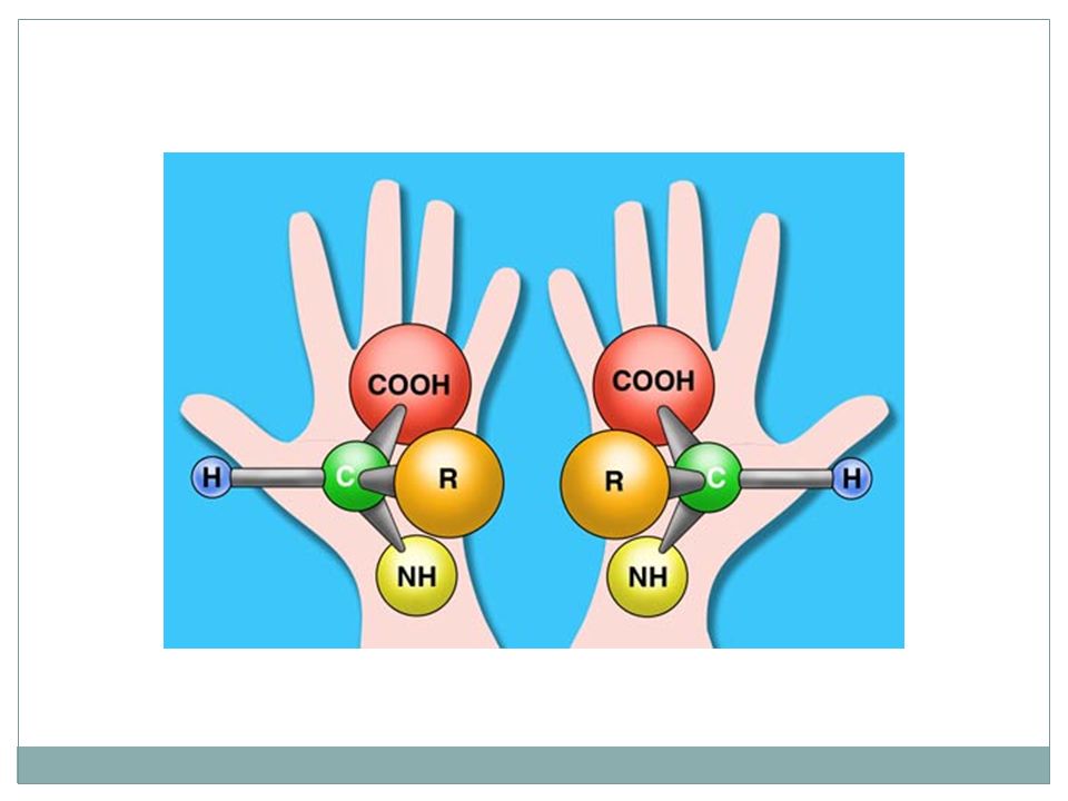
Amino Acid Chirality R CON CA H R NCO CA H L-form D-form (CORN rule) Amino acids in proteins are in the L-form Threonine and Isoleucine have a second optical center which is also identical in all natural amino acids.
 Amino acids in proteins are in the L-form Threonine and Isoleucine have a second optical center which is also identical in all natural amino acids.")
16
The 20 amino acids 1-letter3-letterAmino acid AAlaAlanine CCysCysteine DAspAspartic Acid EGluGlutamic Acid FPhePhenylalanine GGlyGlycine HHisHistidine IIleIsoleucine KLysLysine LLeuLeucine 1-letter3-letterAmino Acid MMetMethionin NAsnAsparagine PProProline QGlnGlutamine RArgArginine SSerSerine TThrThreonin VValValine WTrpTryptophan YTyrTyrosine

17
Amino Acids: Usage

18
Hydrophobic The 20 amino acids Polar, neutral Acidic Basic

19
Polar Amino acids: Cysteine C S Names: Cys, C Occurrence: 1.8 % CH2 CA CB SG CB1 SG1 SG2 CB2 CA1 CA2 Can form disulphide bridges in proteins pKa sidechain: 8.3

20
Polar Amino acids: Histidine C CH2 C CHN NC H H Name: His, H Occurrence: 2.2 % pKa sidechain: 6.04 CA CB CG ND1 CD2 NE2 CE1

21
C CH2 C CHN NC H H C CH2 C CHN NCH C CH2 C CHN NC H H C CH2 C CHN NC H H H H + + H Different ionic states of Histidine

22
C Names: Asp, D Occurrence: 5.2 % CH2 C CA CB CG OD2 Charged Amino acids: Aspartic Acid OO OD1 - pKa sidechain: 3.9

23
C CH2 Names: Lys, K Occurrence: 5.8 % CH2 Charged Amino acids: Lysine pKa sidechain: 9.2 CH2 NH3 + CA CB CG CD CE NZ

24
Unusual Amino Acids: Cyclosporin CH 3 http://purefixion.com/attention/2006_03_26_archive.html Where is the error?

25
Unusual Amino Acids: Cyclosporin http://www.cellsignal.com/products/9973.html Correct!!

26
Structural Bioinformatics: Proteins Proteins: The Molecule of Life Proteins: Building Blocks Proteins: Secondary Structures Proteins: Tertiary and Quartenary Structure Proteins: Geometry

27
CC C N RnRn O H CC C N R O H n+1 H H Peptide bond The Protein: A polymer of Amino acids Nter Cter

28
The Peptide Bond CC C N RnRn O H CC C N R O H n+1 H H Peptide bond CC C O CC N H O C CC CC N H The peptide bond is planar Conformation “Cis” Conformation “Trans”

29
Nter Cter Helices Hydrogen bonds: O (i) N (i+4)
 N (i+4)")
30
Helices 3 10 helix - helix (4 13 ) - helix (5 16 )
 - helix (5 16 )")
31
Helices 3 10 helix - helix (4 13 ) - helix (5 16 ) “Thin”; 3.0 residues /turn; ~ 4 % of all helices “Fat”; 4.2 residues /turn; instable “Right”; 3.6 residues /turn; 5.4 Å /turn; most helices
 - helix (5 16 ) Thin ; 3.0 residues /turn; ~ 4 % of all helices Fat ; 4.2 residues /turn; instable Right ; 3.6 residues /turn; 5.4 Å /turn; most helices")
32
Identify Helix Type 1. Find one hydrogen bond loop 2. Count number of residues (by number of C atoms in the loop). Here : 4 1 2 3 4 3. Count number of atoms in the loop (including first O and last H). Here: 13 1 2 3 4 5 6 7 89 10 11 12 13 4 13 helix = -helix
. Here : Count number of atoms in the loop (including first O and last H). Here: helix = -helix.")
33
The -strand Extended chain is flat “Real -strand is twisted” N-H---O-C Hydrogen bonds

34
Two types of -sheets Parallel Anti-parallel

35
-turns 1 2 3 4 1 2 3 4 Type I O is down Type II O is up The chain changes direction by 180 degrees

36
Favorable /Unfavorable Residues In Turns Turn1234 I Asp, Asn, Ser, Cys Pro Gly II Asp, Asn, Ser, Cys ProGly, AsnGly

37
The -hairpin

38
Structural Bioinformatics: Proteins Proteins: The Molecule of Life Proteins: Building Blocks Proteins: Secondary Structures Proteins: Tertiary and Quartenary Structure Proteins: Geometry

39
Protein Tertiary Structure All proteins All proteins Alpha and beta proteins: - proteins (alternating and ) - proteins
 - proteins")
40
The lone helix The helix-turn-helix motif All-Alpha topologies Glucagon (hormone involved Is regulating sugar metabolism) PDB code : 1GCN The 2 helices are twisted ROP: RNA-binding Protein PDB code: 1ROP
 PDB code : 1GCN The 2 helices are twisted ROP: RNA-binding Protein PDB code: 1ROP")
41
All Beta Topology Beta sandwiches: Fatty acid binding protein PDB code: 1IFB

42
Closed Beta Barrel PDB file: 2POR

43
The Greek Key Topology Folds including the Greek key topology include 4 to 13 strands.

44
The Jellyroll Topology A Greek key with an extra swirl PDB code 2BUK (coat protein of a virus)
")
45
The Beta Propellor Eight-plated propellor: Each plate is a four-stranded anti-parallel sheet PDB code 4AAH

46
The Rossman fold: Alternate beta / alpha motif Always right handed Alpha- Beta Topology

47
The Horseshoe PDB code: 2BNH

48
The alpha/beta barrel In a succession of alpha/beta motifs, if the first strand connects to the last, then the structure resembles a Barrel. PDB code : 1TIM

49
Hemoglobin - 4 chains: 2- chain, 2- chain (Heme- four iron groups) Quaternary Structures Assemblies of Protein Chains
 Quaternary Structures Assemblies of Protein Chains")
50
Structural Bioinformatics: Proteins Proteins: The Molecule of Life Proteins: Building Blocks Proteins: Secondary Structures Proteins: Tertiary and Quartenary Structure Proteins: Geometry

51
Protein Structure Representation CPK: hard sphere modelBall-and-stickCartoon

52
Degrees of Freedom in Proteins 1 2 3 4 + Bond length Bond angle 12 Dihedral angle

53
Protein Structure: Variables Backbone: 3 angles per residue : and Sidechain: 1 to 7 angles, ; each has 3 favored values: 60 o, -60 o, 180 o.

54
Ramachandran Plots All residues, but glycineGlycine Acta Cryst. (2002). D58, 768-776
. D58,")
55
What have we learnt? All proteins are polymers built up from 20 amino acids. All 20 amino acids have a similar structure: they all have a main-chain, consisting of an amino group and an acidic group, attached to a central carbon, named CA; the remaining atoms form the side-chain, that can be hydrophobic, polar or charged (acid or basic). The conformation of the backbone of amino acids is restricted, except for glycine that does not have a sidechain. There are 3 main graphical representations of proteins: space-filling, wireframe and cartoon.
. The conformation of the backbone of amino acids is restricted, except for glycine that does not have a sidechain. There are 3 main graphical representations of proteins: space-filling, wireframe and cartoon..")
56
There are 3 major types of secondary structures: -helices, -sheets and -turns. Most helices are -helices, stabilized through a network of CO (i) --- HN (i+4) hydrogen bonds There are two types of -sheets: parallel and anti-parallel -turns correspond to 180 change in the backbone direction. What have we learnt?
 --- HN (i+4) hydrogen bonds There are two types of -sheets: parallel and anti-parallel -turns correspond to 180 change in the backbone direction. What have we learnt .")
57
There are three main classes of proteins: all Alpha, all Beta and Alpha + Beta. The latter can be divided in two, considering the alternating alpha/beta proteins as defining their own class. Bundles are common alpha-proteins Common beta folds include the greek key and the sandwiches. Immuno- globulins adopt a beta fold. The Rossman fold (alternating alpha/beta) is a common motif in proteins. It is found in the horseshoe, as well as in the TIM fold. What have we learnt?
 is a common motif in proteins. It is found in the horseshoe, as well as in the TIM fold. What have we learnt .")
Similar presentations


 1’ C 5’ PO4 (free) DNA is a linear polymer of nucleotide subunits joined together by phosphodiester bonds - covalent bonds between.>")


 receptors (eg. pain receptors) transport (ions across membranes, oxygen in blood) molecular motors recognition.>")

 Ser Trp Leu O N N N N O O C C C C O O CαCα CαCα CαCα CαCα Lys H H H H H The.>")


