Download presentation
Presentation is loading. Please wait.

1
Solid organ disorders 2008-12 Dept. of Radiology XUE

2
Solid organ disorders Liver Liver Diffuse liver disease Diffuse liver disease Cirrhosis Cirrhosis Fatty infiltration Fatty infiltration hemochromatosis hemochromatosis Localized liver lesion Localized liver lesion Abscesses Abscesses Hepatic cysts Hepatic cysts Cavernous hemangioma Cavernous hemangioma Hepatocellular carcinoma Hepatocellular carcinoma Metastatic disease Metastatic disease

3
Solid organ disorders Biliary tree & gallbladder Biliary tree & gallbladder Congenital disorders Congenital disorders 胆囊炎 胆囊炎 胆系结石 胆系结石 胆系肿瘤 胆系肿瘤 Pancreas Pancreas Acute/chronic pancreatitis Acute/chronic pancreatitis Pancreatic cancer Pancreatic cancer Spleen Spleen

5
Fatty infiltration Lack of mass effect Lack of mass effect Lack of displacement of hepatic blood vessels Lack of displacement of hepatic blood vessels Diffuse low density Diffuse low density Sometimes fatty infiltration can also be localized Sometimes fatty infiltration can also be localized

9
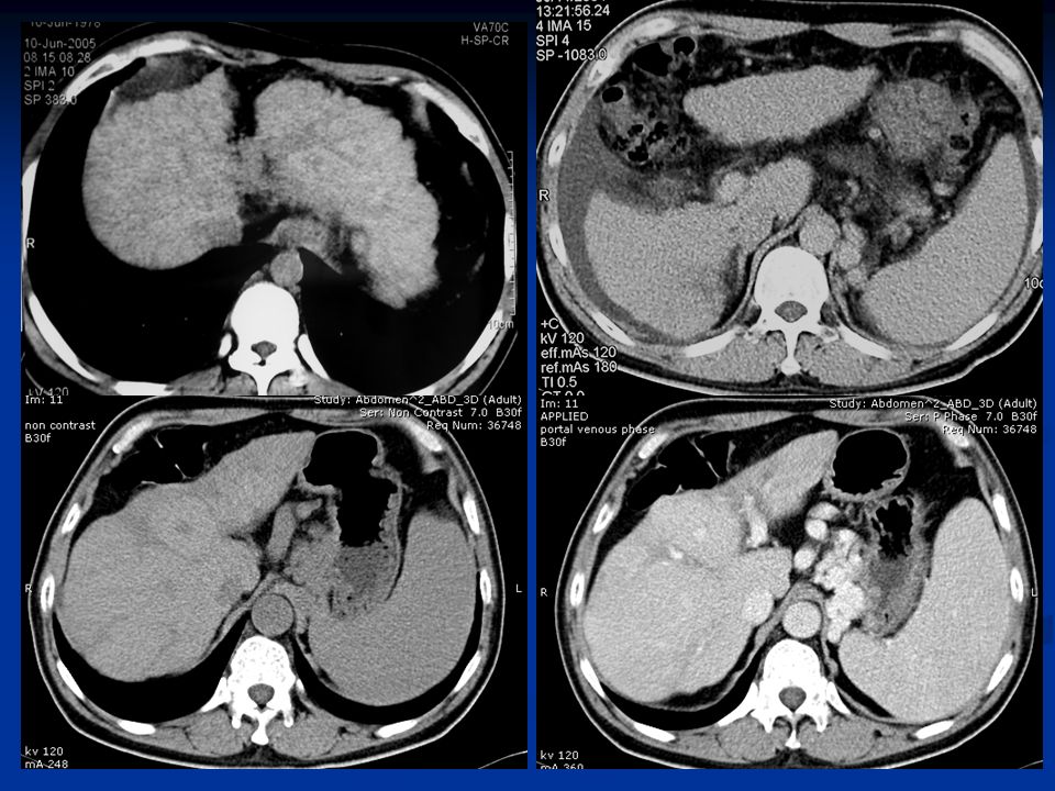
Imaging tools Imaging tools Barium swallow: esophagus varices Barium swallow: esophagus varices CT : shape CT : shape MRI : nodular regeneration differential diagnosis MRI : nodular regeneration differential diagnosis DSA : therapy DSA : therapy Radiographic features: Radiographic features: Hepatomegaly Hepatomegaly Hepatic atrophy Hepatic atrophy Coarsening of hepatic parenchymal texture Coarsening of hepatic parenchymal texture Nodularity of liver surface Nodularity of liver surface Hypertrophy of caudate lobe with atrophy of right lobe Hypertrophy of caudate lobe with atrophy of right lobe nodular regeneration nodular regeneration Cirrhosis

12
Hepatic abscess Thick wall (enhanced) mass with low- attenuation center Thick wall (enhanced) mass with low- attenuation center Pyogenic; fungal; amebic Pyogenic; fungal; amebic Ring – like enhancement Ring – like enhancement Single ring : wall 脓肿壁 Single ring : wall 脓肿壁 Dual ring : wall + edema 脓肿壁+水肿带 Dual ring : wall + edema 脓肿壁+水肿带 Triple ring : necrosis+fibroticogranuloma+edema Triple ring : necrosis+fibroticogranuloma+edema 炎性坏死+纤维肉芽+水肿 炎性坏死+纤维肉芽+水肿
 mass with low- attenuation center Thick wall (enhanced) mass with low- attenuation center Pyogenic; fungal; amebic Pyogenic; fungal; amebic Ring – like enhancement Ring – like enhancement Single ring : wall 脓肿壁 Single ring : wall 脓肿壁 Dual ring : wall + edema 脓肿壁+水肿带 Dual ring : wall + edema 脓肿壁+水肿带 Triple ring : necrosis+fibroticogranuloma+edema Triple ring : necrosis+fibroticogranuloma+edema 炎性坏死+纤维肉芽+水肿 炎性坏死+纤维肉芽+水肿")
13
Hepatic cyst

14
Hepatic hemengingoma

15
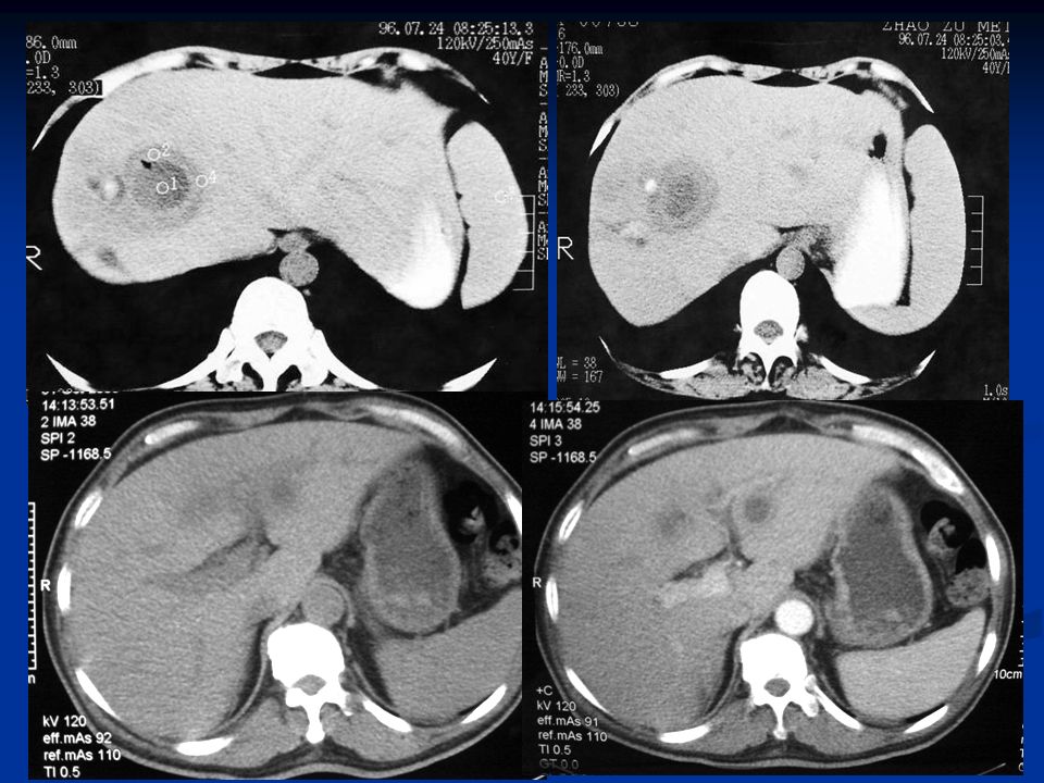
Classification solitary massive >5cm multicentric small nodular <5cm diffuse microscopic <1cm Enhancement during arterial phase (inhomogeneous contrast accumulation), Washout during portal phase
, Washout during portal phase")
18
Metastatic disease

19
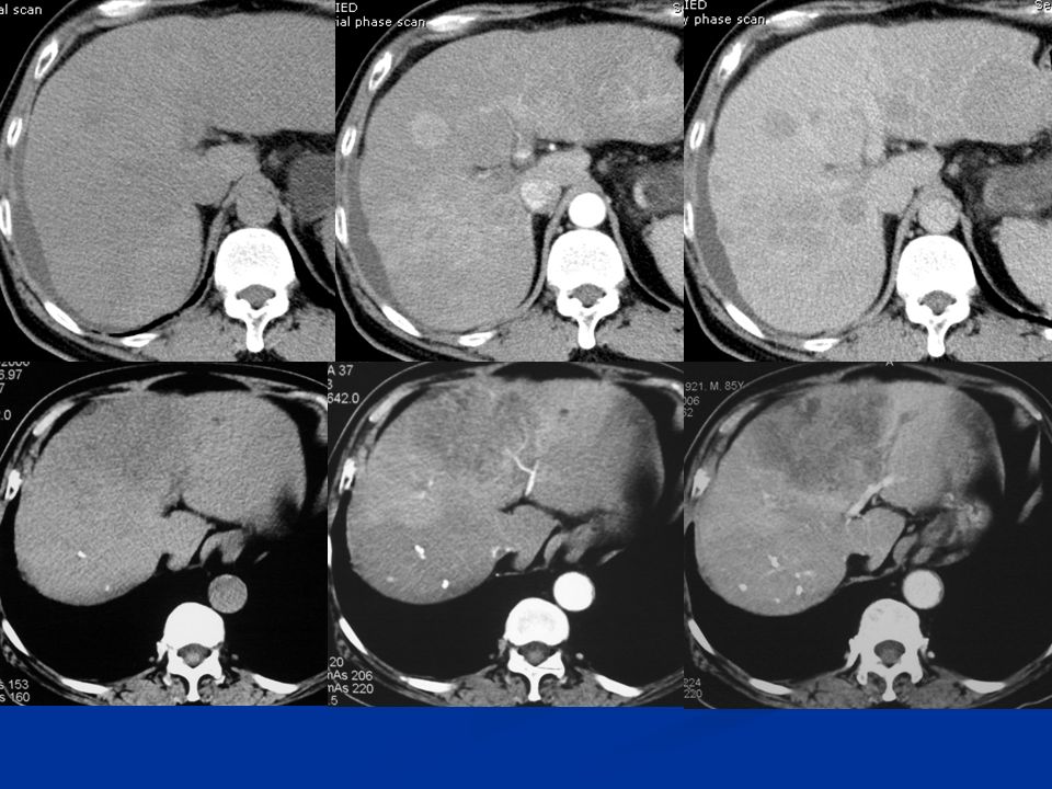
影像学征象 CT 平扫:多发大小不等圆形结节,边缘光整或不光 整,多呈低密度; CT 平扫:多发大小不等圆形结节,边缘光整或不光 整,多呈低密度; 增强扫描:环状或结节状增强,血供丰富的显著强 化,少数增强后变为等密度; 增强扫描:环状或结节状增强,血供丰富的显著强 化,少数增强后变为等密度; MRI : “ 靶征 ” 、 “ 晕圈征 ” ; MRI : “ 靶征 ” 、 “ 晕圈征 ” ; DSA :多血管、等血管、少血管型。 DSA :多血管、等血管、少血管型。 肝转移癌

20
Congenital deformity of bile duct Congenital choledochocyst

21
Cholecystolithiasis ( 胆囊结石 ) Chololithiasis ( 胆管结石 )
 Chololithiasis ( 胆管结石 )")
23
Cholecystitis ( 胆囊炎 ) 胆囊增大 囊壁增厚 囊周水肿 胆囊增大或缩小 囊壁均匀增厚 囊壁可见钙化 AcuteChronic
 胆囊增大 囊壁增厚 囊周水肿 胆囊增大或缩小 囊壁均匀增厚 囊壁可见钙化 AcuteChronic")
24
胆囊癌 Cholecystocarcinoma 隆起型 隆起型 厚壁型 厚壁型 实块型 实块型 混合型 混合型

25
肝门部胆管细胞癌 伴肝内胆管扩张

26
急性胰腺炎 acut pancreatitis 急性水肿型胰腺炎: 平扫胰腺体积弥漫性或局限性增大,密度减低,边缘模糊,肾前及肾周筋膜 增厚,增强扫描胰腺轻度强化,胰周水肿更明显; 急性出血坏死型胰腺炎: 胰腺明显增大,胰腺内由于出血出现不均匀密度增高,增强扫描坏死的胰腺 组织不强化,还可出现胰周积液和腹水;

27
胆源性急性水肿型胰腺炎

29
Chronic pancreatitis 胰腺大小正常或缩小; 胰管串珠状扩张; 胰管结石; 假性囊肿形成。

30
胰腺疾病 胰腺癌 (pancreatic carcinoma) 胰腺癌 (pancreatic carcinoma) 90% 起源于胰腺导管上皮,约 10% 为腺泡细胞癌; 80% 位于胰头,其余在体尾部,少数呈弥漫性生长; 肿瘤以浸润性生长方式向周围扩展。
 胰腺癌 (pancreatic carcinoma) 90% 起源于胰腺导管上皮,约 10% 为腺泡细胞癌; 80% 位于胰头,其余在体尾部,少数呈弥漫性生长; 肿瘤以浸润性生长方式向周围扩展。")
31
Pancreatic cancer

33
Splenic trauma

34
Splenic trauma 脾脏外伤 包膜下血肿 包膜下血肿 挫裂伤 挫裂伤 撕裂伤 撕裂伤 实质内新鲜血肿 实质内新鲜血肿 破裂伴活动性动脉出血 破裂伴活动性动脉出血 血管栓塞脾梗死 血管栓塞脾梗死

35
Hemenginoma

36
脾淋巴瘤 Splenic lymphoma 均匀肿大 均匀肿大 粟粒状结节 粟粒状结节 多发性团块 多发性团块 大的孤立性团块 大的孤立性团块

Similar presentations


 ( Immune System )>")


 — 绪论 定义:研究正常人体微细结构及其相关 定义:研究正常人体微细结构及其相关 功能的学科 功能的学科 内容:细胞、组织、器官和系统 内容:细胞、组织、器官和系统.>")
. 幼鸡的一种急性、高度接触性传染 病。发病率高、病程短。幼鸡感染 后,可导致免疫抑制,并可诱发多 种疫病或多种疫苗免疫失败。 幼鸡的一种急性、高度接触性传染 病。发病率高、病程短。幼鸡感染 后,可导致免疫抑制,并可诱发多.>")


. 本病是由禽腺病毒引起,鸡以产蛋下 降为特征的一种传染病,表现为鸡产蛋骤 然下降,软壳蛋、畸形蛋增加,褐色蛋壳 颜色变淡。 1976 年 Van Eck 首先报道了本病,我国 在 1991 年分离到了病毒。>")
>")

 : 兽医病理生理学是研究动物疾病发生的 原因和条件,研究疾病全过程中患病体的 机能、代谢的动态变化及其机制,揭示疾 病发生、发展和转归的规律,阐明疾病的 本质,为疾病的防治提供理论依据。>")
. 鸡的急性、高度接触传染性的呼吸道疾病。 特征是病鸡咳嗽、喷嚏、气管发生罗音,产蛋 减少、蛋的品质下降。 肾病变型肾肿大、尿酸盐沉积。 腺胃型.>")