Download presentation
Presentation is loading. Please wait.

1
ANATOMY OF THE SMALL INTESTINE
Dr. Jamila El-Medany

2
OBJECTIVES At the end of the lecture, students should:
List the different parts of small intestine. Describe the anatomy of duodenum, jejunum & ileum regarding: the shape, length, site of beginning & termination, peritoneal covering, arterial supply & lymphatic drainage. Differentiate between each part of duodenum regarding the length, level & relations. Differentiate between the jejunum & ileum regarding the characteristic anatomical features of each of them.

3
What is MESENTERY? Anterior abdominal wall Posterior abdominal wall
Loop of intestine Posterior abdominal wall

4
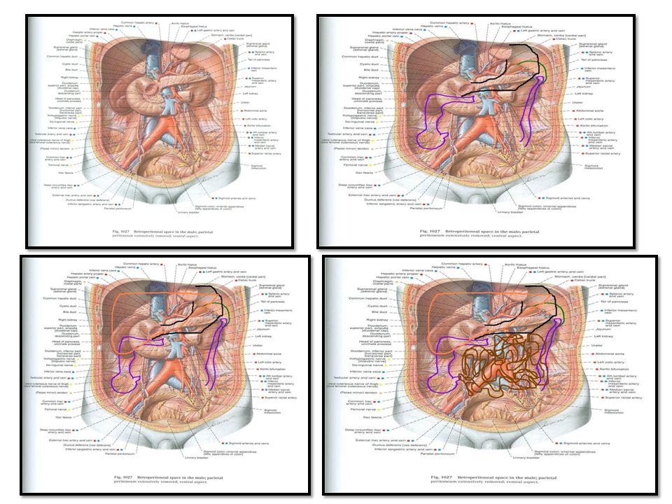
FIXED (Retro peritoneal) PART
FREE (MOVABLE) PART (WITH MESENTERY) JEJUNUM & ILEUM FIXED (Retro peritoneal) PART (NO MESENTERY) DUODENUM 1 2
 PART. (WITH MESENTERY) JEJUNUM & ILEUM. FIXED (Retro peritoneal) PART. (NO MESENTERY) DUODENUM")
5
DUODENUM SHAPE: C-shaped loop LENGTH: 10 inches
BEGINNING: at pyloro-duodenal junction TERMINATION: at duodeno-jejunal flexure PERITONEAL COVERING: retroperitoneal

6
PARTS The duodenum is divided into (4) parts: 1st : Superior.
2nd : Descending (vertical). 3rd : Inferior (Horizontal) 4th : Ascending
. 3rd : Inferior (Horizontal) 4th : Ascending.")
7
LENGTH – SURFACE ANATOMY
PART LENGTH LEVEL FIRST PART (Superior) 2 INCHES L1 (Transpyloric Plane) SECOND PART (Descending 3 INCHES DESCENDS FROM L1 TO L3 THIRD PART (Horizontal) 4 INCHES L3 (SUBCOTAL PLANE) FOURTH PART (Ascending) 1 INCHES ASCENDS FROM L3 TO L2
 2 INCHES. L1. (Transpyloric. Plane) SECOND PART. (Descending. 3 INCHES. DESCENDS FROM L1 TO L3. THIRD PART. (Horizontal) 4 INCHES. L3 (SUBCOTAL PLANE) FOURTH PART. (Ascending) 1 INCHES. ASCENDS FROM L3 TO L2.")
8
Structures Related pancreas psoas

9
RELATIONS OF FIRST PART
3) 2) 1) X X Anterior Liver Posterior 1)Bile duct 2) Gastroduodenal artery 3)Portal vein
 2) 1) X. X. Anterior. Liver. Posterior. 1)Bile duct. 2) Gastroduodenal artery. 3)Portal vein.")
10
RELATIONS OF SECOND PART
Anterior 1)Liver 2)Transverse Colon 3)Small intestine Posterior Right kidney X Lateral R Colic Flexure Medial Pancreas
Liver. 2)Transverse Colon. 3)Small intestine. Posterior. Right kidney. X. Lateral. R Colic. Flexure. Medial. Pancreas.")
11
OPENINGS IN SECOND PART OF DUODENUM
Common opening of bile duct & main pancreatic duct: on summit of major duodenal papilla. Opening of accessory pancreatic duct (one inch higher): on summit of minor duodenal papilla.
: on summit of minor duodenal papilla.")
12
RELATIONS OF THIRD PART
Anterior: a)Small intestine b) Superior mesenteric vessels Posterior: 1) Right psoas major 2) Inferior vena cava 3) Abdominal aorta 4) Inferior mesenteric vessels. 1 2 3
Small intestine. b) Superior mesenteric vessels. Posterior: 1) Right psoas major. 2) Inferior vena cava. 3) Abdominal aorta. 4) Inferior mesenteric vessels")
13
RELATIONS OF FOURTH PART
Anterior: Small intestine Posterior: Left psoas major psoas

15
Blood Supply & Lymph drainage
Because the duodenum is derived from both: Foregut & Midgut, It has its Arterial Supply from : Celiac & Superior mesenteric arteries. Venous Drainage to : Superior mesenteric& Portal veins. LYMPHATIC DRAINAGE: Celiac & Superior mesenteric lymph nodes.

16
JEJUNUM & ILEUM SHAPE: Coiled tube LENGTH: 6 meters (20 feet)
BEGINNING: at Duodeno-jejunal flexure TERMINATION: at Ilieo-caecal junction EMBRYOLOGICAL ORIGIN: Midgut Blood SUPPLY: Superior mesenteric A & V LYMPHATIC DRAINAGE: Superior mesenteric lymph nodes

17
JEJUNUM ILEUM LENGTH Shorter (proximal 2/5) of SI
Longer (distal 3/5) of SI DIAMETER Wider Narrower WALL Thicker (more plicae circulares) Thinner (less plicae circulares) APPEARANCE Dark red (more vascular) Light red (less vascular) VESSELS High & Less arcades (long terminal branches) Low & More arcades (short terminal branches MESENTERIC FAT Small amount & away from intestinal border Large amount & close to intestinal border LYMPHOID TISSUE Few aggregations Numerous aggregations (Peyer’s patches)
 of SI. DIAMETER. Wider. Narrower. WALL. Thicker (more plicae circulares) Thinner (less plicae circulares) APPEARANCE. Dark red (more vascular) Light red (less vascular) VESSELS. High & Less arcades (long terminal branches) Low & More arcades (short terminal branches. MESENTERIC FAT. Small amount & away from intestinal border. Large amount & close to intestinal border. LYMPHOID TISSUE. Few aggregations. Numerous aggregations (Peyer’s patches)")
18
THANK YOU

Similar presentations