Download presentation
Presentation is loading. Please wait.

1
RADIOGRAPHIC TECHNIQUE - I
Lecture 2, 3 Prepared By: Ala’a Ali Tayem Abed

3
Upper-Limbs Positioning

4
Upper-Limb Positioning HAND, FINGERS, THUMB and WRIST Positioning
Remove rings, watches, and other radiopaque objects. Seat the patient at the side or end of the table and place the cassette at a location and angle that allows the patient to be in the most comfortable position. Direct the central ray (CR) at a right angle to the midpoint of the cassette. When performing a bilateral examination of hands or wrists, separately radiograph each side. Shield the patient’s gonads from scattered radiation. Use close collimation. This technique is recommended for all upper-limb radiographs. When placing multiple exposures on one cassette, the side of the unexposed cassette should always be covered with lead. Use right or left markers and any other vital identification markers when appropriate.
 at a right angle to the midpoint of the cassette. When performing a bilateral examination of hands or wrists, separately radiograph each side. Shield the patient’s gonads from scattered radiation. Use close collimation. This technique is recommended for all upper-limb radiographs. When placing multiple exposures on one cassette, the side of the unexposed cassette should always be covered with lead. Use right or left markers and any other vital identification markers when appropriate.")
5
Thumb Projections: AP or PA. Oblique. Lateral.

6
THUMB: Positioning of the thumb is unique because its axis differs from that of the other digits. Basic views of the thumb include Anteroposterior (AP), Posteroanterior (PA), Oblique, and Lateral. Stress views of the first Metacarpo-phalangeal (MCP) Joint may be required for evaluation of injuries of the ligaments of this joint. THUMB Cassette Size: 24x30cm. Cassette Orientation: Landscape. All three thumb images can fit on one film. FFD: 100cm. Centering Point: MCP. Central Ray: Perpendicular to Cassette. Collimation: To include distal tip of thumb and distal carpal bones. Positioning: (AP): Rotate the Hand Medially to make the Thumb in True AP Projection. (PA): Place hand in lateral position with little finger on cassette. (Oblique): Place hand flat with the palmar surface down. This orientate the thumb to an oblique position (Lateral): Flex all Fingers and make the Lateral aspect of thumb resting on the cassette with.
, Posteroanterior (PA), Oblique, and Lateral. Stress views of the first Metacarpo-phalangeal (MCP) Joint may be required for evaluation of injuries of the ligaments of this joint. THUMB. Cassette Size: 24x30cm. Cassette Orientation: Landscape. All three thumb images can fit on one film. FFD: 100cm. Centering Point: MCP. Central Ray: Perpendicular to Cassette. Collimation: To include distal tip of thumb and distal carpal bones. Positioning: (AP): Rotate the Hand Medially to make the Thumb in True AP Projection. (PA): Place hand in lateral position with little finger on cassette. (Oblique): Place hand flat with the palmar surface down. This orientate the thumb to an oblique position. (Lateral): Flex all Fingers and make the Lateral aspect of thumb resting on the cassette with.")
7
THUMB AP PA

8
OBLIQUE THUMB

9
LATERAL THUMB

10
Evaluation criteria of Thumb projections
1. The area from the distal tip of the thumb to the trapezium should be clearly demonstrated. 2. There should be no rotation, and concavity of the phalangeal and metacarpal shafts should be demonstrated with equal amounts of soft tissue on both sides of the phalanges. 3. The first CMC joint should be free of superimposition of the hand or other bony elements. 4. The first metacarpal and trapezium should be clearly demonstrated. 5. There should be open interphalangeal and MCP joint spaces. 6. The soft tissue and bony trabeculation should be clearly present.

11
Fingers ( Digits) Projections:
PA. Oblique. Lateral.

12
Digits (second through fifth)
Fingers (2 – 5) Cassette Size: 24x30cm Cassette Orientation: Landscape. • All three images projections of fingers can fit on one film. FFD: 100cm Central Ray: Perpendicular to Cassette Centering Point: PIP joint Collimation: To include distal tip of finger and distal carpal bones Positioning: PA: • Place hand flat with the palmar surface down. • Separate digits slightly. Oblique: • Rotate palm 45 degrees toward IR until digits are resting on support. Lateral: • Place hand in lateral position (thumb side up) with finger to be examined fully extended and centered to portion of IR being exposed. • Rest Digit on Lateral (2, 3) or Medial (4, 5) Surface as needed to obtain smallest possible OID. • Ensure that long axis of finger is parallel to IR.
 Cassette Size: 24x30cm. Cassette Orientation: Landscape. • All three images projections of fingers can fit on one film. FFD: 100cm. Central Ray: Perpendicular to Cassette. Centering Point: PIP joint. Collimation: To include distal tip of finger and distal carpal bones. Positioning: PA: • Place hand flat with the palmar surface down. • Separate digits slightly. Oblique: • Rotate palm 45 degrees toward IR until digits are resting on support. Lateral: • Place hand in lateral position (thumb side up) with finger to be examined fully extended and centered to portion of IR being exposed. • Rest Digit on Lateral (2, 3) or Medial (4, 5) Surface as needed to obtain smallest possible OID. • Ensure that long axis of finger is parallel to IR.")
13
Lateral PA Oblique

15
PA Oblique Lateral

16
Hand Projections: PA. Oblique. Lateral.

17
Radiographic Positioning of the HAND
Cassette Size: 24x30cm Cassette Orientation: Landscape (Crosswise). FFD: 100cm Central Ray: Perpendicular to cassette Centering Point: Entering hand at 3rd MCP Jt. Collimation: To include entire hand and Distal Forearm. Positioning: PA : Place affected hand/finger palmar side down on cassette. OBLIQUE: Place affected hand/finger palmar side down on a 45º sponge/angle thumb side raised. LATERAL : Place affected hand with thumb raised. To properly visualize the phalanges the fingers should be positioned in a fan like arrangement.
. FFD: 100cm. Central Ray: Perpendicular to cassette. Centering Point: Entering hand at 3rd MCP Jt. Collimation: To include entire hand and Distal Forearm. Positioning: PA : Place affected hand/finger palmar side down on cassette. OBLIQUE: Place affected hand/finger palmar side down on a 45º sponge/angle thumb side raised. LATERAL : Place affected hand with thumb raised. To properly visualize the phalanges the fingers should be positioned in a fan like arrangement.")
18
PA Oblique Lateral

19
PA HAND Evaluation criteria
The entire hand, wrist, and about 2.5 cm of the distal forearm should be visible. MCP and interphalangeal joints should appear open. No rotation of hand. The digits should be separated slightly with soft tissues and should not be overlapping.

20
OBLIQUE HAND Evaluation criteria
Entire hand, wrist, and about 2.5 cm of the distal forearm should be visible in oblique view. MCP and interphalangeal joints should be open. A 45° oblique is evidenced by the following: Midshafts of third, fourth, and fifth metacarpals should not overlap; some overlap of the distal heads of third, fourth, and fifth metacarpals but no overlap of distal second and third metacarpals should occur; excessive overlap of metacarpals indicates over rotation, and too much separation indicates under rotation.

21
Lateral Hand Evaluation Criteria
Entire hand, wrist, and about 2.5 cm of the distal forearm should be visible. Fingers should appear equally separated, with phalanges in the lateral position and joint spaces open. Thumb should appear in a slightly oblique position completely free of superimposition, with joint spaces open. Hand and wrist should be in a true-lateral position evidenced by the following: 1. Distal radius and ulna superimposed. 2. metacarpals are superimposed.

22
Wrist Projections: PA. Oblique. Lateral.

23
Radiographic Positioning of the WRIST
Cassette Size: 24x30cm Cassette Orientation: Landscape (Crosswise). • All three Wrist images can usually fit on one film. FFD: 100cm Central Ray: Perpendicular to the cassette. Centering Point: PA: Midway between the radial and ulnar styloid processes. OBLIQUE: Radial Styloid Process. LATERAL: Radial Styloid Process. Collimation: To include the distal 1/3 of the forearm and metacarpal bones. Positioning: PA: Forearm resting with anterior aspect on the table, with cassette under wrist. OBLIQUE: • Forearm resting with anterior aspect on the table, with cassette under wrist. • Rotate wrist 45º with thumb side raised and rest on sponge if required. LATERAL: • Forearm resting with ulnar side on the table, with cassette under wrist.
. • All three Wrist images can usually fit on one film. FFD: 100cm. Central Ray: Perpendicular to the cassette. Centering Point: PA: Midway between the radial and ulnar styloid processes. OBLIQUE: Radial Styloid Process. LATERAL: Radial Styloid Process. Collimation: To include the distal 1/3 of the forearm and metacarpal bones. Positioning: PA: Forearm resting with anterior aspect on the table, with cassette under wrist. OBLIQUE: • Forearm resting with anterior aspect on the table, with cassette under wrist. • Rotate wrist 45º with thumb side raised and rest on sponge if required. LATERAL: • Forearm resting with ulnar side on the table, with cassette under wrist.")
24
PA OBLIQUE LATERAL

25
PA WRIST Evaluation criteria for PA wrist:
True PA is evidenced by the following: 1. separation of the distal radius and ulna is present, except for possible minimal superimposition at the distal radioulnar joint. Soft tissue and bony trabeculation should be visible.

26
OBLIQUE WRIST Evaluation criteria
Distal radius, ulna, carpals, and at least the midmetacarpal area should be visible. The trapezium and Scaphoid should be well visualized, with only slight superimposition of other carpals on their medial aspects. 45° oblique of the wrist should be evident by ulnar head being partially superimposed by distal radius. Soft tissue and bony trabeculation should be visible.

27
LATERAL WRIST Evaluation criteria
Distal radius and ulna, carpals, and at least the midmetacarpal area should be visible. True-lateral position is evidenced by the following: 1. Ulnar head should be superimposed over distal radius. 2. proximal second through fifth metacarpals all should appear aligned and superimposed. Soft tissue and bony trabeculation should be visible.

28
WRIST – ADDITIONAL VIEW – SCAPHOID ULNAR DEVIATION
Cassette Size: 24x30cm Cassette Orientation: Landscape (Crosswise). FFD: 100cm Central Ray: Perpendicular to cassette. Centering Point: Perpendicular to Scaphoid. Collimation: To include carpal bones. Positioning: As for PA wrist but with the wrist in ulnar deviation A) PA wrist in ulnar deviation. B) PA wrist in radial deviation. C) AP oblique wrist (lateral rotation). D) PA axial wrist for Scaphoid (Stecher method).
. FFD: 100cm. Central Ray: Perpendicular to cassette. Centering Point: Perpendicular to Scaphoid. Collimation: To include carpal bones. Positioning: As for PA wrist but with the wrist in ulnar deviation. A) PA wrist in ulnar deviation. B) PA wrist in radial deviation. C) AP oblique wrist (lateral rotation). D) PA axial wrist for Scaphoid. (Stecher method).")
29
SCAPHOID – ULNAR DEVIATION PA axial wrist for Scaphoid
Evaluation criteria Extreme radial deviation should be clearly demonstrated. Soft tissue and bony trabeculation should be visible. SCAPHOID – ULNAR DEVIATION PA axial wrist for Scaphoid (Stecher method)
")
30
Forearm Projections: AP. Lateral.

31
Radiographic Positioning of the Forearm
Cassette Size: 35 x 35 cm or 35 x 43 cm depending on Patient size. Cassette Orientation: Portrait or Diagonal if required. FFD: 100cm. Central Ray: Perpendicular to cassette. Centering Point: Midshafts of forearm. Collimation: To include both wrist and elbow within field. Positioning: AP : Posterior aspect of Forearm on cassette with both wrist and elbow in AP position. LATERAL: Medial side of Forearm on cassette with both wrist and elbow in lateral position. Elbow flexed at 90.

32
Forearm AP LATERAL

33
AP FOREARM Evaluation criteria
The entire radius and ulna should be visible, with pertinent soft tissues. The wrist and distal humerus (Elbow) should be clearly demonstrated. No rotation as evidenced by humeral Epicondyle visualized in profile with slight superimposition of the radial head, neck, and tuberosity over the proximal ulna. Similar radiographic densities of the proximal and distal forearm.
 should be clearly demonstrated. No rotation as evidenced by humeral Epicondyle visualized in profile with slight superimposition of the radial head, neck, and tuberosity over the proximal ulna. Similar radiographic densities of the proximal and distal forearm.")
34
LATERAL FOREARM Evaluation criteria 1. No rotation as evidenced by:
Superimposition of the radius and ulna at their distal end. Superimposition by the radial head over the Coronoid process. Radial tuberosity facing anteriorly. Superimposed humeral Epicondyle. 2. Elbow should be flexed 90؛. 3. Pertinent soft tissues and bony trabeculation should be visible.

35
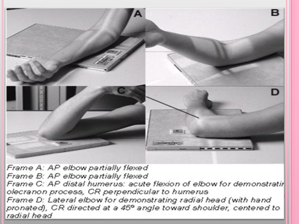
Elbow Projections: AP. Oblique medial (internal) rotation and lateral (external) rotation. Lateral.
 rotation and lateral (external) rotation. Lateral.")
36
Radiographic Positioning of the Elbow
Cassette Size: 24x30cm. Cassette Orientation: Landscape. FFD: 100cm. Central Ray: Perpendicular to Cassette. Centering Point: Elbow Joint. Collimation: To include Distal third of humerus and proximal third of forearm. Positioning: AP: • Elbow as close to straight as the patient is able, with posterior aspect on cassette. • Humerus and forearm should both be in contact with the cassette in order to ensure a open joint space. OBLIQUE: • Elbow as close to straight as the pt is able, with posterior aspect on cassette. • Rotate entire arm laterally as far as pt will tolerate. • Humerus and forearm should both be in contact with the cassette. LATERAL: • Elbow flexed at 900, with wrist in lateral orientation. • Forearm, Humerus and cassette all parallel.

37
Elbow AP LATERAL

38
Elbow O B L I Q U E Lateral (External) Rotation Medial (Internal)
 Rotation Medial (Internal)")
39
AP ELBOW Evaluation criteria
• Distal humerus, elbow joint space, proximal radius, and ulna should be clearly demonstrated. • No rotation is evidenced by appearance of bilateral Epicondyle seen in profile. • Elbow joint appears open with fully extended arm. • Soft tissue and bony trabeculation should be visible.

40
LATERAL ELBOW Evaluation criteria
• Distal humerus, proximal forearm should be clearly demonstrated. • Open elbow joint centered to the central ray. • Elbow flexed 90º. • Superimposed humeral Epicondyle. • Radial tuberosity facing anteriorly. • Radial head partially superimposing the Coronoid process. • Olecranon process seen in profile.

41
AP oblique projection medial (internal) rotation
Evaluation criteria • Coronoid process in profile should be clearly demonstrated. • Elongated medial humeral Epicondyle. • Ulna superimposed by the radial head and neck. • Olecranon process within the Olecranon fossa. • Soft tissue and bony trabeculation visible.

42
AP oblique projection lateral (external) rotation
Evaluation criteria • Correct 45؛ lateral oblique should project the radial head, neck, and tuberosity, free of superimposition by ulna. • Open elbow joint should be clearly demonstrated. • Soft tissue and bony trabeculation should be visible.

44
Thank You Best Wishes For All

Similar presentations


 Lecture 3 Myology of the Elbow.>")


 –____ bones __________ –Phalanx Metacarpals (Hand) –_____ bones.>")

