Download presentation
Presentation is loading. Please wait.

1
Vertebral Column, Spinal Cord & Nerves George Salter, PH
Vertebral Column, Spinal Cord & Nerves George Salter, PH.D Dental and Optometry--2009

2
The nervous system consists of the CNS- brain and spinal cord &
The PNS which is = to everything else Lumbar Plexus

5
Body Pedicle Lamina Processes: Transverse (2) Articular (4)
Spinous (1) Pedicle Lamina
 Pedicle. Lamina.")
6
Boundaries of vertebral foramen
Lamina

7
Spina Bifida

8
Typical intervertebral joints 3 between adjacent vertebrae
2 facet joints 1 symphysis (disc) 3 between adjacent vertebrae Superior articular process Facet joint (zygapophysis) Intervertebral disc Inferior articular process Intervertebral foramen
 3 between adjacent vertebrae. Superior articular. process. Facet joint. (zygapophysis) Intervertebral. disc. Inferior articular. process. Intervertebral. foramen.")
9
CERVICAL VERTEBRAE Transverse Process: With Foramen & Tubercles Bifid Spinous Process

10
Uncinate Process

11
Of Luschka -

12
Ligaments Ligamentum Flavum
Interspinous & Supraspinous & Ligamentum Nuchae Anterior Longitudinal Posterior Longitudinal Atlanto-occipital

13
supraspinous ligament
Interspinous Lig supraspinous ligament Ligamentum Flavum

14
nuchal ligament C7

15
Anterior Longitudinal Ligament (ALL) Posterior (PLL)
 Posterior (PLL)")
16
PLL Pedicle

17
AOM PAO LIG. FLAVUM ALL

18
SPINAL CORD & NERVES

20
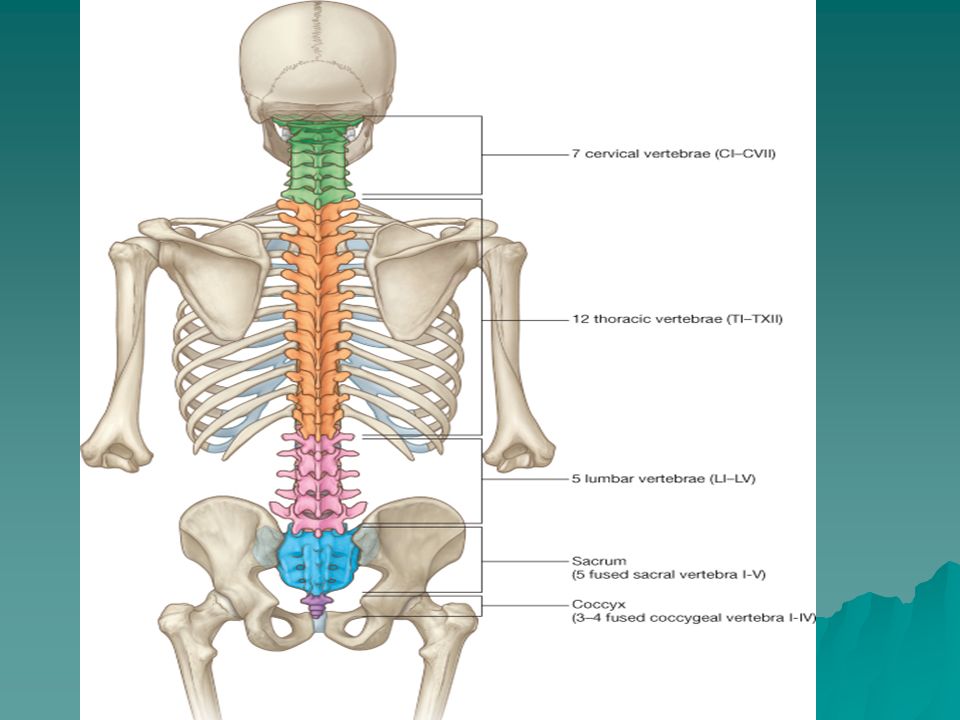
31 PAIR OF SPINAL NERVES: 8 CERVICAL; 12 THORACIC 5 LUMBAR 5 SACRAL
1 COCCYGEAL Spinal cord segment definintion Spinal Cord segments correspond to vertebral column segments

21
Lumbosacral enlargement (cord segments L2 – S3)
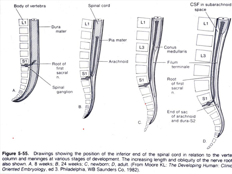
1st cervical n. (C1) exits above C1 vertebra Base of skull Cervical enlargement (cord segments C4 – T1) 8th cervical n. exits below C7 vertebra Cervical nerves Thoracic nerves Thoracic nerves Lumbar nerves Sacral & coccygeal nerves Lumbosacral enlargement (cord segments L2 – S3) Conus medullaris (termination of cord) 1. The spinal cord extends from the f. magnum at the base of the skull (where it is continuous w/the medulla of the brain) to the LV1/LV2 level. 2. There are 31 prs. of spinal nerves (8C., 12T,5L,5S, & 1Co). The part of the cord to which a pair of spinal nerves attaches is a spinal cord segment (hence there are also 31 spinal cord segments). 3. CN1-CN7 emerge superior to their respective vertebrae (ie, CN1 exits above CV1 and below skull). CN8 emerges between C7 & T1 vertebrae. Beginning w/T1 spinal nerve, spinal nerves emerge inferior to their respective vertebrae. 4. Since the cord ends at LV1/LV2, the roots of the spinal nerves become increasingly longer and more oblique as they descend to exit their respective foramina. Hence the formation of the cauda equina. 5. The spinal cord shows two enlargements (cervical and lumbosacral) for the origin of the nerves innervating the upper and lower limbs. 6. The tapered terminal part of the spinal cord is the conus medullaris. Distal to the conus the cord is prolonged as a nonnervous strand of tissue (pia mater) termed the internal filum terminale. 7. Also note that the dural sac surrounding the cord ends at SV2. Cauda equina Internal filum terminale of pia mater Termination of dural sac
 exits above C1 vertebra. Base of skull. Cervical enlargement. (cord segments C4 – T1) 8th cervical n. exits below C7 vertebra. Cervical nerves. Thoracic nerves. Thoracic nerves. Lumbar nerves. Sacral & coccygeal nerves. Lumbosacral enlargement. (cord segments L2 – S3) Conus medullaris (termination of cord) 1. The spinal cord extends from the f. magnum at the base of the skull (where it is continuous w/the medulla of the brain) to the LV1/LV2 level. 2. There are 31 prs. of spinal nerves (8C., 12T,5L,5S, & 1Co). The part of the cord to which a pair of spinal nerves attaches is a spinal cord segment. (hence there are also 31 spinal cord segments). 3. CN1-CN7 emerge superior to their respective vertebrae (ie, CN1 exits above CV1 and below skull). CN8 emerges between C7 & T1 vertebrae. Beginning w/T1 spinal nerve, spinal nerves emerge inferior to their respective vertebrae. 4. Since the cord ends at LV1/LV2, the roots of the spinal nerves become increasingly longer and more oblique as they descend to exit their respective. foramina. Hence the formation of the cauda equina. 5. The spinal cord shows two enlargements (cervical and lumbosacral) for the origin of the nerves innervating the upper and lower limbs. 6. The tapered terminal part of the spinal cord is the conus medullaris. Distal to the conus the cord is prolonged as a nonnervous strand of tissue. (pia mater) termed the internal filum terminale. 7. Also note that the dural sac surrounding the cord ends at SV2. Cauda equina. Internal filum terminale. of pia mater. Termination of dural sac.")
23
Spinal Cord is shorter than
vertebral column

25
The nervous system consists of the CNS- brain and spinal cord &
The PNS which is = to everything else Lumbar Plexus

27
Additional terms: Cutaneous Brs. & sympathetic trunk & its communicating branches (rami communicantes)
 .")
29
Boundaries of intervertebral foramen

31
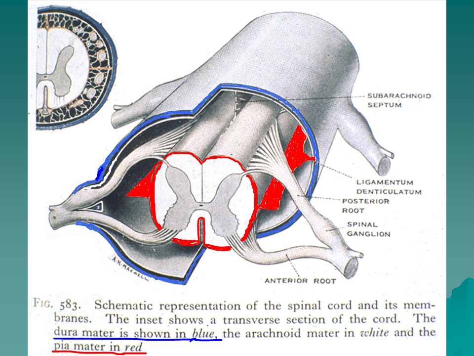
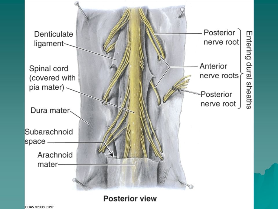
MENINGES: Dura Mater Arachnoid Mater Pia Mater

33
Dura Mater Arachnoid Mater and Pia Mater Dura Mater=Pachymenix Arachnoid and Pia =Leptomeninges

36
“MENINGEAL SPACES” Epidural---between vertebra and dura mater and contains fat and epidural (internal) vertebral plexus Subdural---only a potential space Subarachnoid---contains a web-like connective tissue plexus AND cerebrospinal fluid (CSF) plus vessels and spinal nerve rootlets
 plus vessels and spinal nerve rootlets.")
37
Subarachnoid Subdural & Epidural SPACES

38
Spinal Cord ends at one level & meninges at another
1st cervical n. (C1) exits above C1 vertebra Base of skull Cervical enlargement (cord segments C4 – T1) 8th cervical n. exits below C7 vertebra Cervical nerves Thoracic nerves Thoracic nerves Lumbar nerves Sacral & coccygeal nerves Lumbosacral enlargement (cord segments L2 – S3) Conus medullaris (termination of cord) 1. The spinal cord extends from the f. magnum at the base of the skull (where it is continuous w/the medulla of the brain) to the LV1/LV2 level. 2. There are 31 prs. of spinal nerves (8C., 12T,5L,5S, & 1Co). The part of the cord to which a pair of spinal nerves attaches is a spinal cord segment (hence there are also 31 spinal cord segments). 3. CN1-CN7 emerge superior to their respective vertebrae (ie, CN1 exits above CV1 and below skull). CN8 emerges between C7 & T1 vertebrae. Beginning w/T1 spinal nerve, spinal nerves emerge inferior to their respective vertebrae. 4. Since the cord ends at LV1/LV2, the roots of the spinal nerves become increasingly longer and more oblique as they descend to exit their respective foramina. Hence the formation of the cauda equina. 5. The spinal cord shows two enlargements (cervical and lumbosacral) for the origin of the nerves innervating the upper and lower limbs. 6. The tapered terminal part of the spinal cord is the conus medullaris. Distal to the conus the cord is prolonged as a nonnervous strand of tissue (pia mater) termed the internal filum terminale. 7. Also note that the dural sac surrounding the cord ends at SV2. Cauda equina Internal filum terminale of pia mater Spinal Cord ends at one level & meninges at another Termination of dural sac
 exits above C1 vertebra. Base of skull. Cervical enlargement. (cord segments C4 – T1) 8th cervical n. exits below C7 vertebra. Cervical nerves. Thoracic nerves. Thoracic nerves. Lumbar nerves. Sacral & coccygeal nerves. Lumbosacral enlargement. (cord segments L2 – S3) Conus medullaris (termination of cord) 1. The spinal cord extends from the f. magnum at the base of the skull (where it is continuous w/the medulla of the brain) to the LV1/LV2 level. 2. There are 31 prs. of spinal nerves (8C., 12T,5L,5S, & 1Co). The part of the cord to which a pair of spinal nerves attaches is a spinal cord segment. (hence there are also 31 spinal cord segments). 3. CN1-CN7 emerge superior to their respective vertebrae (ie, CN1 exits above CV1 and below skull). CN8 emerges between C7 & T1 vertebrae. Beginning w/T1 spinal nerve, spinal nerves emerge inferior to their respective vertebrae. 4. Since the cord ends at LV1/LV2, the roots of the spinal nerves become increasingly longer and more oblique as they descend to exit their respective. foramina. Hence the formation of the cauda equina. 5. The spinal cord shows two enlargements (cervical and lumbosacral) for the origin of the nerves innervating the upper and lower limbs. 6. The tapered terminal part of the spinal cord is the conus medullaris. Distal to the conus the cord is prolonged as a nonnervous strand of tissue. (pia mater) termed the internal filum terminale. 7. Also note that the dural sac surrounding the cord ends at SV2. Cauda equina. Internal filum terminale. of pia mater. Spinal Cord ends. at one level & meninges. at another. Termination of dural sac.")
42
WHAT ELSE PASSES THROUGH INTER- VERTEBRAL FORAMEN?

44
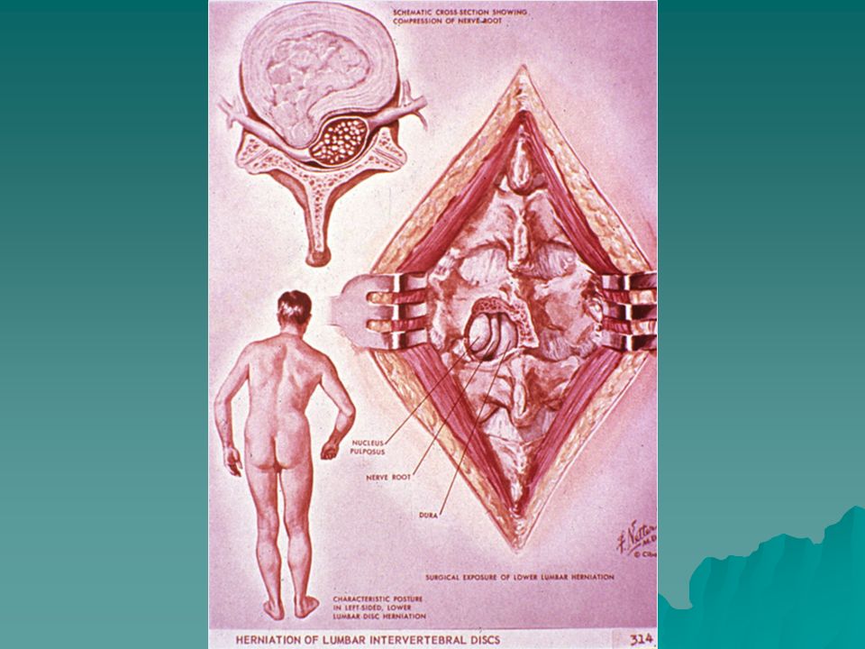
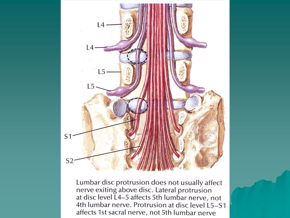
Intervertebral discs

46
Position of Spinal nerve in interver- tebral foramen

48
DERMATOME- STRIP OF SKIN SUPPLIED BY A PAIR OF DORSAL ROOTS

49
S1

50
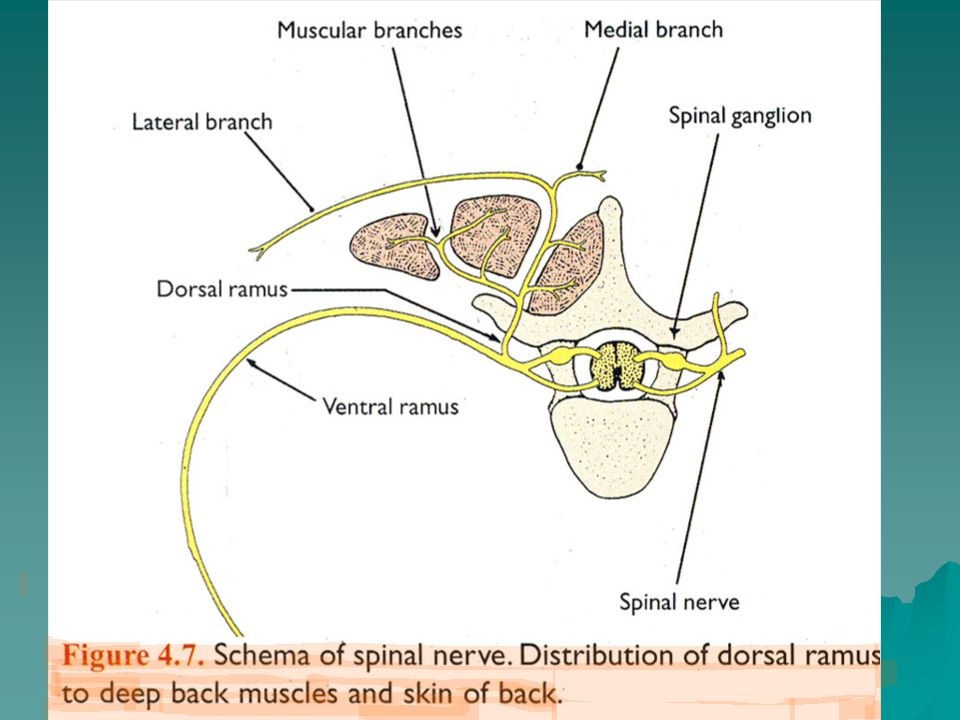
A Typical Spinal Nerve

51
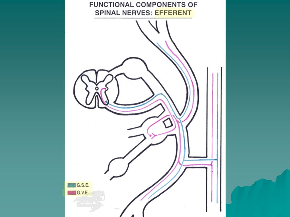
FUNCTIONAL COMPONENTS OF SPINAL NERVES

54
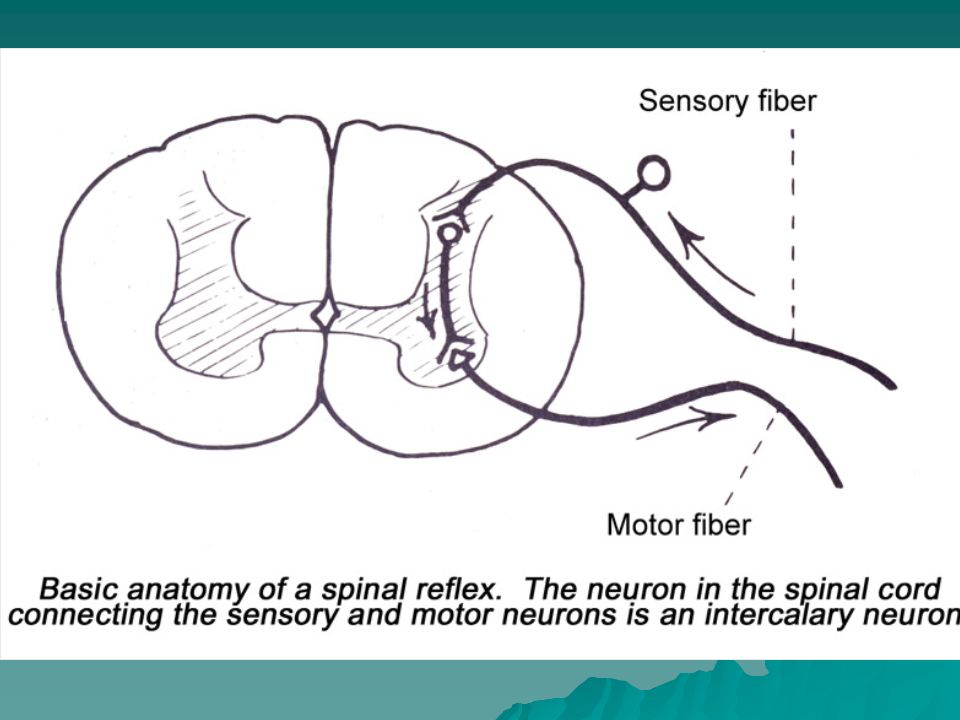
All Spinal nerves have ______ functional components. All are general
All Spinal nerves have ______ functional components? All are general. When talking about the cranial nerves and their functional components, one has to take all the possible spinal nerve functional components and add the possibility of three more. NOW REMEMBER, all spinal nerves have both sensory and motor fibers in them, but some of the cranial nerves are, indeed mixed, but some are totally sensory and some totally motor. The additional 3 functional components are designated special instead of general.

55
Back Muscles Superficial: Trapezius
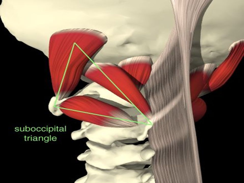
Latissimus Dorsi, Levator Scapulae, Rhomboids, Serratus Muscles Native (or Deep) : Splenius Erector Spinae Transversospinalis Other: interspinalis, intertransversarium, levator costarum, suboccipital muscles
 : Splenius. Erector Spinae. Transversospinalis. Other: interspinalis, intertransversarium, levator costarum, suboccipital muscles.")
57
VA

59
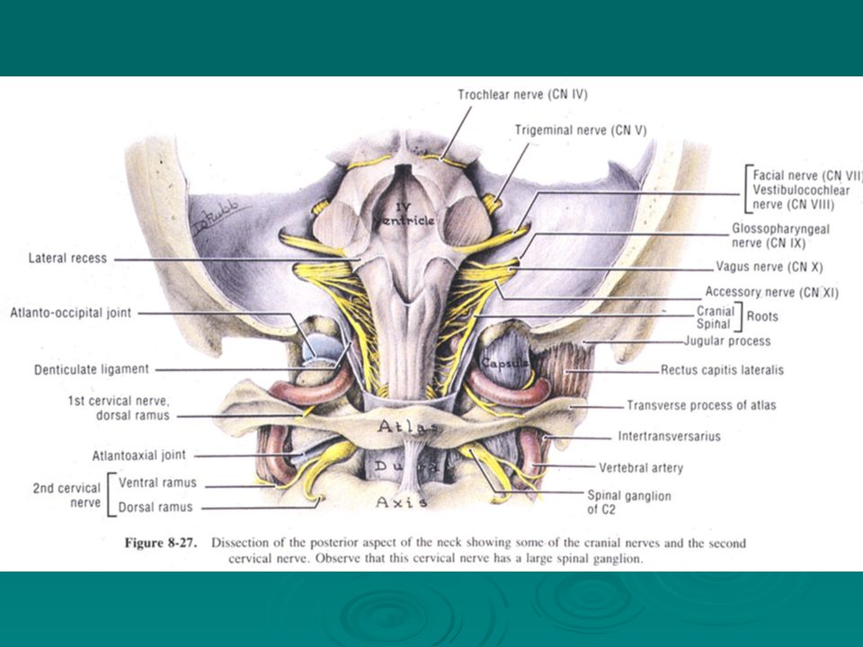
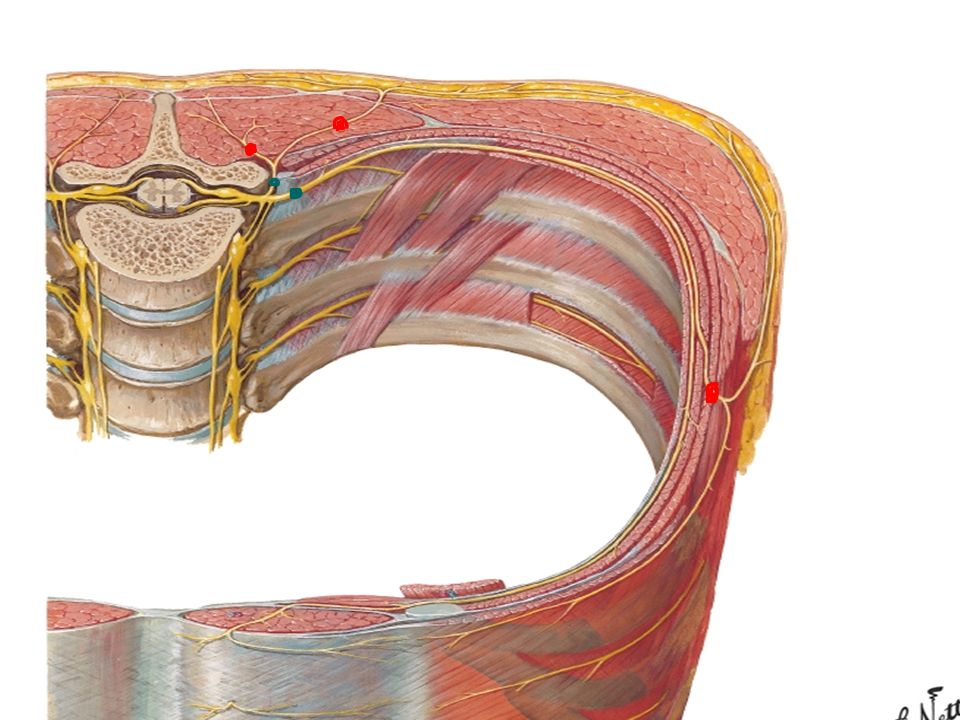
Greater Occipital Nerve
Suboccipital Nerve

60
VA

61
THE END

Similar presentations


 and Nerves. NERVOUS SYSTEM 1.Collect sensory input 2.Integrate sensory input 3.Motor output Functions of Nervous System.>")


: Brain, spinal cord 2.PERIPHERAL.>")


