Download presentation
Presentation is loading. Please wait.

1
Microscopic Structure of Bone

2
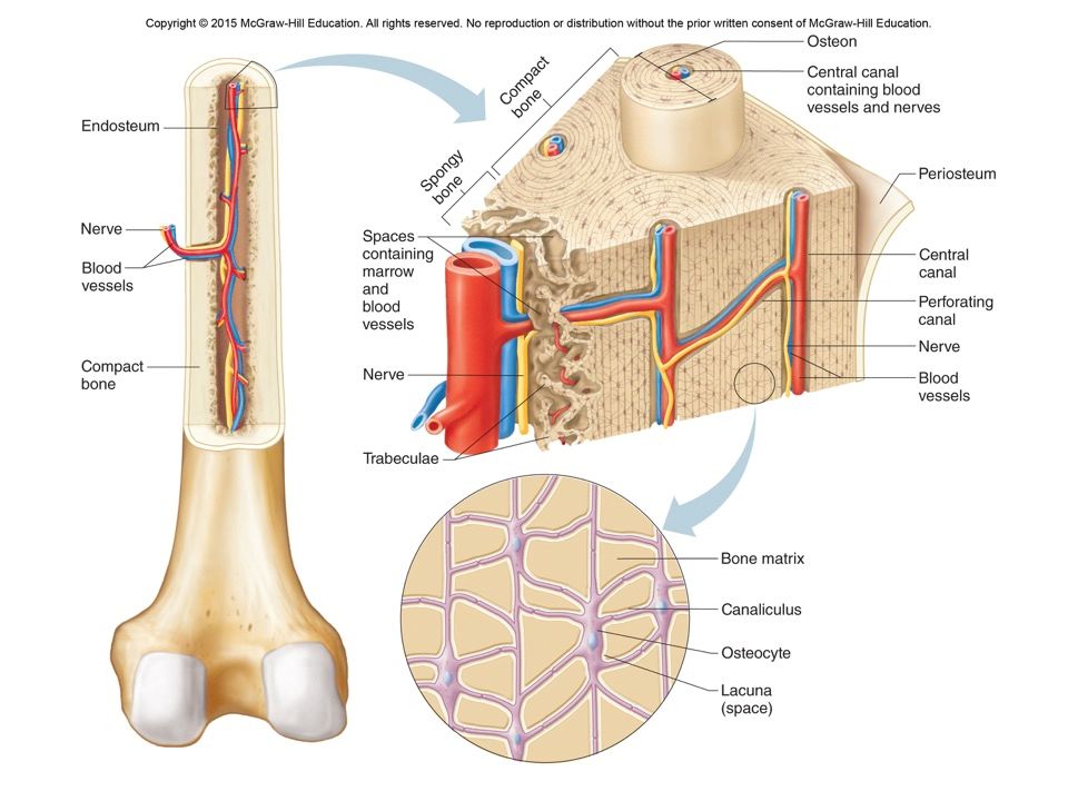
STRUCTURE A. COMPACT BONE
-coats all bones, especially present in the diaphysis of long bones -strong, weight-bearing, stress-resisting bone with few spaces -dynamic (change in response to demand)
")
4
STRUCTURE A. COMPACT BONE – strong, weight-bearing, stress-resisting bone with few spaces 1. OSTEON “HAVERSIAN SYSTEM”– repeating unit, run length of shaft

5
STRUCTURE COMPACT BONE – strong, weight-bearing, stress-resisting bone with few spaces 2. HAVERSIAN (CENTRAL) CANALS – present in each osteon; lengthwise canals supplying nerves & vascular tissue
 CANALS – present in each osteon; lengthwise canals supplying nerves & vascular tissue.")
6
STRUCTURE COMPACT BONE – strong, weight-bearing, stress-resisting bone with few spaces 3. LAMELLAE – rings of hard calcified matrix surrounding haversion canals

7
STRUCTURE COMPACT BONE – strong, weight-bearing, stress-resisting bone with few spaces 4. PERFORATING (VOLKMANN’S) CANALS – cross-sectional canals supplying nerves & blood vessels; connect w/ blood vessels of periosteum, central canal & marrow cavity
 CANALS – cross-sectional canals supplying nerves & blood vessels; connect w/ blood vessels of periosteum, central canal & marrow cavity.")
8
STRUCTURE COMPACT BONE – strong, weight-bearing, stress-resisting bone with few spaces 5. LACUNAE – small openings containing osteocytes -between lamellae

9
STRUCTURE COMPACT BONE – strong, weight-bearing, stress-resisting bone with few spaces 6. CANALICULI – (“tiny canals”) containing osteocyte extensions and fluids connecting both lacunae and haversion canals together (creates branching network for transportation of nutrients and wastes) osteocytes communicate with one another
 containing osteocyte extensions and fluids connecting both lacunae and haversion canals together (creates branching network for transportation of nutrients and wastes) osteocytes communicate with one another.")
10
B. SPONGY BONE irregular latticework of branches, trabeculae (“little beams”; found where bone not heavily stressed but precisely oriented along lines of stress coming from many directions)
")
11
B. SPONGY BONE many spaces containing red marrow (blood cell production) light wt.; found in flat bones, the ends of long bones, and short and irregular bones

12
Red vs. Yellow Marrow In children red marrow fills most marrow cavities In adults: most of red marrow turns to fatty yellow marrow Red marrow limited to skull, vertebrae, ribs sternum, part of hip and proximal heads of humerus and femur In event of severe or chronic anemia or blood loss, yellow marrow can transform back into red

13
STRUCTURE

14
STRUCTURE

15
STRUCTURE A. D. E. B. F. D. F. C. B. A. COMPACT BONE
1. OSTEON “HAVERSION SYSTEM” 2. HAVERSION (CENTRAL) CANALS 3. LAMELLAE 4. PERFORATING (VOLKMANN’S) CANALS 5. LACUNAE 6. CANILICULI D. E. B. F. D. F. C. B.
 CANALS. 3. LAMELLAE. 4. PERFORATING (VOLKMANN’S) CANALS. 5. LACUNAE. 6. CANILICULI. D. E. B. F. D. F. C. B.")
16
Ossification: Bone Formation
Bones form by replacing connective tissues in the fetus. Some form within sheet-like layers of connective tissue (intramembranous bones), while others replace masses of cartilage (endochondral bones). begins week 6 of development
, while others replace masses of cartilage (endochondral bones). begins week 6 of development.")
17
Ossification

18
Intramembranous Bone development
The broad, flat bones of the skull form as intramembranous bones. Osteoblasts deposit a bony matrix around themselves in all directions, forming spongy bone. Once the deposited bony matrix completely surrounds the osteoblasts, they are then called osteocytes.

19
Intramembranous Ossification

20
Intramembranous Bone development
4. Cells of the membranous tissue that lie outside the developing bone give rise to the periosteum. 5. Osteoblasts on the inside of the periosteum form a layer of compact bone over the spongy bone. 6. The formation of bone is referred to as ossification.

21
Intramembranous Bone Development

22
Ossification: Bone Formation
Development of bone: Video: Intramembranous vs Endochondral: How to grow a bone:

23
Endochondral Bones 23

24
Endochondral bone development
Most of the bones of the skeleton fall into this category. They first develop as hyaline cartilage models shaped like the future bones and are then replaced with bone. Cartilage calcifies and is broken down in the diaphysis Disintegrating cartilage is invaded by blood vessels and osteoblasts that first form spongy bone at the primary ossification center in the diaphysis.

26
Endochondral bones, cont.
4. Secondary ossification centers appear later in the epiphyses. A band of hyaline cartilage, the epiphyseal plate, remains between the two ossification centers. Layers of cartilage cells undergoing mitosis make up the epiphyseal plate. 26

27
Endochondral bones, cont.
7. Osteoclasts break down the calcified matrix and are replaced with bone-building osteoblasts that deposit bone in place of calcified cartilage. 8. A long bone continues to lengthen while the cartilaginous cells of the epiphyseal plate are active. Once the plate ossifies, the bone is done growing in length. The medullary cavity forms in the diaphysis due to the activity of osteoclasts. 27

28
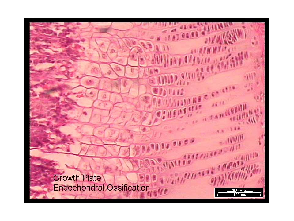
2. ENDOCHONDRAL OSSIFICATION
*replacement of original hyaline cartilage models by bone (continues until approx. age 25) Continual mitosis of epiphyseal plate cartilage & replacement by diaphysis bone = Elongation
 Continual mitosis of epiphyseal plate cartilage & replacement. by diaphysis bone = Elongation.")
30
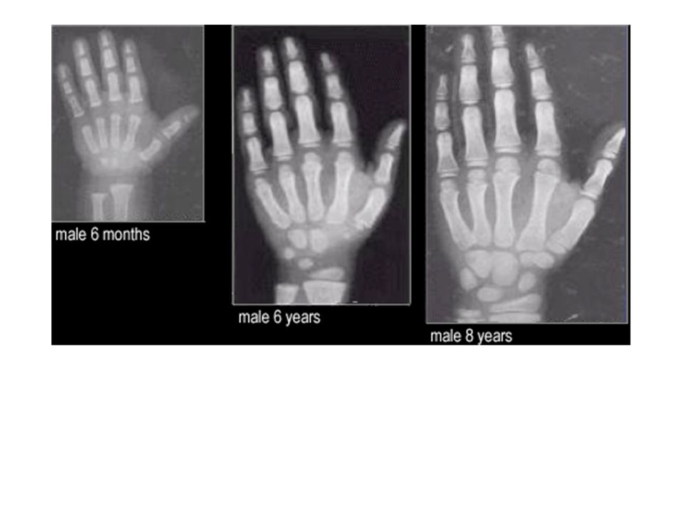
Bone Elongation Animation
To show not listen to (good intro image) X-Rays of Epiphysial plate:
 X-Rays of Epiphysial plate: v=Plvd7eslg-Q.")
31
Bone Elongation Not all bones lengthen
Takes place at the Epiphysial Plate Made of cartilage –fills in with bone Osteoclasts remove outer layer of calcified cartilage Additional cartilage fills in Older cartilage nearest diaphysis calcifies to create new bone Epiphysial Plate becomes solid (calcified) Epiphysial Line by adulthood. Plate = with cartialage Line = NO cartilage, just bone
 Epiphysial Line by adulthood. Plate = with cartialage. Line = NO cartilage, just bone.")
32
BONE GROWTH

34
Epiphyseal Plate

35
Epiphyseal Plate

36
Epiphyseal Line

37
Epiphyseal Plate or Line
Adult or Child? Images are from Answer Epiphyseal Plate or Line Adult or Child

38
Achondroplastic Dwarfism

39
Achondroplastic Dwarfism
Long bones stop growing in childhood Short in stature but generally large head Usually about 4 feet Failure of chondrocytes of metaphysis to multiply and enlarge Autosomal Dominant disorder

40
Osteitis Deformans (Paget disease)
")
41
Paget disease Bone is excessively broken down (osteoclast activity) and reformed (osteoblast activity) Resulting bone is structurally unstable and immature Osteoclasts are 5x larger than normal (resorbing bone too fast), in response, normal size osteoblasts deposit new bone quickly, but its poorly formed
, in response, normal size osteoblasts deposit new bone quickly, but its poorly formed.")
42
Bone Widening

43
Bone Widening Bone built up from outside: Osteoblasts
Bone broken down from inside: Osteoclasts Megullary Cavity increases in size Osteocytes secrete bone around a blood vessel Claude

44
Bone Widening Animation
45
Bone Widening Animation
46
Rickets

47
Rickets Caused by Vitamin D deficiency in childhood
Vit D crucial for calcium and phosphorus absorption in digestive tract Not enough minerals so bones are too flexible During Industrial Revolution, a lot of children who worked in factories (little exposure to sunlight and malnourished) Starting to increase again in urban US because spending so much time in doors and drinking soda instead of milk
 Starting to increase again in urban US because spending so much time in doors and drinking soda instead of milk.")
Similar presentations


 Joints ► Cartilages Ligaments ► Divided.>")


 Joints Cartilages Ligaments Divided into two divisions Axial skeleton –>")


