Download presentation
Presentation is loading. Please wait.

1
Bioinformatics in Vaccine Design

2
Vaccination Administration of a substance to a person with the purpose of preventing a disease Vaccination works by creating a type of immune response that enables the memory cells to later respond to a similar organism before it can cause disease Vaccines traditionally composed of attenuated or killed micro-organisms More recently, parts of micro-organisms have been used (subunit vaccines) e.g. a single purified protein, or a mixture of such proteins
 e.g. a single purified protein, or a mixture of such proteins.")
3
Vaccines work by preparing the immune system for attack

4
Vaccine Design Strategies
Classical Approaches Killed Vaccines (e.g. typhoid) Live Attenuated Strains (e.g. BCG) Subunit Vaccines (e.g. tetanus) Modern Approaches Genetically modified attenuated strains “Naked” and recombinant DNA vaccines Subunit vaccines: a bioinformatics approach
 Live Attenuated Strains (e.g. BCG) Subunit Vaccines (e.g. tetanus) Modern Approaches. Genetically modified attenuated strains. Naked and recombinant DNA vaccines. Subunit vaccines: a bioinformatics approach.")
5
Antigen Processing T cell Binding Proteasomal Cleavage Cathepsin
MHC class I Peptide Binding Proteasomal Cleavage T cell Binding Cathepsin TAP Transporter Binding MHC class II

6
Transporter associated with antigen processing (TAP)
TAP is a member of the ATP-binding cassette transporter family. It delivers cytosolic peptides into the endoplasmic reticulum (ER), where they bind to nascent MHC class I molecules. The TAP structure is formed of two proteins: TAP-1 and TAP-2, which have one hydrophobic region and one ATP-binding region each. They assemble into a heterodimer, which results in a four-domain transporter.
, where they bind to nascent MHC class I molecules. The TAP structure is formed of two proteins: TAP-1 and TAP-2, which have one hydrophobic region and one ATP-binding region each. They assemble into a heterodimer, which results in a four-domain transporter.")
7
Subunit Vaccine Characteristics
A protein that is a good subunit vaccine candidate… readily visible to the host immune system cleaved into peptide bind to MHC molecules transport to the cell surface form stable complexes with T-cell receptors and elicit a T-cell immune response conserved between bacterial / viral strains

8
Software for Signal Prediction
PROGRAM FUNCTION METHOD SignalP Predicts the presence and location of signal peptide cleavage sites Neural network / Hidden Markov model SPScan Scans protein for the presence of secretary signal peptides Weight Matrix SigCleave Reports protein signal cleavage sites Matrix

9
Predicting Inner Membrane Proteins
Proteins span cytoplasmic membranes with helical sequences Transmembrane helices are segments of about 20 predominantly hydrophobic amino acids There are many accurate programs for predicting transmembrane helices e.g. TMpred

10
TMpred can rule out inner membrane proteins
The first transmembrane segment is the signal sequence Proteins with more than one “helical segment” will stay in the inner membrane These proteins will only be readily accessible to the immune system in Gram positive bacteria TMpred plot: Bovine rhodopsin

11
Predicting Proteasomal Cleavage
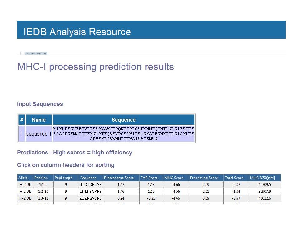
Relevant in Class I pathway only ~20% of all peptide bonds are cleaved Average peptide length 6-8 amino acids Not all peptide bonds are equally likely cleaved Cleavage more likely after hydrophobic than after hydrophilic amino acids NetChop is one program for predicting cleavage points It uses neural network methods

12
Peptide Binding to the MHC
Class I MHC: Binds peptides from the cytosol or nuclear compartment Most often from viruses Some bacterial peptides (e.g. from Mycobacterium tuberculosis) Complexes bind CD8 receptors on “killer” T cells Class II MHC: Binds peptides from intracellular vesicles The most important pathway for most bacterial peptides Complexes bind CD4 receptors on “helper” T cells This stimulates antibody production by B cells
 Complexes bind CD8 receptors on killer T cells. Class II MHC: Binds peptides from intracellular vesicles. The most important pathway for most bacterial peptides. Complexes bind CD4 receptors on helper T cells. This stimulates antibody production by B cells.")
13
The MHC locus is POLYGENIC:
DO The MHC locus is POLYGENIC: Each individual’s immune system has 8 different MHC class II molecules. The MHC locus is also highly POLYMORPHIC: These 8 MHC class II molecules vary between members of a given population.

14
Predicting MHC Binding
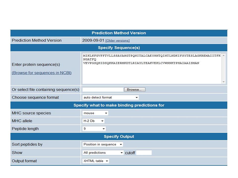
Many programs predict binding from sequence patterns Most use “matrix” type methods, assuming amino acid binding pockets are independent MHC Class I binding is easier to predict Less interaction between binding pockets More amino acid specificity But the class II pathway is the more important for bacterial infection

15
Epitopes/ Antigenic determinants
The portions of the antigen molecules which are responsible for specificity of the antigens in antigen-antibody (Ag-Ab) reactions Types of Epitopes Sequential / Continuous epitopes: recognized by Th cells linear peptide fragments amphipathic helical 9-12 mer Conformational / Discontinuous epitopes: recognized by both Th & B cells non-linear discrete amino acid sequences, come together due to folding exposed mer
 reactions. Types of Epitopes. Sequential / Continuous epitopes: recognized by Th cells. linear peptide fragments. amphipathic helical 9-12 mer. Conformational / Discontinuous epitopes: recognized by both Th & B cells. non-linear discrete amino acid sequences, come together due to folding. exposed mer.")
16
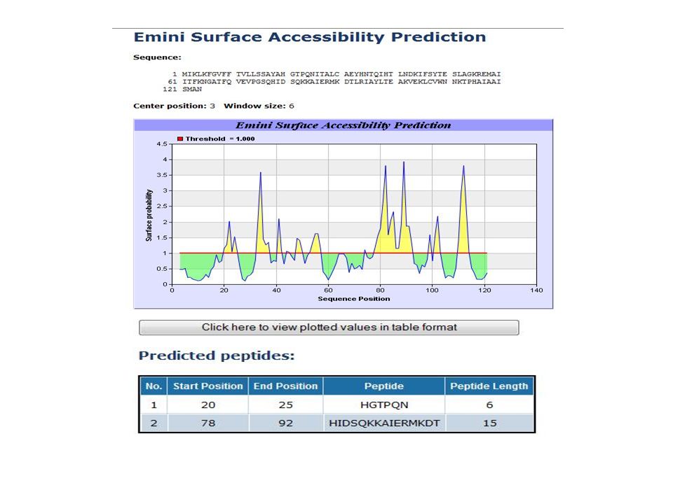
Properties of Epitopes
They occur on the surface of the protein and are more flexible than the rest of the protein They have high degree of exposure to the solvent They have beta-turns in the secondary structure The amino acids making the epitope are usually charged and hydrophilic

17
Epitope prediction algorithms
B cell: Hopp and Woods –1981 Welling et al –1985 Parker & Hodges Kolaskar & Tongaonkar – 1990 Kolaskar & Urmila Kulkarni T cell: Margalit, Spouge et al Rothbard & Taylor – 1988 Stille et al –1987 Tepitope -1999

18
Hopp & Woods method Pioneering work Based on the fact that only the hydrophilic nature of amino acids is essential for an sequence to be an antigenic determinant Local hydrophilicity values are assigned to each amino acid by the method of repetitive averaging using a window of six Not very accurate

19
Welling’s method Based on the percentage of each amino acid present in known epitopes compared with the percentage of amino acid in the average composition of a protein. Assigns an antigenicity value for each amino acid from the relative occurrence of the amino acid in an antigenic determinant site. Regions of 7 amino acid with relatively high antigenicity are extended to amino acid depending on the antigenicity values of neighboring residues.

20
Parker & Hodges method Utilizes 3 parameters : Hydrophilicity Accessibility Flexibility Hydrophilicity parameter was calculated using HPLC from retention co-efficients of model synthetic peptides. Surface profile was determined by summing the parameters for each residue of a seven-residue segment and assigning the sum to the fourth residue. One of the most useful prediction algorithms

21
Kolaskar & Tongaonkar’s method
Semi-empirical method which uses physiological properties of amino acid residues. Frequencies of occurrence of amino acids in experimentally known epitopes. Data of 169 epitopes from 34 different proteins was collected of which 156 which have less than 20 aa per determinant were used.

22
T-cell epitope prediction algorithms
Considers amphipathic helix segments, tetramer and pentamer motifs (charged residues or glycine) followed by 2-3 hydrophobic residues and then a polar residue. Sequence motifs of immunodominant secondary structure capable of binding to MHC with high affinity. Virtual matrices which are used for predicting MHC polymorphism and anchor residues.
 followed by 2-3 hydrophobic residues and then a polar residue. Sequence motifs of immunodominant secondary structure capable of binding to MHC with high affinity. Virtual matrices which are used for predicting MHC polymorphism and anchor residues.")
23
Ab-binding sites: Sequential & Conformational Epitopes!
Paratope Sequential Conformational Ab-binding sites

24
VAXIPRED: A software package for predicting subunit vaccine targets

25
Immune epitope tools

Similar presentations


>")

