Download presentation
Presentation is loading. Please wait.

1
Coagulation Disorders and Disseminated Intravascular Coagulation (DIC)
Jianzhong Sheng MD, PhD Department of Pathophysiology School of Medicine Zhejiang University

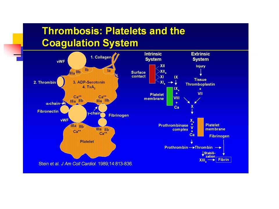
2
Normal Hemostasis First step in hemostasis is formation of a platelet aggregate At the molecular level interaction of coagulation factors takes place on the surface of activated platelets The Tissue Factor–FVIIa complex is the physiological activator of normal hemostasis

3
Hemostasis Endothelial cell Subendothelial matrix Nitric oxide
Hemostasis refers to the prevention of blood loss, and is accomplished by vasoconstriction and coagulation by cellular and coagulation factors. Undue bleeding is controlled and the fluidity of the blood is maintained by counterbalances within the coagulation and fibrinolytic systems. Blood vessel injury or disruption, platelet defects, abnormalities of the normally circulating anticoagulants and fibrinolytic mechanisms may upset the balance between fibrinolysis and coagulation. Blood normally circulates through endothelium-lined vessels without coagulation or platelet activation occurring and without appreciable hemorrhage. Injury to the endothelial cells triggers the hemostatic process, which typically begins with the attachment of platelets (“Adhesion”) to the damaged endothelium or exposed subendothelial proteins such as collagen and von Willebrand factor (vWf). The platelets then change form (“Activate”) and release factors that stimulate the clotting process. They also bind together (“Aggregate”). At the same time, plasma proteins may react with elements in the subendothelium, activating the “contact” phase of coagulation. Exposed fibroblasts and macrophages present tissue factor, a membrane protein, to the blood at the injured site, thereby triggering the “Extrinsic “phase of blood coagulation. Under normal conditions, hemostasis protects the individual from massive bleeding secondary to trauma. In abnormal states, life-threatening bleeding can occur or thrombosis can occlude the vascular tree. Hemostasis is influenced by a number of different factors including: (a) vascular extracellular matrix and alterations in endothelial reactivity, (b) platelets, (c) coagulation proteins, (d) inhibitors of coagulation, and (e) fibrinolysis. Cotran RS, Kumar V, Robbins SL, eds. Robbins pathologic basis of disease, 5th ed. Philadelphia: W.B. Saunders, 1994 pp Goodnight S. Physiology of coagulation and the role of vitamin K. In: Ansell JE, Oertel LB, Wittkowsky AK, eds. Managing oral anticoagulation therapy, Gaithersburg: Aspen Publishers, 1997 pp 1B-1:1-5.
 to the damaged endothelium or exposed subendothelial proteins such as collagen and von Willebrand factor (vWf). The platelets then change form ( Activate ) and release factors that stimulate the clotting process. They also bind together ( Aggregate ). At the same time, plasma proteins may react with elements in the subendothelium, activating the contact phase of coagulation. Exposed fibroblasts and macrophages present tissue factor, a membrane protein, to the blood at the injured site, thereby triggering the Extrinsic phase of blood coagulation. Under normal conditions, hemostasis protects the individual from massive bleeding secondary to trauma. In abnormal states, life-threatening bleeding can occur or thrombosis can occlude the vascular tree. Hemostasis is influenced by a number of different factors including: (a) vascular extracellular matrix and alterations in endothelial reactivity, (b) platelets, (c) coagulation proteins, (d) inhibitors of coagulation, and (e) fibrinolysis. Cotran RS, Kumar V, Robbins SL, eds. Robbins pathologic basis of disease, 5th ed. Philadelphia: W.B. Saunders, 1994 pp Goodnight S. Physiology of coagulation and the role of vitamin K. In: Ansell JE, Oertel LB, Wittkowsky AK, eds. Managing oral anticoagulation therapy, Gaithersburg: Aspen Publishers, 1997 pp 1B-1:1-5.")
4
Initiation of coagulation

5
Protein C, Protein S, Antithrombin III
Coagulation Pathways Intrinsic Pathway Extrinsic Pathway IX Tissue Factor + VII Contact TF Pathway X XI TF-VIIa Common Pathway XIIa HKa PL Prothrombin XIa PL (Tenase) IXa VIIIa PL Xa Coagulation may be initiated by vascular injury, however, multiple coagulation pathways are involved in the actual formation of clot. Vasoconstriction occurs immediately following vascular injury and is followed by platelet adhesion to collagen in the vessel wall exposed by injury. Subsequently platelet aggregation results in a platelet plug which is later strengthened by fibrin. Fibrin production may begin with the conversion of factor X to factor Xa. Factor X can be activated by means of two reaction sequences. One requires tissue factor (TF) which is exposed to the blood as a result of vascular injury. Because TF is not in the blood, it is an extrinsic element in coagulation, hence the name "extrinsic" pathway for this sequence. The catalytic action of TF is the central precipitating event in the clotting cascade. TF acts in concert with factor VIla and phospholipid (PL) to convert factor IX to IXa and factor X to Xa. The "intrinsic" pathway is initiated by the "contact" activation of factor XI by the XIIa/activated high molecular weight kininogen (HKa) complex. Factor XIa also converts factor IX to IXa and factor IXa in turn converts factor X to Xa, in concert with factors VIIIa and phospholipid (the “tenase complex”). However factor Xa is formed, it is the active catalytic ingredient of the "Prothrombinase” complex, which includes factor Va and PL and converts prothrombin to thrombin. Thrombin cleaves fibrinopeptides (FPA, FPB) from fibrinogen, allowing the resultant fibrin monomers to polymerize, and converts factor XIII to XIIIa which crosslinks the fibrin clot. Thrombin accelerates the clotting cascade by its potential to activate factors V and VIII, but continued proteolytic action also activates protein C which degrades Va and VIIIa. Adapted from: Colman RW, Hirsh J, Marder VJ, Salzman EW. Overview of hemostasis.Overview of the thrombotic process and its therapy. In: Colman RW, Hirsh J, Marder VJ, Salzman EW, eds. Hemostasis and thrombosis, 3rd ed. Philadelphia: J.B. Lippincott, 1994 p Colman RW, Hirsh J, Marder VJ, Salzman EW. Overview of the thrombotic process and its therapy. In: Colman RW, Hirsh J, Marder VJ, Salzman EW, eds. Hemostasis and thrombosis, 3rd ed. Philadelphia: J.B. Lippincott, 1994 pp Goodnight S. Physiology of coagulation and the role of vitamin K. In: Ansell JE, Oertel LB, Wittkowsky AK, eds. Managing oral anticoagulation therapy, Gaithersburg: Aspen Publishers, 1997 pp 1-7. XIII Va (Prothrombinase) Thrombin Protein C, Protein S, Antithrombin III XIIIa Fibrinogen Fibrin (weak) Fibrin (strong)
 IXa. VIIIa. PL. Xa. Coagulation may be initiated by vascular injury, however, multiple coagulation pathways are involved in the actual formation of clot. Vasoconstriction occurs immediately following vascular injury and is followed by platelet adhesion to collagen in the vessel wall exposed by injury. Subsequently platelet aggregation results in a platelet plug which is later strengthened by fibrin. Fibrin production may begin with the conversion of factor X to factor Xa. Factor X can be activated by means of two reaction sequences. One requires tissue factor (TF) which is exposed to the blood as a result of vascular injury. Because TF is not in the blood, it is an extrinsic element in coagulation, hence the name extrinsic pathway for this sequence. The catalytic action of TF is the central precipitating event in the clotting cascade. TF acts in concert with factor VIla and phospholipid (PL) to convert factor IX to IXa and factor X to Xa. The intrinsic pathway is initiated by the contact activation of factor XI by the XIIa/activated high molecular weight kininogen (HKa) complex. Factor XIa also converts factor IX to IXa and factor IXa in turn converts factor X to Xa, in concert with factors VIIIa and phospholipid (the tenase complex ). However factor Xa is formed, it is the active catalytic ingredient of the Prothrombinase complex, which includes factor Va and PL and converts prothrombin to thrombin. Thrombin cleaves fibrinopeptides (FPA, FPB) from fibrinogen, allowing the resultant fibrin monomers to polymerize, and converts factor XIII to XIIIa which crosslinks the fibrin clot. Thrombin accelerates the clotting cascade by its potential to activate factors V and VIII, but continued proteolytic action also activates protein C which degrades Va and VIIIa. Adapted from: Colman RW, Hirsh J, Marder VJ, Salzman EW. Overview of hemostasis.Overview of the thrombotic process and its therapy. In: Colman RW, Hirsh J, Marder VJ, Salzman EW, eds. Hemostasis and thrombosis, 3rd ed. Philadelphia: J.B. Lippincott, 1994 p Colman RW, Hirsh J, Marder VJ, Salzman EW. Overview of the thrombotic process and its therapy. In: Colman RW, Hirsh J, Marder VJ, Salzman EW, eds. Hemostasis and thrombosis, 3rd ed. Philadelphia: J.B. Lippincott, 1994 pp Goodnight S. Physiology of coagulation and the role of vitamin K. In: Ansell JE, Oertel LB, Wittkowsky AK, eds. Managing oral anticoagulation therapy, Gaithersburg: Aspen Publishers, 1997 pp 1-7. XIII. Va. (Prothrombinase) Thrombin. Protein C, Protein S, Antithrombin III. XIIIa. Fibrinogen. Fibrin. (weak) Fibrin. (strong)")
6
TF X Normal Hemostasis II Xa IIa (Thrombin) VIIa Va TF-Bearing Cell
Current data suggest that high-dose FVIIa can enhance thrombin generation when normal levels of all of the coagulation factors are present. FVIIa on the platelet surface generates additional FX (and probably FIXa), so that thrombin generation is significantly increased. This observation may account for the efficacy of FVIIa in patients with thrombocytopenia. With high-dose FVIIa, each platelet can produce more thrombin than it would normally. So even if there are fewer platelets at the site of an injury, each platelet that does localize is more efficient at generating thrombin. The following slides illustrate the interactive steps involved in hemostatic activation associated with TF-FVIIa activation. Hoffman M et al. Blood Coagul Fibrinolysis 1998;9(suppl 1):S61–S65.
, so that thrombin generation is significantly increased. This observation may account for the efficacy of FVIIa in patients with thrombocytopenia. With high-dose FVIIa, each platelet can produce more thrombin than it would normally. So even if there are fewer platelets at the site of an injury, each platelet that does localize is more efficient at generating thrombin. The following slides illustrate the interactive steps involved in hemostatic activation associated with TF-FVIIa activation. Hoffman M et al. Blood Coagul Fibrinolysis 1998;9(suppl 1):S61–S65.")
7
Normal Hemostasis II VIIIa IIa X VIII/vWF TF VIIa Xa Va
TF-Bearing Cell Va TF VIIa Xa VIII/vWF IIa VIIIa

8
Normal Hemostasis II IIa X TF-Bearing Cell Va TF VIIa Xa VIII/vWF
VIIIa V Va Platelet

9
Normal Hemostasis II X TF-Bearing Cell Va TF VIIa Xa VIII/vWF IIa
VIIIa V Va Platelet Activated Platelet

10
Normal Hemostasis II X TF-Bearing Cell Va TF VIIa Xa VIII/vWF IIa
VIIIa TF VIIa V Va IX Platelet IXa Activated Platelet

11
Normal Hemostasis II II IIa X TF-Bearing Cell Va TF VIIa Xa VIII/vWF
VIIIa TF VIIa V Va IX Platelet II IXa X Xa IIa IXa VIIIa Va Activated Platelet

12
Normal Hemostasis II II IIa X VIII/vWF TF VIIa Xa Va IIa
TF-Bearing Cell VIIIa TF VIIa V Va IX Platelet II IXa X Normal Hemostasis Current data suggest that high-dose FVIIa can enhance thrombin generation when normal levels of all of the coagulation factors are present. FVIIa on the platelet surface generates additional FX (and probably FIXa), so that thrombin generation is significantly increased. This observation may account for the efficacy of FVIIa in patients with thrombocytopenia. With high-dose FVIIa, each platelet can produce more thrombin than it would normally. So even if there are fewer platelets at the site of an injury, each platelet that does localize is more efficient at generating thrombin. Hoffman M et al. Blood Coagul Fibrinolysis 1998;9(suppl 1):S61–S65. Xa IIa IXa VIIIa Va Activated Platelet Hoffman et al. Blood Coagul Fibrinolysis 1998;9(suppl 1):S61.
, so that thrombin generation is significantly increased. This observation may account for the efficacy of FVIIa in patients with thrombocytopenia. With high-dose FVIIa, each platelet can produce more thrombin than it would normally. So even if there are fewer platelets at the site of an injury, each platelet that does localize is more efficient at generating thrombin. Hoffman M et al. Blood Coagul Fibrinolysis 1998;9(suppl 1):S61–S65. Xa. IIa. IXa. VIIIa. Va. Activated Platelet. Hoffman et al. Blood Coagul Fibrinolysis 1998;9(suppl 1):S61.")
13
Normal Hemostasis: Pivotal role of TF/VIIa
X VIII/vWF VIIa TF Xa Va IIa TF-Bearing Cell VIIIa TF VIIa V Va IX Platelet II IXa X This slide illustrates the pivotal role of FVIIa/tissue factor activation in producing hemostasis. This slide represents a schematic model of normal hemostasis that requires activation of both FX and FIX. FVIIa/tissue factor (TF)-activated FXa and FIXa play distinct roles in coagulation. FXa cannot move to the platelet surface because of the presence of normal plasma inhibitors, but instead remains on the TF-bearing cell and activates a small amount of thrombin. This thrombin is not sufficient for fibrinogen cleavage but is critical for hemostasis since it can activate platelets, activate and release FVIII from von Willebrand factor (vWF), activate platelet and plasma FV, and activate FXI. FIXa moves to the platelet surface, where it forms a complex with FVIIIa and activates FX on the platelet surface. This platelet surface FXa is relatively protected from normal plasma inhibitors and can complex with platelet surface FVa, where it activates thrombin in quantities sufficient to provide for fibrinogen cleavage. Hoffman M et al. Blood Coagul Fibrinolysis 1998;9(suppl 1):S61–S65. Xa IIa IXa VIIIa Va Activated Platelet VIIa IXa Va VIIIa Xa IIa IX II X Hoffman et al. Blood Coagul Fibrinolysis 1998;9(suppl 1):S61.
-activated FXa and FIXa play distinct roles in coagulation. FXa cannot move to the platelet surface because of the presence of normal plasma inhibitors, but instead remains on the TF-bearing cell and activates a small amount of thrombin. This thrombin is not sufficient for fibrinogen cleavage but is critical for hemostasis since it can activate platelets, activate and release FVIII from von Willebrand factor (vWF), activate platelet and plasma FV, and activate FXI. FIXa moves to the platelet surface, where it forms a complex with FVIIIa and activates FX on the platelet surface. This platelet surface FXa is relatively protected from normal plasma inhibitors and can complex with platelet surface FVa, where it activates thrombin in quantities sufficient to provide for fibrinogen cleavage. Hoffman M et al. Blood Coagul Fibrinolysis 1998;9(suppl 1):S61–S65. Xa. IIa. IXa. VIIIa. Va. Activated Platelet. VIIa. IXa. Va. VIIIa. Xa. IIa. IX. II. X. Hoffman et al. Blood Coagul Fibrinolysis 1998;9(suppl 1):S61.")
14
Platelet Activation Pathways
ADP Adrenaline COLLAGEN THROMBIN ADP Hemophilia GpIIb/IIIa Aggregation GpIIb/IIIa GpIIb/IIIa Aggregation GpIIb/IIIa Aggregation Adhesion Multiple pathways are responsible for platelet activation. Platelets adhere to damaged blood vessels via cell surface adhesion molecules and their membrane receptors such as glycoprotein Ib/IX (GP Ib/IX), the ligand for von Willebrand factor (VWF), which in turn can activated platelets and cause conformational changes. Further, other activators including thrombin, adrenaline, ADP, and collagen can also activate platelets. When activation occurs, the glycoprotein IIb/IIIa membrane receptor (GP IIb/IIIa) is exposed. This receptor forms bridges using fibrinogen resulting in aggregation. Platelet activation also exposes a phospholipid surface (meeting place) upon which coagulation proteins carry out their reactions. The sequential activation of these coagulation factors ultimately leads to the formation of fibrin, which is a critical component in stabilizing the hemostatic plug. Thrombin when generated, plays a pivotal role in hemostasis, via both fibrin conversion and platelet activation. Platelet GpIb Adrenaline Adhesion vWF Exposed Collagen Endothelium Gp Glycoprotein
, the ligand for von Willebrand factor (VWF), which in turn can activated platelets and cause conformational changes. Further, other activators including thrombin, adrenaline, ADP, and collagen can also activate platelets. When activation occurs, the glycoprotein IIb/IIIa membrane receptor (GP IIb/IIIa) is exposed. This receptor forms bridges using fibrinogen resulting in aggregation. Platelet activation also exposes a phospholipid surface (meeting place) upon which coagulation proteins carry out their reactions. The sequential activation of these coagulation factors ultimately leads to the formation of fibrin, which is a critical component in stabilizing the hemostatic plug. Thrombin when generated, plays a pivotal role in hemostasis, via both fibrin conversion and platelet activation. Platelet. GpIb. Adrenaline. Adhesion. vWF. Exposed Collagen. Endothelium. Gp Glycoprotein.")
15
Factor VIIa Bleeding through a Cut in a Vessel Wall Tissue Factor
The first step in all coagulation: The Tissue Factor- Factor VIIa complex formation

16
TissueFactor- Factor VIIa
Bleeding through a Cut in a Vessel Wall TissueFactor- Factor VIIa Complex The first step in all coagulation: The Tissue Factor- Factor VIIa complex formation This catalysis the coagulation cascade in normal persons and in patients with bleeding disorders

17
TissueFactor- Factor VIIa
Recombinant Factor VIIa Platelet Binding TissueFactor- Factor VIIa Complex rFactorVIIa Platelets Recombinant Factor VIIa (rFVIIa) in high concentration binds to platelets; this complex catalysis further coagulation. The local coagulation activation is greatly enhanced
 in high concentration. binds to platelets; this complex catalysis further coagulation. The local coagulation activation is greatly enhanced.")
18
rFVIIa Further Formation of a Hemostatic Plug TissueFactor- rFVIIa
Complex rFVIIa Platelets High peak levels of recombinant Factor VIIa (rFVIIa) induces formation of a strong fibrin network. This network cross-binds and forms a solid hemostatic plug
 induces formation of a strong fibrin network. This network cross-binds and forms a solid. hemostatic plug.")
19
Disseminated Intravascular Coagulation (DIC)
")
20
DIC Primarily a thrombotic process
Systemic process producing both thrombosis and hemorrhage Also called consumption coagulopathy and defibrination syndrome1 Its clinical manifestation may be widespread hemorrhage in acute, fulminant cases2. -Background -Pathophysiology -Etiology -Clinical Manifestations -Diagnosis -Treatment -Xigris

22
DIC Basic pathophysiology
Entry into the circulation of procoagulant substances Trigger systemic activation of the coagulation system and platelets Lead to the disseminated deposition of fibrin-platelet thrombi. Procoagulant stimulus is tissue factor (most cases) Lipoprotein Not normally exposed to blood. Tissue factor gains access to blood by Tissue injury, Malignant cells, Expression on the surfaces of monocytes and endothelial cells by inflammatory mediators. -Background -Pathophysiology -Etiology -Clinical Manifestations -Diagnosis -Treatment
 Lipoprotein. Not normally exposed to blood. Tissue factor gains access to blood by. Tissue injury, Malignant cells, Expression on the surfaces of monocytes and endothelial cells by inflammatory mediators. -Background. -Pathophysiology. -Etiology. -Clinical Manifestations. -Diagnosis. -Treatment.")
24
DIC Tissue factor triggers Other procoagulants Thrombin
Protease Induces fibrin formation and platelet activation Other procoagulants Cysteine protease Mucin(粘液素) Trypsin -Background -Pathophysiology -Etiology -Clinical Manifestations -Diagnosis -Treatment
 Trypsin. -Background. -Pathophysiology. -Etiology. -Clinical Manifestations. -Diagnosis. -Treatment.")
25
DIC Acute DIC Coagulation factors are consumed at a rate in excess of the capacity of the liver to synthesize them, Platelets are consumed in excess of the capacity of bone marrow megakaryocytes to release them. -Background -Pathophysiology -Etiology -Clinical Manifestations -Diagnosis -Treatment

26
DIC Laboratory manifestations Prolonged prothrombin time (PT)
Prolonged Activated partial thromboplastin time (aPTT) Thrombocytopenia. Increased fibrin formation Stimulates compensatory process of secondary fibrinolysis, Plasminogen activators generate plasmin to digest fibrin (and fibrinogen) into fibrin(ogen) degradation products (FDPs). FDPs are potent circulating anticoagulants that contribute further to the bleeding manifestations of DIC. Intravascular fibrin deposition can cause fragmentation of red blood cells and lead to the appearance of schistocytes in blood smears Hemolytic anemia is unusual in DIC. Microvascular thrombosis in DIC can compromise the blood supply to some organs and lead to multiorgan failure -Background -Pathophysiology -Etiology -Clinical Manifestations -Diagnosis -Treatment
 Thrombocytopenia. Increased fibrin formation. Stimulates compensatory process of secondary fibrinolysis, Plasminogen activators generate plasmin to digest fibrin (and fibrinogen) into fibrin(ogen) degradation products (FDPs). FDPs are potent circulating anticoagulants that contribute further to the bleeding manifestations of DIC. Intravascular fibrin deposition can cause fragmentation of red blood cells and lead to the appearance of schistocytes in blood smears. Hemolytic anemia is unusual in DIC. Microvascular thrombosis in DIC can compromise the blood supply to some organs and lead to multiorgan failure. -Background. -Pathophysiology. -Etiology. -Clinical Manifestations. -Diagnosis. -Treatment.")
27
DIC -Background -Pathophysiology -Etiology -Clinical Manifestations
-Diagnosis -Treatment

28
DIC DIC always has an underlying etiology
Must be identified and eliminated to treat the coagulopathy(凝血病) successfully. The development of DIC in many of these disorders is associated with an unfavorable outcome1. Occurs in 1% of hospitalized patients Mortality rate approaches 40-80% -Background -Pathophysiology -Etiology -Clinical Manifestations -Diagnosis -Treatment
 successfully. The development of DIC in many of these disorders is associated with an unfavorable outcome1. Occurs in 1% of hospitalized patients. Mortality rate approaches 40-80% -Background. -Pathophysiology. -Etiology. -Clinical Manifestations. -Diagnosis. -Treatment.")
29
DIC Causes Infection Most common cause of DIC.
The syndrome particularly is associated with gram-negative or gram-positive sepsis Can be triggered by a variety of other Bacterial Fungal Viral Rickettsial, and protozoal microorganisms. -Background -Pathophysiology -Etiology -Clinical Manifestations -Diagnosis -Treatment

30
DIC Obstetrics The placenta and uterine contents are rich sources of
Tissue factor Other procoagulants that normally are excluded from the maternal circulation -Background -Pathophysiology -Etiology -Clinical Manifestations -Diagnosis -Treatment

31
DIC Clinical manifestations of DIC may accompany obstetric complications, especially in the third trimester. These syndromes range from Acute, fulminant, and often fatal DIC in amniotic fluid embolism Blood is exposed to large amounts of tissue factor in a short period of time creating large amounts of thrombin Multiorgan failure Chronic or subacute DIC with a retained dead fetus. Exposure to small amounts of tissue factor -Background -Pathophysiology -Etiology -Clinical Manifestations -Diagnosis -Treatment

32
DIC Other obstetric problems associated with DIC include
Abruptio placentae(胎盘剥离) Toxemia Septic abortion. -Background -Pathophysiology -Etiology -Clinical Manifestations -Diagnosis -Treatment
 Toxemia. Septic abortion. -Background. -Pathophysiology. -Etiology. -Clinical Manifestations. -Diagnosis. -Treatment.")
33
DIC Clinical manifestations Determined by Chronicity Enhanced by
Nature Intensity Duration of the underlying stimulus. Chronicity Low-grade DIC is often asymptomatic Diagnosed only by laboratory abnormalities. Bleeding is most common clinical finding Generalized or widespread ecchymoses Chronic disease Thrombotic complications Trousseau's syndrome in cancer Gangrene of the digits or extremities Hemorrhagic necrosis of the skin Purpura fulminans Enhanced by Coexistence of liver disease -Background -Pathophysiology -Etiology -Clinical Manifestations -Diagnosis -Treatment

34
DIC Diagnosis of severe, acute (easy)
Prolongation of PT, aPTT and Thrombin time Due to consumption and inhibition of clotting factors Thrombocytopenia Fibrin degradatin products Increased due to secondary fibrinolysis Measured by latex agglutination or D-dimer assays. Schistocytes may be seen in the peripheral blood smear Neither sensitive nor specific for DIC. -Background -Pathophysiology -Etiology -Clinical Manifestations -Diagnosis -Treatment

35
DIC Chronic or compensated forms of DIC
Highly variable patterns of abnormalities in "DIC screen" coagulation tests. Increased FDPs and prolonged PT are generally more sensitive measures than are abnormalities of the aPTT and platelet count. Overcompensated synthesis of consumed clotting factors and platelets in some chronic forms Cause shortening of the PT and aPTT and/or thrombocytosis Though, elevated levels of FDPs indicate secondary fibrinolysis in such cases. -Background -Pathophysiology -Etiology -Clinical Manifestations -Diagnosis -Treatment

36
DIC Treatment Identify underlying cause and treat
All other therapies are temporizing -Background -Pathophysiology -Etiology -Clinical Manifestations -Diagnosis -Treatment

37
DIC Asymptomatic patients with self-limited DIC
Have only laboratory manifestations of the coagulopathy No treatment may be necessary. -Background -Pathophysiology -Etiology -Clinical Manifestations -Diagnosis -Treatment

38
DIC Actively bleeding or who are at high risk of bleeding,
Blood component treatments of choice Transfusions of platelets Improve the thrombocytopenia Fresh-frozen plasma (FFP) Replace all consumed coagulation factors and correct the prolonged PT and aPTT. Large volumes of plasma in severe cases The theoretical concern that these blood products may "fuel the fire" and exacerbate the DIC has not been supported by clinical experience -Background -Pathophysiology -Etiology -Clinical Manifestations -Diagnosis -Treatment
 Replace all consumed coagulation factors and correct the prolonged PT and aPTT. Large volumes of plasma in severe cases. The theoretical concern that these blood products may fuel the fire and exacerbate the DIC has not been supported by clinical experience. -Background. -Pathophysiology. -Etiology. -Clinical Manifestations. -Diagnosis. -Treatment.")
39
DIC Special cases Profound hypofibrinogenemia Sepsis
Additional transfusion of cryoprecipitate, Plasma concentrate enriched in fibrinogen Sepsis Infusion of antithrombin III concentrate may be considered as an adjunctive measure -Background -Pathophysiology -Etiology -Clinical Manifestations -Diagnosis -Treatment

40
DIC Pharmacologic inhibitors of coagulation and fibrinolysis Heparin
Theoretical benefit It blocks thrombin and the secondary fibrinolysis. Might exacerbate the bleeding tendency Usually reserved for Forms manifested by Thrombosis Acrocyanosis (发绀) Cancer Vascular malformations Retained dead fetus Acute promyelocytic leukemia. 早幼粒细胞 -Background -Pathophysiology -Etiology -Clinical Manifestations -Diagnosis -Treatment
 Cancer. Vascular malformations. Retained dead fetus. Acute promyelocytic leukemia. 早幼粒细胞. -Background. -Pathophysiology. -Etiology. -Clinical Manifestations. -Diagnosis. -Treatment.")
41
DIC Antifibrinolytic agents, ε-aminocaproic acid and tranexamic acid
Generally are contraindicated May precipitate thrombosis May be effective in decreasing life-threatening bleeding -Background -Pathophysiology -Etiology -Clinical Manifestations -Diagnosis -Treatment

42
Thank you

Similar presentations


 and recombinant Factor VIIa Mechanism of Action Jerrold H. Levy, MD Emory University School of Medicine and Emory Healthcare Atlanta,>")


>")


 and plasma (the liquid in which the cells.>")
