Download presentation
Presentation is loading. Please wait.

2
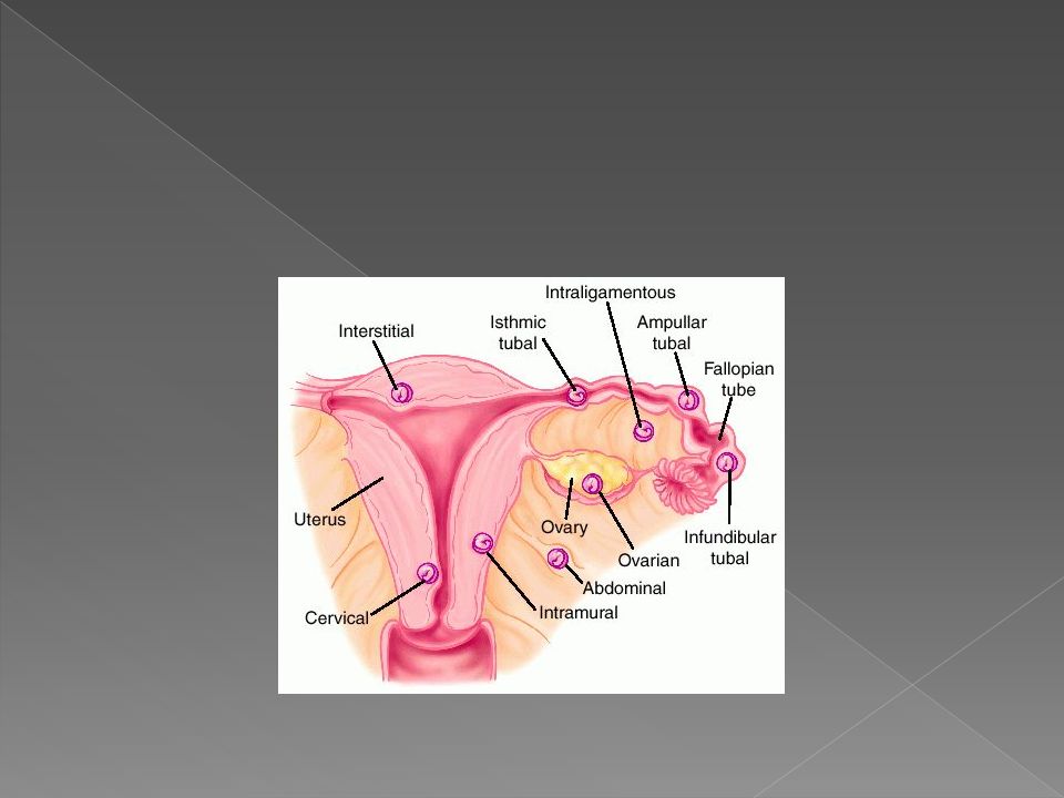
An ectopic pregnancy, is a complication of pregnancy in which the embryo implants outside the uterine cavity. [1] With rare exceptions, ectopic pregnancies are not viable. Furthermore, they are dangerous for the parent, since internal haemorrhage is a life threatening complication. Most ectopic pregnancies occur in the Fallopian tube (so- called tubal pregnancies ), but implantation can also occur in the cervix, ovaries, and abdomen. An ectopic pregnancy is a potential medical emergency, and, if not treated properly, can lead to death.complication of pregnancyuterine cavity [1]Fallopian tubecervix ovariesabdomenmedical emergency In a normal pregnancy, the fertilized egg enters the uterus and settles into the uterine lining where it has plenty of room to divide and grow. About 1% of pregnancies are in an ectopic location with implantation not occurring inside of the womb, and of these 98% occur in the Fallopian tubes. Detection of ectopic pregnancy in early gestation has been achieved mainly due to enhanced diagnostic capability. Despite all these notable successes in diagnostics and detection techniques ectopic pregnancy remains a source of serious maternal morbidity and mortality worldwide, especially in countries with poor prenatal care. [2] [2]
, but implantation can also occur in the cervix, ovaries, and abdomen. An ectopic pregnancy is a potential medical emergency, and, if not treated properly, can lead to death.complication of pregnancyuterine cavity [1]Fallopian tubecervix ovariesabdomenmedical emergency In a normal pregnancy, the fertilized egg enters the uterus and settles into the uterine lining where it has plenty of room to divide and grow. About 1% of pregnancies are in an ectopic location with implantation not occurring inside of the womb, and of these 98% occur in the Fallopian tubes. Detection of ectopic pregnancy in early gestation has been achieved mainly due to enhanced diagnostic capability. Despite all these notable successes in diagnostics and detection techniques ectopic pregnancy remains a source of serious maternal morbidity and mortality worldwide, especially in countries with poor prenatal care. [2] [2].")
4
Tubal pregnancy The vast majority of ectopic pregnancies implant in the Fallopian tube. Pregnancies can grow in the fimbrial end (5% of all ectopics), the ampullary section (80%), the isthmus (12%), and the cornual and interstitial part of the tube (2%). [3] Mortality of a tubal pregnancy at the isthmus or within the uterus (interstitial pregnancy) is higher as there is increased vascularity that may result more likely in sudden major internal hemorrhage [3]interstitial pregnancy
, the ampullary section (80%), the isthmus (12%), and the cornual and interstitial part of the tube (2%). [3] Mortality of a tubal pregnancy at the isthmus or within the uterus (interstitial pregnancy) is higher as there is increased vascularity that may result more likely in sudden major internal hemorrhage [3]interstitial pregnancy.")
6
Non-tubal ectopic pregnancy : While a fetus of ectopic pregnancy is typically not viable, very rarely, a live baby has been delivered from an abdominal pregnancy. In such a situation the placenta sits on the intra-abdominal organs or the peritoneum and has found sufficient blood supply. This is generally bowel or mesentery, but other sites, such as the renal (kidney), liver or hepatic (liver) artery or even aorta have been described. Support to near viability has occasionally been described, but even in third world countries, the diagnosis is most commonly made at 16 to 20 weeks gestation. Such a fetus would have to be delivered by laparotomy. Maternal morbidity and mortality from extrauterine pregnancy is high as attempts to remove the placenta from the organs to which it is attached usually lead to uncontrollable bleeding from the attachment site.abdominal pregnancyplacentaperitoneumlaparotomy
, liver or hepatic (liver) artery or even aorta have been described. Support to near viability has occasionally been described, but even in third world countries, the diagnosis is most commonly made at 16 to 20 weeks gestation. Such a fetus would have to be delivered by laparotomy. Maternal morbidity and mortality from extrauterine pregnancy is high as attempts to remove the placenta from the organs to which it is attached usually lead to uncontrollable bleeding from the attachment site.abdominal pregnancyplacentaperitoneumlaparotomy.")
7
Heterotopic pregnancy In rare cases of ectopic pregnancy, there may be two fertilized eggs, one outside the uterus and the other inside. This is called a heterotopic pregnancy Although rare, heterotopic pregnancies are becoming more common, likely due to increased use of IVF

8
An ectopic pregnancy occurs when a pregnancy starts outside the womb (uterus). The most common site for an ectopic pregnancy is within one of the tubes through which the egg passes from the ovary to the uterus (fallopian tube). An ectopic pregnancy is often caused by a condition that blocks or slows the movement of a fertilized egg through the fallopian tube to the uterus. This may be caused by a physical blockage in the tube by hormonal factors and by other factors, such as smoking. Most cases of scarring are caused by: Past ectopic pregnancy Past infection in the fallopian tubes Surgery of the fallopian tubes Up to 50% of women who have ectopic pregnancies have had swelling (inflammation) of the fallopian tubes (salpingitis) or pelvic inflammatory disease (PID).PID Some ectopic pregnancies can be due to: Birth defects of the fallopian tubes Complications of a ruptured appendixruptured appendix Endometriosis Endometriosis Scarring caused by previous pelvic surgery
. An ectopic pregnancy is often caused by a condition that blocks or slows the movement of a fertilized egg through the fallopian tube to the uterus. This may be caused by a physical blockage in the tube by hormonal factors and by other factors, such as smoking. Most cases of scarring are caused by: Past ectopic pregnancy Past infection in the fallopian tubes Surgery of the fallopian tubes Up to 50% of women who have ectopic pregnancies have had swelling (inflammation) of the fallopian tubes (salpingitis) or pelvic inflammatory disease (PID).PID Some ectopic pregnancies can be due to: Birth defects of the fallopian tubes Complications of a ruptured appendixruptured appendix Endometriosis Endometriosis Scarring caused by previous pelvic surgery.")
9
The following may also increase the risk of ectopic pregnancy: Age over 35 Having had many sexual partners In vitro fertilization In vitro fertilization In a few cases, the cause is unknown. Sometimes, a woman will become pregnant after having her tubes tied (tubal sterilization).tubal sterilization Ectopic pregnancy is also more likely in women who have: Had surgery to reverse tubal sterilization in order to become pregnant Had an intrauterine device (IUD) and became pregnant (very unlikely when IUDs are in place) Ectopic pregnancies occur in 1 in every 40 to 1 in every 100 pregnancies.
.tubal sterilization Ectopic pregnancy is also more likely in women who have: Had surgery to reverse tubal sterilization in order to become pregnant Had an intrauterine device (IUD) and became pregnant (very unlikely when IUDs are in place) Ectopic pregnancies occur in 1 in every 40 to 1 in every 100 pregnancies..")
10
The classic signs and symptoms of ectopic pregnancy include: abdominal pain, the absence of menstrual periods (amenorrhea), and vaginal bleeding or intermittent bleeding (spotting). The woman may not be aware that she is pregnant. These characteristic symptoms occur in ruptured ectopic pregnancies (those accompanied by severe internal bleeding) and non-ruptured ectopic pregnancies. However, while these symptoms are typical for an ectopic pregnancy, they do not mean an ectopic pregnancy is necessarily present and could represent other conditions. In fact, these symptoms also occur with a threatened abortion (miscarriage) i The signs and symptoms of an ectopic pregnancy typically occur six to eight weeks after the last normal menstrual period, but they may occur later if the ectopic pregnancy is not located in the Fallopian tube. Other symptoms of pregnancy (for example, nausea and breast discomfort, etc.) may also be present in ectopic pregnancy.
 and non-ruptured ectopic pregnancies. However, while these symptoms are typical for an ectopic pregnancy, they do not mean an ectopic pregnancy is necessarily present and could represent other conditions. In fact, these symptoms also occur with a threatened abortion (miscarriage) i The signs and symptoms of an ectopic pregnancy typically occur six to eight weeks after the last normal menstrual period, but they may occur later if the ectopic pregnancy is not located in the Fallopian tube. Other symptoms of pregnancy (for example, nausea and breast discomfort, etc.) may also be present in ectopic pregnancy..")
11
Pulse rate, blood pressure, shoulder pain. Low blood pressure, fainting, dizziness & rapid heart rate noted typically in ruptured ectopic pregnancy (intra-abdominal bleeding) Gynecological examination: bimanual or speculum examination may provoke the rupture the rupture of the tube. Bulky uterus, fullness in the right or left adnexia, fullness in the pouch of doughlas. Mobilization of the cervix laterally may provoke pain(cervical exitation is positive).
 Gynecological examination: bimanual or speculum examination may provoke the rupture the rupture of the tube. Bulky uterus, fullness in the right or left adnexia, fullness in the pouch of doughlas. Mobilization of the cervix laterally may provoke pain(cervical exitation is positive)..")
12
Laboratory investigation : Hb, blood group & save for crossmatch & BHCG. HCG : glycoprotein produced by the placenta. It has a half-life of up to 24hours & peak around 10weeks. BHCG level less than 5mIU/ml negative. Above 25mIU/ml is positive. In normal pregnancy, BHCG level almost double every 48 hours in a normally developing pregnancy. In patients with ectopic, the rise of HCG is often suboptimal.

13
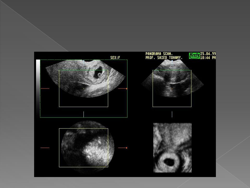
An intra-uterine gestational sac should be visualized at about 4,5weeks of gestation. The corresponding BHCG is around 1500mlIU/ml. At5weeks gestation, fetal cardiac pulsation corresponding to 3000mlIU/ml. Thus if there were discrepancy between BHCG concentration & that seen on ultrasound scan, the differential diagnosis of EP must be made. Identification of an intrauterine pregnancy on TVS effectively excludes the possibility of an EP except with rare heterotopic pregnancy. The presence of free fluid during TVS is suggestive of ruptured EP. Laparoscopy : can be used in diagnosis & treatment of EP.

14
1. The US finding of an extra-uterine sac with embryo is the most reliable diagnosis of EP 2. An empty ectopic sac or a heterogeneous adnexial mass is a more common US feature 3. The presence of fluid in the pouch of doughlas is a non-specific sign of EP 4. In 20-30per cent of EP, pseudo-gestational sac is seen as a small, centrally located endometrial fluid collection surrounded by a single echogenic rim of endometrial tissue undergoing decidual reaction

16
1. abortion 2. GTN 3. Appendicitis 4. Ovarian cyst & other causes of acute abdomen. MANAGEMENT: Surgical : laproscopical surgery is superior to laparotomy. The laproscopical approach is the gold standard in hemodynamic ally stable patient. Laparotomy remain the main stay in ruptured EP & unstable patient. Salpingectomy, partial or total removal of the tube if severely damaged tube or ruptured EP. Salpingotomy: removal of EP& suturing of the tube. This carry risk of recurrent EP. salpingostomy: removal of EP & left site of tube open to heal by secondary healing

17
Medical treatment: By using methotrexate 1mg/kg body weight either given systemically or injected locally under laproscopical or US guide Criteria for methotrexate treatment of EP: Indications: 1. Hemodynamic ally stable, no active bleeding, no or minimal pain. 2. No contraindication to methotrexate 3. Unruptured adnexial mass less than 4cm in size on scan 4. No fetal cardiac activity 5. HCG does not exceed 5000iu/l 6. Able to return for follow up care for several weeks

18
Breast feeding, immune deficiency, chronic liver disease, active pulmonary disease, active peptic ulcer or colitis, blood disorders, hepatic, renal or hematological dysfunctions. Side effects: 1. Nausea & vomiting 2. Stomatitis 3. Diarrhea, abdominal discomfort 4. photosensitivity skin reaction 5. Impaired liver function 6. Pneumonitis (rare) 7. Sever neutropenia (rare) 8. Reversible alopecia 9. Hematosalpinx
 7. Sever neutropenia (rare) 8. Reversible alopecia 9. Hematosalpinx.")
19
1. Diagnosis as guidelines 2. Check criteria for medical management with the consultant responsible for patient. 3. Check full blood count, urea, electrolyte, LFT, blood group 4. Counsel patient, give written information & obtain written consent 5. Obtain patient height & weight to calculate body surface area 6. Calculate methotrexate dose(50mg/m2) 7. Arrange follow up appointment between day 5&7 post injection (more than 15per cent decrease check weekly until less than 25iu/l ). If less than that, consider repeat dose &recheck in 5days.
 7. Arrange follow up appointment between day 5&7 post injection (more than 15per cent decrease check weekly until less than 25iu/l ). If less than that, consider repeat dose &recheck in 5days..")
20
Most EP resolve spontaneously The overall efficacy of this management is 69per cent. The management of these cases can be greatly facilitated by using serum HCG in conjunction with serum progesterone S.progesterone less 25nmol/l & BHCG less than 25iu/l non viable pregnancy ----reassurance S.progesterone 20-60nmol/l& BHCG more than 25 high risk of EP so repeat test in 2 days S.prog. More than 60& BHCG less than 1000---low risk of EP S.proges. More than 60 & BHCG more than 1000 ---EP so repeat scan as soon as possible+_ laproscopy

22
Thank you

Similar presentations


 Medical therapy method terexate.>")


 is a process by which egg cells are manually fertilized by sperm outside of the womb. IVF is a major treatment.>")
>")

 is a generic term for inflammation of the uterus( (endmetritis), fallopian tubes (salpingitis), and/or.>")
