Download presentation
Presentation is loading. Please wait.

1
Pectoral Region: Lecture 13
Dr. Farhat Aamir

2
Objectives At the end of this lecture the students should be able to:
Enumerate different features of the skin of upper limb. Identify area of distribution of major cutaneous nerves in upper limb. Define fascia of the upper limb. Discuss the superficial and deep fascia of the upper limb. Describe various modifications of deep fascia. Describe muscles, vessels, nerves and lymphatics of pectoral region. Identify clinical application.

3
Skin Skin is lax and it has Langer's lines which run transversely
Langer’s lines are those lines which carry considerable amount of collagen fibers

4
The major cutaneous nerves of the upper limb
1. Supraclavicular nerves. 2. Superior/upper lateral cutaneous nerve of arm. 3. Intercostobrachial and medial cutaneous of arm (median). 4. Inferior/lower lateral cutaneous nerve of arm (radial). 5. lateral cutaneous nerve of forearm. (musculocutaneous) 6. Medial cutaneous nerve of forearm. 7. Ulnar nerve. 8. Superficial branch of radial nerve. 9. Median nerve.
. 4. Inferior/lower lateral cutaneous nerve of arm (radial). 5. lateral cutaneous nerve of forearm. (musculocutaneous) 6. Medial cutaneous nerve of forearm. 7. Ulnar nerve. 8. Superficial branch of radial nerve. 9. Median nerve.")
5
Cont. 1. supraclavicular nerves.
2. superior lateral cutaneous nerve of arm (axillary nerve). 3. Intercostobrachial and medial cutaneous of arm (median). 4. Posterior cutaneous nerve of arm, post. Cutaneous of forearm. 5. lateral cutaneous nerve of forearm. (musculocutaneous) 6. Medial cutaneous nerve of forearm (median). 7. Ulnar nerve. 8. Superficial branch of radial. 9. Median nerve.
. 3. Intercostobrachial and medial cutaneous of arm (median). 4. Posterior cutaneous nerve of arm, post. Cutaneous of forearm. 5. lateral cutaneous nerve of forearm. (musculocutaneous) 6. Medial cutaneous nerve of forearm (median). 7. Ulnar nerve. 8. Superficial branch of radial. 9. Median nerve.")
6
Fascia of upper limb Superficial Fascia: expand to accommodate the mammary gland in the female Deep fascia: tough and dense connective tissue Deep fascia: is continuous above with the deep investing fascia of neck Below it is continuous with the membranous superficial fascia of the anterior abdominal wall

7
Modifications of Deep Fascia
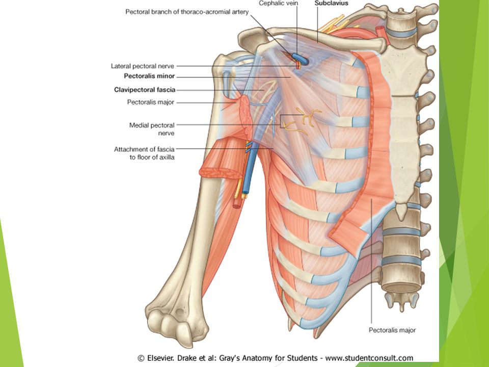
Clavipectoral Fascia: From clavicle to axillary fascia Lateral: coracoid Medial: external intercostal membrane of first two space Three structures piercing clavipectoral fascia

9
Fascial modifications
Extensor retinaculum: thickening of the deep fascia on the dorsum of the wrist.

10
Fascial modifications
Brachial Fascia: tubular investment of arm Attached to humerus via lateral and medial intermuscular septum Bicipital aponeurosis: extension of tendon of biceps brachii

11
Fascial modifications
Fascia, antebrachial: forms a tubular investment of the forearm muscles Attached to the radius via the lateral intermuscular septum; it is attached to the subcutaneous border of the ulna

12
Fascial modifications
Axillary Fascia: forms the floor of the axilla inferiorly Attached to the suspensory ligament of the axilla Continuous with the pectoral fascia

13
Fascial modifications
Palmar aponeurosis: palm of the hand composed of very dense connective tissue that extends out into each of the fingers

14
Fascial modifications
Flexor retinaculum: ventral surface of the wrist osseofibrous tunnel for passage of the flexor tendons tendons are surrounded by synovial tendon sheathes where they pass deep to retinacula

15
Pectoralis Major Origin: clavicle, sternum the sixth or seventh costal cartilage; the first to the seventh costal cartilages and the aponeurosis of external oblique. Insertion: lateral lip of the bicipital grove or intertubercular sulcus of the humerus by means of a thick bilaminar tendon which is about 5cm in length. Innervation: lateral pectoral nerve and medial pectoral nerve Action: adduction and medial rotation of the humerus

16
Pectoralis minor O: 3rd to 5th rib I: coracoid process
Innervation: Medial pectoral nerve and lateral pectoral nerve Action: draw scapula forward, accessory muscle of respiration

17
Subclavius O: 1st rib, close to the costochondral junction
I: subclavian groove on clavicle Innervation: Nerve to sub clavius Action: pulls point of shoulder down

18
SERRATUS ANTERIOR O: upper eight ribs
I: costal surface of medial border of scapula Innervation: long thoracic nerve (C5,6,C7) Action: protract scapula, abduction of arm above 90 degree
 Action: protract scapula, abduction of arm above 90 degree.")
19
4. Anterior perforating branch of internal thoracic a
The vessels and nerves of the pectoral region are branches of the subclavian and axillary arteries and the intercostal nerves. 1 . Thoracoacromial a. 2. Cephalic v. 3. lateral thoracic a. 4. Anterior perforating branch of internal thoracic a 5. lateral cutaneous branches intercostal n. 6. Anterior cutaneous branches intercosta nerve 7. Medial and lateral pectoral nerve

20
Superficial veins The veins start on the back of the hand in a dorsal plexus and become two major veins The cephalic vein empties into the last part of the axillary vein in the pectoral region basilic vein joins the brachial vein to become the axillary vein in the mid-arm region

21
1. cephalic v. 2. basilic v. 3. median cubital v. 4. medial cutaneous nerve of forearm 5. lateral cutaneous nerve of forearm

22
Clinical application For e.g. compartment syndromes, fibromyalgia (chronic widespread pain and a heightened and painful response to pressure), Dupuytren's contracture is a fixed flexion contracture of the hand due to a palmar fibromatosis, where the fingers bend towards the palm and cannot be fully extended (straightened). and fasciitis Fasciitis : inflammation of fascia The superficial veins are used for intravenous injections They are also abused by drug users
, Dupuytren s contracture is a fixed flexion contracture of the hand due to a palmar fibromatosis, where the fingers bend towards the palm and cannot be fully extended (straightened). and fasciitis. Fasciitis : inflammation of fascia. The superficial veins are used for intravenous injections. They are also abused by drug users.")
23
Clinical Anatomy Bicipital aponeurosis: deep to the superficial veins of the upper limb provides some protection for the deeper structures during venipuncture Carpal tunnel syndrome is pressure on the median nerve Deep fascia involved in the spread or containment of infections or pus, oedematous effusions

24
Clinical application Winging of scapula.
Injury to long thoracic nerve.

Similar presentations