Download presentation
Presentation is loading. Please wait.

1
Neonatal metabolic disorders Dr. Mohamed Haseen Basha Assistant professor (Pediatrics) Faculty of Medicine Al Maarefa College of Science and Technology
 Faculty of Medicine Al Maarefa College of Science and Technology.")
2
Inborn errors of metabolism (IEM) are disorders in which there is a block at some point in the normal metabolic pathway caused by a genetic defect of a specific enzyme. IEM are now often referred to as Congenital Metabolic Diseases or Inherited Metabolic Diseases.

3
Clinical Presentation: Severe illness in the newborn, regardless of the underlying cause, tends to manifest with non-specific findings, such as Poor feeding Drowsiness Lethargy Hypotonia Failure to thrive.

4
Clinical pointers towards an underlying IEM include: Deterioration after a period of apparent normalcy Parental consanguinity Family history of neonatal deaths Rapidly progressive encephalopathy and seizures of unexplained cause Severe metabolic acidosis Persistent vomiting Peculiar odor Acute fatty liver or HELLP (hemolysis, elevated liver enzymes & low platelet counts) during pregnancy: seen in women carrying fetuses with long- chain-3- hydroxyacyl-coenzyme dehydrogenase deficiency (LCHADD).
 during pregnancy: seen in women carrying fetuses with long- chain-3- hydroxyacyl-coenzyme dehydrogenase deficiency (LCHADD).")
5
Examination findings that may provide a clue to the underlying IEM Examination findings that may provide a clue to the underlying IEM Clinical pointers towards specific IEM’s Clinical findingDisorder Coarse faciesLysosomal disorders CataractGalactosemia, Zellweger syndrome Retinitis pigmentosaMitochondrial disorders Cherry red spotLipidosis HepatomegalyStorage disorders urea cycle defects Renal enlargementZellweger syndrome Eczema/alopeciaBiotinidase deficiency Abnormal kinky hairMenke disease Decreased pigmentation Phenylketonuria

6
Patterns of presentation of IEM include Encephalopathy with or without metabolic acidosis: Encephalopathy, seizures, and tone abnormalities are predominant presenting features of organic acidemias, urea cycle defects and congenital lactic acidosis. Intractable seizures are prominent in pyridoxine dependency, non-ketotic hyperglycinemia, molybdenum co-factor defect and folinic-acid responsive seizures.

7
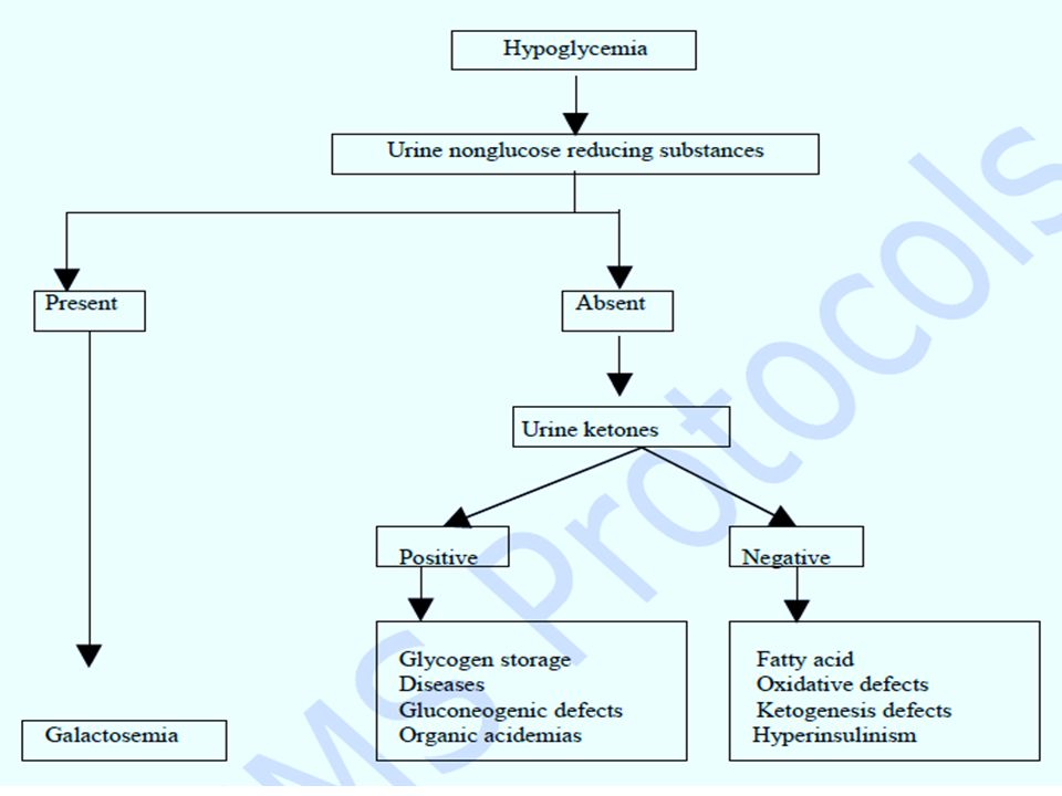
Acute liver disease: Jaundice alone: Gilbert syndrome, Criggler-Najjar syndrome Hepatic failure (jaundice, ascites, hypoglycemia, coagulopathy): Tyrosinemia, Galactosemia, neonatal hemochromatosis, glycogen storage disease type IV. Neonatal cholestasis: alpha-1 antitrypsin deficiency, Niemann-Pick disease type C. Hypoglycemia: persistent and severe hypoglycemia may be an indicator of an underlying IEM. Hypoglycemia is a feature of galactosemia, fatty acid oxidation defects, organic acidemias, glycogen storage disorders and disorders of gluconeogenesis.

8
Dysmorphic features: Peroxisomal disorders Pyruvate dehydrogenase deficiency Congenital disorders of glycosylation (CDG) Lysosomal storage diseases. Some IEMs may present with non-immune hydrops fetalis; these include Lysosomal storage disorders Congenital disorders of glycosylation (CDG).
..")
9
Cardiac disease: cardiomyopathy is a prominent feature in some IEM including Fatty acid oxidation defects, Glycogen storage disease type II Mitochondrial electron transport chain defects.

10
Differential diagnosis IEM should be considered in the differential diagnosis of any sick neonate along with common acquired causes such as Sepsis Hypoxic-ischemic encephalopathy Duct-dependant cardiac lesions Congenital adrenal hyperplasia Congenital infections.

11
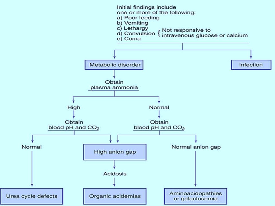
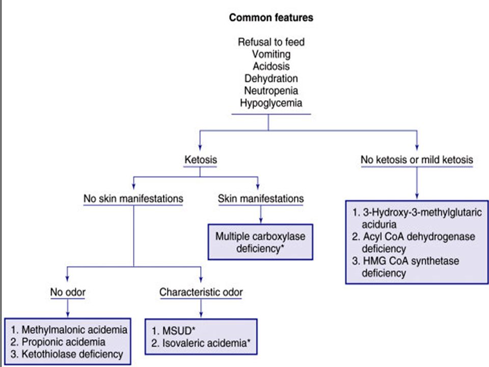
Investigations First line investigations (metabolic screen): Complete blood count: (neutropenia and thrombocytopenia seen in propionic and methylmalonic academia) Arterial blood gases and electrolytes Blood glucose Plasma ammonia (Normal values in newborn: 90-150 mg/dl or 64-107 mmol/L) Arterial blood lactate (Normal values: 0.5-1.6 mmol/L) Liver function tests Urine ketones Urine reducing substances. Serum uric acid (low in molybdenum cofactor deficiency).
..")
15
Second line investigations (ancillary and confirmatory tests) Gas chromatography mass spectrometry (GCMS) of urine: for diagnosis of organic acidemias. Plasma amino acids and acyl carnitine profile by tandem mass spectrometry (TMS): for diagnosis of organic acidemias, urea cycle defects, aminoacidopathies and fatty acid oxidation defects. High performance liquid chromatography (HPLC): for quantitative analysis of amino acids in blood and urine; required for diagnosis of organic acidemias and aminoacidopathies. Lactate/pyruvate ratio: in cases with elevated lactate.
: for diagnosis of organic acidemias, urea cycle defects, aminoacidopathies and fatty acid oxidation defects. High performance liquid chromatography (HPLC): for quantitative analysis of amino acids in blood and urine; required for diagnosis of organic acidemias and aminoacidopathies. Lactate/pyruvate ratio: in cases with elevated lactate..")
16
Urinary orotic acid: in cases with hyperammonemia for classification of urea cycle defect. Enzyme assay: This is required for definitive diagnosis. Available enzyme assays include: Biotinidase assay- Suspected biotinidase deficiency(intractable seizures, seborrheic rash, alopecia) GALT (galactose 1-phosphate uridyl transferase) assay- suspected galactosemia (hypoglycemia, cataracts, reducing sugars in urine). Plasma very long chain fatty acid (VLCFA) levels: elevated in peroxisomal disorders.
 GALT (galactose 1-phosphate uridyl transferase) assay- suspected galactosemia (hypoglycemia, cataracts, reducing sugars in urine). Plasma very long chain fatty acid (VLCFA) levels: elevated in peroxisomal disorders..")
17
Neuroimaging: MRI may provide helpful pointers towards etiology while results of definitive investigations are pending. Some IEM may be associated with structural malformations e.g. Zellweger syndrome - diffuse cortical migration and sulcation abnormalities. Propionic & methylmalonic acidemia - Basal ganglia signal change Maple syrup urine disease (MSUD) - Brainstem and cerebellar edema
 - Brainstem and cerebellar edema.")
18
Agenesis of corpus callosum - Menke’s disease, pyruvate decarboxylase deficiency and nonketotic hyperglycinemia. Glutaric aciduria - Frontotemporal atrophy, Subdural hematomas.

19
Magnetic resonance spectroscopy (MRS): may be helpful in selected disorders E.g. Lactate peak elevated - mitochondrial disorders Leucine peak elevated in MSUD. Electroencephalography (EEG): Comb-like rhythm - MSUD, Burst suppression in NKH and Holocarboxylase synthetase deficiency. Mutation analysis: CSF amino acid analysis: CSF Glycine levels elevated in NKH.
: Comb-like rhythm - MSUD, Burst suppression in NKH and Holocarboxylase synthetase deficiency. Mutation analysis: CSF amino acid analysis: CSF Glycine levels elevated in NKH..")
20
Precautions to be observed while collecting samples Should be collected before specific treatment is started or feeds are stopped, as may be falsely normal if the child is off feeds. Samples for blood ammonia and lactate should be transported in ice and immediately tested. Lactate sample should be arterial and should be collected after 2 hrs fasting in a pre-heparinized syringe. Ammonia sample is to be collected approximately after 2 hours of fasting in EDTA vacutainer. Avoid air mixing. Sample should be free flowing. Detailed history including drug details should be provided to the lab. (sodium valproate therapy may increase ammonia levels).
..")
21
Samples to be obtained in infant with suspected IEM when diagnosis is uncertain and death seems inevitable (Metabolic autopsy) Blood: 5-10 ml; frozen at -20 0 C; both heparinized for chromosomal studies and EDTA for DNA studies Urine: frozen at –20 o C CSF: store at –20 o C Skin biopsy: including dermis in culture medium or saline with glucose. Store at 4-8 0 C. Do not freeze. Liver, muscle, kidney and heart biopsy: as indicated. Clinical photograph (in cases with dysmorphism) Infantogram (in cases with skeletal abnormalities)
 Infantogram (in cases with skeletal abnormalities).")
22
Treatment Treatment of genetic disorders of metabolism is complex and requires medical and technical expertise. The therapeutic regimen often needs to be tailored to the individual patient because of large phenotypic variations in the severity of the disease, even within a single family. Providing education and support for the family is the key to successful long-term therapy. Effective treatment is best achieved by a team of specialists (physician metabolic genetics specialist, nutritionist, geneticist, neurologist, and psychologist) in a major medical center.
 in a major medical center..")
23
1. Special diets play an important role in the treatment of affected children. Dietary changes should be tailored to the pathophysiology of the condition and vary greatly among disorders. 2. Peritoneal dialysis or hemodialysis for expeditious removal of accumulated noxious compounds. This is a very effective modality for treatment of the acute phase of the condition. 3. Administration of the deficient metabolite. 4. Administration of the cofactor or coenzyme to maximize the residual enzyme activity.

24
5. Activation of alternate pathways to reduce the noxious compounds accumulated because of the genetic mutation. 6. Administration of the deficient enzyme. 7. Bone marrow transplantation. 8. Liver transplantation.

25
Aims of treatment To reduce the formation of toxic metabolites by decreasing substrate availability (by stopping feeds and preventing endogenous catabolism) To provide adequate calories To enhance the excretion of toxic metabolites. To institute co-factor therapy for specific disease and also empirically if diagnosis not established. Supportive care- Treatment of seizures (avoid sodium valproate – may increase ammonia) Maintain euglycemia and normothermia Fluid, electrolyte & acid-base balance Treatment of infection and mechanical ventilation if required.
 Maintain euglycemia and normothermia Fluid, electrolyte & acid-base balance Treatment of infection and mechanical ventilation if required..")
26
Management of hyperammonemia Discontinue all feeds. Provide adequate calories by intravenous glucose and lipids. Maintain glucose infusion rate 8-10mg/kg/min. Start intravenous lipid 0.5g/kg/day (up to 3g/kg/day). After stabilization gradually add protein 0.25 g/kg till 1.5 g/kg/day. Dialysis is the only means for rapid removal of ammonia, and hemodialysis is more effective and faster than peritoneal dialysis.
. After stabilization gradually add protein 0.25 g/kg till 1.5 g/kg/day. Dialysis is the only means for rapid removal of ammonia, and hemodialysis is more effective and faster than peritoneal dialysis..")
27
Alternative pathways for nitrogen excretion-: Sodium benzoate (IV or oral)- loading dose 250 mg/kg then 250- 400 mg/kg/day in 4 divided doses. Sodium phenyl butyrate -loading dose 250 mg/kg followed by 250-500 mg/kg/day. L-arginine (oral or IV)- 300 mg/kg/day L-carnitine (oral or IV)- 200 mg/kg/day Supportive care: treatment of sepsis, seizures, ventilation. Avoid sodium valproate.
- 300 mg/kg/day L-carnitine (oral or IV)- 200 mg/kg/day Supportive care: treatment of sepsis, seizures, ventilation. Avoid sodium valproate..")
28
Acute management of newborn with suspected organic acidemia The patient is kept nil per orally and intravenous glucose is provided. Carnitine: 100 mg/kg/day IV or oral. Treat acidosis: Sodium bicarbonate 0.35-0.5mEq/kg/hr (max 1- 2mEq/kg/hr) Start Biotin: 10 mg/day orally. Start Vitamin B12: 1-2 mg/day I/M (useful in B12 responsive forms of methylmalonic acidemias) Start Thiamine: 300 mg/day (useful in Thiamine-responsive variants of MSUD). Supportive care: hydration, treatment of sepsis, seizures, ventilation.
 Start Biotin: 10 mg/day orally. Start Vitamin B12: 1-2 mg/day I/M (useful in B12 responsive forms of methylmalonic acidemias) Start Thiamine: 300 mg/day (useful in Thiamine-responsive variants of MSUD). Supportive care: hydration, treatment of sepsis, seizures, ventilation..")
29
Management of congenital lactic acidosis Supportive care: hydration, treatment of sepsis, seizures, ventilation. Avoid sodium valproate. Treat acidosis: sodium bicarbonate 0.35-0.5mEq/kg/hr (max 1-2mEq/kg/hr) Thiamine: up to 300 mg/day in 4 divided doses. Riboflavin: 100 mg/day in 4 divided doses. Add co-enzyme Q: 5-15 mg/kg/day L-carnitine: 50-100 mg/kg orally.
 Thiamine: up to 300 mg/day in 4 divided doses. Riboflavin: 100 mg/day in 4 divided doses. Add co-enzyme Q: 5-15 mg/kg/day L-carnitine: mg/kg orally..")
30
Treatment of newborn with refractory seizures with no obvious etiology (suspected metabolic etiology) If patient persists to have seizures despite 2 or 3 antiepileptic drugs in adequate doses, consider trial of pyridoxine 100 mg intravenously. If intravenous preparation not available, oral pyridoxine can be given (15 mg/kg/day). If seizures persist despite pyridoxine, give trial of biotin 10 mg/day and folinic acid 15 mg/day (folinic acid responsive seizures). Rule out glucose transporter defect: measure CSF and blood glucose. In glucose transporter defect, CSF glucose level is equal to or less than 1/3rd of the blood glucose level. This disorder responds to the ketogenic diet.
. If seizures persist despite pyridoxine, give trial of biotin 10 mg/day and folinic acid 15 mg/day (folinic acid responsive seizures). Rule out glucose transporter defect: measure CSF and blood glucose. In glucose transporter defect, CSF glucose level is equal to or less than 1/3rd of the blood glucose level. This disorder responds to the ketogenic diet..")
31
Management of asymptomatic newborn with a history of sibling death with suspected IEM: After baseline metabolic screen, start oral dextrose feeds (10% dextrose). After 24 hours, repeat screen. If normal, start breast feeds. Monitor sugar, blood gases and urine ketones, blood ammonia 6 hourly. Some authorities recommend starting medium chain triglycerides (MCT oil) before starting breast feeds After 48 hours, repeat metabolic screen. Obtain samples for TMS and urine organic acid tests. The infant will need careful observation and follow-up for the first few months, as IEM may present in different age groups in members of the same family.
 before starting breast feeds After 48 hours, repeat metabolic screen. Obtain samples for TMS and urine organic acid tests. The infant will need careful observation and follow-up for the first few months, as IEM may present in different age groups in members of the same family..")
32
Long term treatment of IEM Dietary treatment: This is the mainstay of treatment in phenylketonuria, maple syrup urine disease, homocystinuria, galactosemia, and glycogen storage disease Type I & III. Special diets for PKU and MSUD are commercially available, however very expensive Some disorders like urea cycle disorders and organic acidurias require dietary modification (protein restriction) in addition to other modalities. Enzyme replacement therapy (ERT): Pompe’s disease (Glycogen storage disorder Type II)which may present in the newborn period and for which ERT is now available.
 in addition to other modalities. Enzyme replacement therapy (ERT): Pompe’s disease (Glycogen storage disorder Type II)which may present in the newborn period and for which ERT is now available..")
33
Cofactor replacement therapy: The catalytic properties of many enzymes depend on the participation of non protein prosthetic groups, such as vitamins and minerals, as obligatory cofactors. The following co-factors may be beneficial in certain IEM. Thiamine: mitochondrial disorders, thiamine responsive variants of MSUD, PDH deficiency & complex I deficiency) Riboflavin: Glutamic acidemia Type I, Type II, mild variants of ETF, ETFDH, complex I deficiency. Biotin: Biotinidase deficiency, holocarboxylase synthetase deficiency
 Riboflavin: Glutamic acidemia Type I, Type II, mild variants of ETF, ETFDH, complex I deficiency. Biotin: Biotinidase deficiency, holocarboxylase synthetase deficiency.")
34
Pyridoxine: 50% of cases of homocystinuria due to cystathionine β- synthetase deficiency, pyridoxine dependency with seizures, xanthurenic aciduria, primary hyperoxaluria type I, Hyperornithemia with gyrate atrophy Cobalamin: Methylmalonic academia (cblA, cblB), Homocystinuria and methylmalonic academia (cblC, cblD, cblF) Folinic acid: Hereditary orotic aciduria, Methionine synthase deficiency, Cerebral folate transporter deficiency, hereditary folate malabsorption, Kearns-Sayre syndrome
, Homocystinuria and methylmalonic academia (cblC, cblD, cblF) Folinic acid: Hereditary orotic aciduria, Methionine synthase deficiency, Cerebral folate transporter deficiency, hereditary folate malabsorption, Kearns-Sayre syndrome")
35
Prevention Genetic counselling and prenatal diagnosis: Most of the IEM are single gene defects, inherited in an autosomal recessive manner, with a 25% recurrence risk. Therefore when the diagnosis is known and confirmed in the index case, prenatal diagnosis can be offered. The samples required are chorionic villus tissue or amniotic fluid. Modalities available are: Substrate or metabolite detection: Phenylketonuria, Peroxisomal defects. Enzyme assay: Lysosomal storage disorders like Niemann-Pick disease, Gaucher disease. DNA based (molecular) diagnosis: Detection of mutation in proband/ carrier parents is a prerequisite.
 diagnosis: Detection of mutation in proband/ carrier parents is a prerequisite..")
36
Neonatal screening Tandem mass spectrometry (TMS) is used in some countries for neonatal screening for IEM Aminoacidopathies - phenylketonuria, MSUD, Homocystinuria, Citrullinemia, Argininosuccinic aciduria, hepatorenal tyrosinemia. fatty acid oxidation defects. Organic acidemias - glutaric aciduria, propionic acidemia, methylmalonic acidemia, isovaleric acidemia. The cost of this procedure is high Test is highly sensitive, the specificity is relatively low; and there are difficulties in interpretation of abnormal test results in apparently healthy infants.

37
Categorization of neonatal IEM using metabolic screening tests AcidosisKetosisLactateAmmoniaDiagnosis - + - -Maple syrup urine Disease + +/- - Organic aciduria + +/- + -Lactic acidosis - - - +Urea cycle Disorder - - - -Non - ketotic hyperglyceminuria Sulfite oxidase deficiency Peroxisomal Phenylketonuria Galactosemia

38
Commercially available formulations used in IEM Pyridoxine Hydroxycobalamin Thiamine Riboflavin Biotin Carnitine Folinic acid Sodium Benzoate Arginine Coenzyme Q

39
Thank You

Similar presentations


.>")

