Download presentation
Presentation is loading. Please wait.

1
Teaching point #2 Choose and utilize a standardized way to view each radiograph Alphabet method BIO (Between, inside, outside the lungs) Top down Other My approach: Abnormalities, right lung, left lung, compare the two lungs, trachea, mediastinum, heart, outside the lungs (including abdomen) and bones
 and bones.")
2
Now that you have a search pattern
What structures should you normally see? Anatomy important for lines and tubes Trachea and carina Aortic arch Cavoatrial junction Subclavian vein/artery Internal jugular vein

3
Trachea Carina Left main bronchus Right main bronchus
In the normal chest radiograph only airways within the mediastinum are apparent

4
SVC

5
Aortic arch Aortopulmonary recess Main Pulmonary Artery Cavoatrial junction

6
Cavoatrial junction Our perspective on most central lines
Is it central? Yes = good Is it flopping around hitting the AV Valve? Yes = bad Close to the CAJ is fine CAJ landmarks About 2 cm below the bulge of the right atrial appendage About 2 vertebral bodies below the carina (better for peds)
")
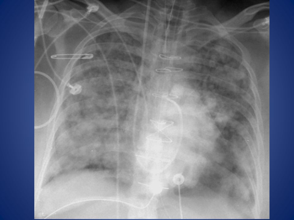
7
Appropriately placed IJ line?
Good placement or not? Yep

8
Lobar anatomy Lesson for today – the upper lobes aren’t just at the top of the CXR, and the lower lobes aren’t just at the bottom.

9
Right Lung: Lobes and Fissures

10
Right Upper & Right Middle Lobes
Lobar Anatomy: Right Upper & Right Middle Lobes RUL RUL RML RML

11
Lobar Anatomy: Right Lower Lobe RLL RLL

12
Lobar Anatomy: Left Upper Lobe LUL LUL

13
Lobar Anatomy: Left Lower Lobe LLL LLL

14
Which lobe is collapsed?

15
Which lobe has a pneumonia?

16
Pattern and Distribution
Abnormalities have two important imaging clues: Pattern of disease Distribution of disease location, location, location!

17
Patterns Consolidation Ground Glass
Lines (interstitial or septal thickening) Reticulation Peripheral Lace-like opacities Cysts Nodules Tree-in-Bud or Budding Tree opacities
 Reticulation. Peripheral Lace-like opacities. Cysts. Nodules. Tree-in-Bud or Budding Tree opacities.")
18
Patterns *Infiltrate is not one of these patterns* Consolidation
Ground Glass Lines (interstitial or septal thickening) Reticulation Cysts Nodules
 Reticulation. Cysts. Nodules.")
19
We can do better “Infiltrate”: A vague term at best, used to describe any abnormality. Avoid it! Lung “fields”: Fields are for cows! They are lungs or lobes “Poor inspiratory effort”: Low lung volumes is at least more appropriate “Nonspecific”: Earn your paycheck

20
Fields are for cows

21
Patterns Consolidation Ground Glass
Lines (interstitial or septal thickening) Reticulation Cysts Nodules Tree-in-Bud or Budding Tree opacities
 Reticulation. Cysts. Nodules. Tree-in-Bud or Budding Tree opacities.")
22
Distribution of Disease
Focal (or multifocal) versus diffuse Dependent distribution (varies with position!) Upper lobe Bronchovascular Peripheral Random
 versus diffuse. Dependent distribution (varies with position!) Upper lobe. Bronchovascular. Peripheral. Random.")
23
Normal versus consolidation

24
Acute Consolidation Infection Water Blood
Indeterminate White Blob (IWB) Infection Bacterial pneumonia Water Pulmonary edema Blood Pulmonary hemorrhage, contusion
 Infection. Bacterial pneumonia. Water. Pulmonary edema. Blood. Pulmonary hemorrhage, contusion.")
25
Acute? Think Infection, Water, Blood

26
Normal Ground Glass

28
Acute Ground Glass Opacity
Indeterminate White Blob (IWB) Infection PCP, viral pneumonia Water Pulmonary edema Blood Pulmonary hemorrhage
 Infection. PCP, viral pneumonia. Water. Pulmonary edema. Blood. Pulmonary hemorrhage.")
29
17 yo Male: Sudden Onset Dyspnea

30
Acute Ground Glass Opacity (2 Weeks of symptoms) & Upper Lobe Distribution
 & Upper Lobe Distribution")
31
Diffuse GGO versus focal consolidation

32
Questions?

33
Teaching points Name the 5 densities that are seen on standard radiographs Choose and utilize a standardized way to view each radiograph Locate the following anatomic structures: Trachea, carina, subclavian artery and vein, SVC, cavoatrial junction Name the 6 patterns seen on chest imaging

34
Teaching points Name the 5 densities that are seen on standard radiographs Air Fat Densities Water/tissue Bone Metal

35
Teaching points Choose and utilize a standardized way to view each radiograph Alphabet method (Airspaces, Bones, Cardiac, etc.) BIO (Between, inside, outside the lungs) Top down Other
 Top down. Other.")
36
Teaching points Locate important anatomic structures:
SVC begins at approximately the 1st anterior rib space Cavoatrial junction: 2 cm below the initial SVC-right atrium bump, or about 2 vertebral bodies below the carina Carina is the branch point of the trachea, it’s usually directly identifiable If it’s not, it should be around T6

37
Teaching Points Consolidation Ground Glass
Lines (interstitial or septal thickening) Reticulation Cysts Nodules Tree-in-Bud or Budding Tree opacities
 Reticulation. Cysts. Nodules. Tree-in-Bud or Budding Tree opacities.")
Similar presentations
 Infections (pneumonia, airways disease)>")


 –Partial.>")


