Download presentation
Presentation is loading. Please wait.

1
CLINICO PATHOLOGICAL CONFERENCE 12 10 2007 Prof T Rogers, Prof J O’Leary, Prof O Sheils

2
. CASE HISTORY: A 67 year-old gentleman of no fixed abode who suffers from chronic alcoholism is found collapsed by Heuston Railway station. He is taken as an emergency to St James’s Hospital. On examination he is found to be semi- conscious but rousable and there is a smell of alcohol from his breath. He is also dehydrated.

3
Initial investigations reveal the following: White blood count: 13 x10 9 /L Proportion neutrophils 70% Haemoglobin: 10g.dl Chest X-ray reveals the abnormalities shown below

4
Radiographic Changes

5
A mass-like, 3 cm density overlies the left hilum (arrow) on the frontal chest radiograph. On the lateral projection it is shown to be in the superior segment of the LLL (arrow), posterior to the tortuous aorta. Q: At this stage what is you differential diagnosis (name 3 options)?: 1 2 3 Q: What investigations would you order/carry out next?: 1 2 3
, posterior to the tortuous aorta. Q: At this stage what is you differential diagnosis (name 3 options) : Q: What investigations would you order/carry out next :")
6
Progress: in the meantime in view of the patient’s poor physical state, he is admitted to the Care of the Elderly ward and placed on intravenous antibiotics. Q: Do you consider this to be appropriate management so far? Further investigations: Blood cultures are negative at 48 hours incubation, sputum microscopy (gram stain) and culture yield no predominant organism, just “normal flora”. The patient is now conscious but somewhat disorientated. He has an intermittent fever up to 38°C.
 and culture yield no predominant organism, just normal flora . The patient is now conscious but somewhat disorientated. He has an intermittent fever up to 38°C..")
7
A chest CT-scan is performed (shown below):
:")
8
CT shows a 3-cm mass in the superior segment of the LLL. The mass has central low attenuation consistent with necrosis. Note the positive bronchus sign--a subsegmental bronchus leading directly to the lesion (arrow). This sign is often associated with increased diagnostic yield at bronchoscopy. It is decided to perform a bronchoscopy and transbronchial biopsy. The sample is sent to Histopathology and part of it is sent to Microbiology.
. This sign is often associated with increased diagnostic yield at bronchoscopy. It is decided to perform a bronchoscopy and transbronchial biopsy. The sample is sent to Histopathology and part of it is sent to Microbiology..")
9
Comment on the histology picture series shown below: 1

10
Comment on the histology picture series shown below: 2

11
At low power, nodular collections of confluent granulomas are surrounded by dark blue lymphoid cells. Note the small, rounded granulomas at the edge of the central nodule. At the centre of the confluent granulomas is a pink zone of necrosis.

12
At the edge of the necrotic zone are lymphocytes, epithelioid cells, and multinucleated giant cells but no well-formed granulomas. Note the two types of multinucleated giant cells: the Langhans' cell with nuclei at the edge and the foreign body type giant cell with nuclei scattered throughout the cytoplasm.

13
The individual, rounded granulomas are composed of activated, epithelioid histiocytes with abundant cytoplasm, multinucleated giant cells, which represent fused epithelioid cells, and lymphocytes.

14
Differential diagnosis: The differential diagnosis includes mycobacterial or fungal granulomas, and special stains for organisms were performed. No fungi were found. Most fungi, with the exception of Pneumocystis jiroveci, can be seen with ordinary H&E stains. The Gomori methenamine silver stain is performed, nevertheless, to find rare organisms and to help identify them. In this particular biopsy, two acid-fast organisms were found on the Ziehl-Neelsen stain. The organisms were found in the necrotic areas and not in the cellular rim. Three weeks later, the culture of the tissue was positive for M. tuberculosis. Non-infectious diseases in the differential diagnosis include Wegener's granulomatosis and nodular sarcoidosis

15
This Ziehl-Neelsen-stained section from another case shows many acid-fast tubercle bacilli that are associated with cells undergoing lysis.

16
Diagnosis: Active tuberculosis, superior segment of left lower lobe Q: what is the incidence of tuberculosis in Ireland per 100,000 population?

18
Further progress: 3 early morning sputum samples reveal the presence of acid and alcohol fast bacilli consistent with M tuberculosis Q: What Microbiology tests will be performed on the sputum samples to confirm the diagnosis and guide treatment? The patient is started on 3 anti-tuberculous antibiotics Q: what are these likely to be? Q: What infection control and public health measures need to be taken in this case? The patient self-discharges and is lost to follow-up.

19
16 weeks later the patient is re-admitted in poor general condition, with significant weight loss and productive cough from last admission with extensive bi-lobar infiltrates. He claims he continued to take his medication but only intermittently as he didn’t have enough tablets. A test is performed on his urine Q: what test is this? Sputum smear is strongly positive for acid fast bacilli.

20
A test is performed in the Tuberculosis laboratory which reveals that the organism is resistant to one of the antituberculous antibiotics: Q: which antibiotic is this? Q: What infection control measure should be implemented? Further results from the TB laboratory show that the strain of Mtb is resistant to two of the antibiotics the patient was originally treated with.

21
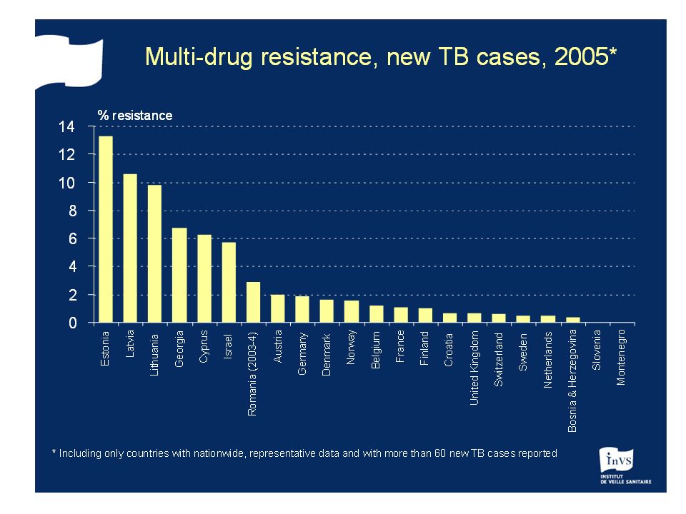
The patient is now treated by DOT Q: What is DOT? Q: What antituberculous antibiotics are available to treat him? Q: what type of tuberculosis does he have? Q: What is the incidence of this type of TB in Ireland? Q: what is its incidence in other parts of Europe eg,former USSR states?

Similar presentations

 Active TB Routine; FBE WCC (Infection) Hb (Anaemic of chronic disease) U&Es (baseline) LFTs (baseline) ESR/CRP (inflammation/infection)>")


 Gram (+) rod (bacilli). Acid-fast Pulmonary.>")


 The incidence of.>")

