Download presentation
Presentation is loading. Please wait.

1
In the name of God

2
Pelvic floor anatomy in female & SUI Dr. Reza Aghelnezhad Endourologist Assistant professor of urology Kermanshah University of Medical Sciences

3
Pelvic floor anatomy in female

11
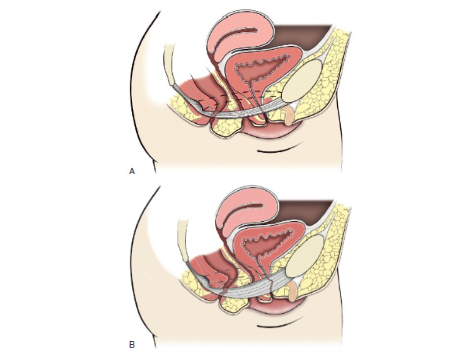
Pelvic organ prolapse (POP)
")
12
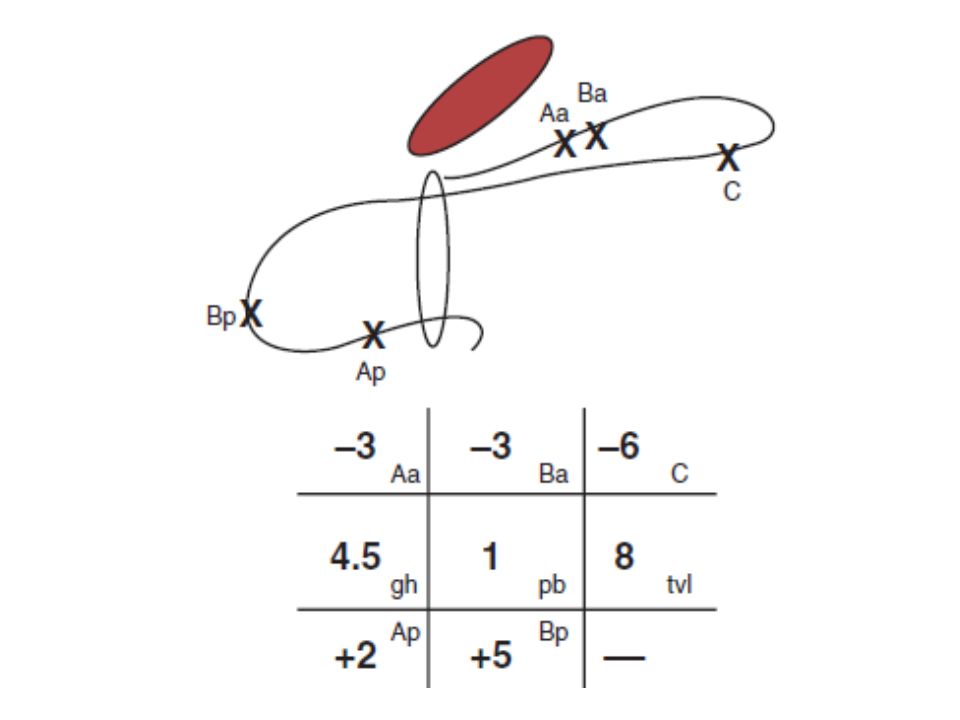
POP-Q staging

13
POP-Q criteria

16
Stress urinary incontinence (SUI)
")
17
Stress UI is the: symptomatic complaint of involuntary leakage on effort or exertion, or on sneezing or coughing.

18
Risk Factors 1 Age: Virtually all studies have found an increase of UI prevalence with age. As a result, while stress incontinence predominates in younger and middle-aged women, urgency and mixed incontinence are more common in older women.

19
Risk Factors 2 Pregnancy, labor, and vaginal delivery versus caesarean section are significant risk factors for later UI, but the strength of this association diminishes substantially with age (level 1 evidence). The vast majority of studies have found an association between parity and later UI.

20
Risk Factors 2 cont. Although several specific parturition factors such as instrumental delivery and birth weight are risk factors for UI in the postpartum period, their association with UI in later life is weak or nonexistent, suggesting that changes in birthing practices in developed countries are unlikely to affect UI in older age (level 2 evidence).
..")
21
Risk Factors 3 Additional evidence has now established body mass as important, modifiable risk factors for UI (level 1 evidence).
.")
22
Risk Factors 4 Physical function also appears to be an independent risk factor for UI in older women. Whether improvement in physical function leads to a reduction in UI remains to be established (level 2 evidence).
..")
23
Risk Factors 5 Evidence from two blinded, randomized controlled trials indicate that oral estrogen, with or without progestogen, is a significant risk factor for UI in women age 55 and older (level 1 evidence).
.")
24
Risk Factors 6 Diabetes is a risk factor for UI in most studies. Although diabetic neuropathy and/or vasculopathy are possible mechanisms by which diabetes could lead to UI, no mechanism has been established, nor is it clear whether prevention or treatment of diabetes, separate from weight reduction, will reduce the risk of UI (level 2 evidence).
..")
25
Risk Factors 7 Menopause, as generally defined, does not appear to be an independent risk factor for stress UI (level 2 evidence).
.")
26
Risk Factors 8 Hysterectomy remains a possible risk factor for later UI, but the evidence is inconsistent (level 2 evidence).
.")
27
Risk Factors 9 Moderate to severe dementia in older women is a moderate to strong independent risk factor for UI (level 2 evidence). Mild loss of cognitive function separated from other factors, increases the risk of UI slightly, if at all, but may increase the impact of UI (level 2 evidence).
..")
28
Risk Factors 10 Smoking, cough, and chronic lung disease: Conflicting data from cross- sectional studies and lack of association between smoking and incident UI in prospective studies suggest that smoking per se is probably not an independent risk factor for UI.( level 2 evidence)
")
29
Risk Factors 11 Data from twin studies suggests that there is a substantial genetic component to UI (level 1 evidence). Several family history studies have found a twofold to threefold greater prevalence of stress UI among first-degree relatives of women with stress UI compared with first- degree relatives of continent women.

30
Risk Factors 12 Diet: Studying diet as a risk factor for UI is challenging. Although some dietary constituents such as coffee, alcohol, or carbonated beverages have been suspected as worsening UI, there is little reason to suspect other dietary constituents as causing, or protecting from, UI.

31
Risk Factors 13 Other potential risk factors including smoking, diet, depression,constipation, UTIs, and exercise, although associated with UI, have not been established as etiologic risk factors and are, in fact, difficult to study with observational data because of the potential for unmeasured confounding and questions of direction of the association (level 3 evidence).
.")
32
ISD Vs. Hypermobility In women with stress incontinence it is likely that there is a degree of sphincter weakness in all cases. The sphincter abnormality will lie on a spectrum from a mild degree where the predominant pathophysiologic abnormality is urethral hypermobility (urethral support defect), to those where irrespective of the urethral support defect there is severe intrinsic sphincteric insufficiency (ISD). Urethral hypermobility and support defects in women are most commonly associated with pregnancy and vaginal delivery, pelvic surgery, and chronic abdominal straining
, to those where irrespective of the urethral support defect there is severe intrinsic sphincteric insufficiency (ISD). Urethral hypermobility and support defects in women are most commonly associated with pregnancy and vaginal delivery, pelvic surgery, and chronic abdominal straining.")
33
Hypermobility

34
h

35
ISD The principles underlying normal sphincteric function are integrated interactions among a number of factors

36
Factor 1:Watertight apposition of the urethral lumen Four urethral wall factors promote continence: (1) wall tension or external compression, (2) inner wall softness, (3) a filler material beneath the mucosa that helps to deform the mucosal folds into apposition, and (4) a lining of mucus provides the stickiness that enables cooptation of these mucosal folds
 wall tension or external compression, (2) inner wall softness, (3) a filler material beneath the mucosa that helps to deform the mucosal folds into apposition, and (4) a lining of mucus provides the stickiness that enables cooptation of these mucosal folds")
37
Factor2:Compression of the wall around the lumen External compression of the urethral lumen is achieved by (1) smooth and striated muscle tone, (2) phasic contractions of the smooth and striated musculature, (3) elastic and viscoelastic properties of the extracellular matrix, (4) mechanical factors related to transmission of abdominal pressure, and (5) structural (anatomic) support of the posterior urethral wall.
 smooth and striated muscle tone, (2) phasic contractions of the smooth and striated musculature, (3) elastic and viscoelastic properties of the extracellular matrix, (4) mechanical factors related to transmission of abdominal pressure, and (5) structural (anatomic) support of the posterior urethral wall.")
38
Other 3 factors: 3. Structural support to keep the proximal urethra from moving during increases in pressure 4. A means of compensating for abdominal pressure changes (pressure transmission) 5. Neural control
 5. Neural control.")
Similar presentations


 is a condition in which a woman's uterus (womb) sags or slips out of its normal position. The uterus.>")


