Download presentation
Presentation is loading. Please wait.

1
THE ROLE OF GENETICS IN MEDICINE & Chromosomal Basis of Heredity Dr S.M.B.Tabei

2
THE ROLE OF GENETICS IN MEDICIN 1- Genetics as a Medical Specialty

4
Relevance of Genetics to All Medical Practice

6
Disciplines within Human and Medical Genetics Human genetics, Human genetics: is the science of variation and heredity in human beings, Medical genetics : Medical genetics :deals with the subset of human genetic variation that is of significance in the practice of medicine and in medical research. Within human and medical genetics, there are many fields of interest : 1- cytogenetics 2- molecular and biochemical genetics 3- genomics 4- population genetics 5- developmental genetics 5- clinical genetics 6- Genetic counseling

7
CLASSIFICATION OF GENETIC DISORDERS genetic variation and mutation in the etiology of a large number of disorders Three main types of disorders are recognized: 1- Single gene disorders : Caused by individual mutant genes. may be present on only one chromosome or on both chromosomes. 2- Chromosome disorders : 2- Chromosome disorders : an excess or a deficiency of the genes contained in whole chromosomes or chromosome segments. 3- Multifactorial disorders : 3- Multifactorial disorders : Multifactorial inheritance is responsible for a number of developmental disorders resulting in congenital malformations and for many common disorders of adult life

8
Chromosomal Basis of Heredity

12
THE HUMAN CHROMOSOMES

14
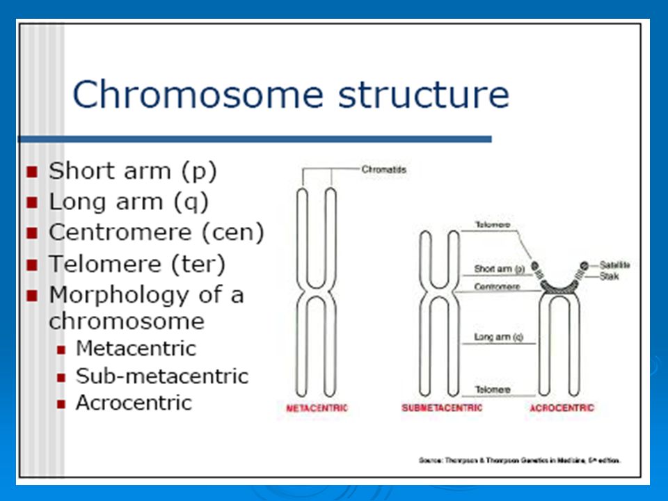
The classification of chromosomes by the position of the centromere. A telocentric chromosome has its centromere at one end; when the chromosome moves toward one pole of the cell during the anaphase of cellular division, it appears as a simple rod. An acrocentric chromosome has its centromere somewhere between the end and the middle of the chromosome; during anaphase movement, the chromosome appears as a J. A metacentric chromosome has its centromere in the middle and appears as a V during anaphase. The classification of chromosomes by the position of the centromere. A telocentric chromosome has its centromere at one end; when the chromosome moves toward one pole of the cell during the anaphase of cellular division, it appears as a simple rod. An acrocentric chromosome has its centromere somewhere between the end and the middle of the chromosome; during anaphase movement, the chromosome appears as a J. A metacentric chromosome has its centromere in the middle and appears as a V during anaphase. (d) Sub metacentric P q
 Sub metacentric P q.")
16
Banding pattern of human chromosomes

17
THE LIFE CYCLE OF A SOMATIC CELL Phases of the cell cycle The division cycle of most eukaryotic cells is divided into four discrete phases: M, G1, S and G2. M phase (mitosis) is usually followed by cytokinesis. S phase is the period during which DNA replication occurs. The cell grows throughout interphase, which includes G1, S and G2.The relative lengths of the cell cycle phases shown here are typical of rapidly replicating mammalian cells. Phases of the cell cycle : The division cycle of most eukaryotic cells is divided into four discrete phases: M, G1, S and G2. M phase (mitosis) is usually followed by cytokinesis. S phase is the period during which DNA replication occurs. The cell grows throughout interphase, which includes G1, S and G2.The relative lengths of the cell cycle phases shown here are typical of rapidly replicating mammalian cells.
 is usually followed by cytokinesis. S phase is the period during which DNA replication occurs. The cell grows throughout interphase, which includes G1, S and G2.The relative lengths of the cell cycle phases shown here are typical of rapidly replicating mammalian cells. Phases of the cell cycle : The division cycle of most eukaryotic cells is divided into four discrete phases: M, G1, S and G2. M phase (mitosis) is usually followed by cytokinesis. S phase is the period during which DNA replication occurs. The cell grows throughout interphase, which includes G1, S and G2.The relative lengths of the cell cycle phases shown here are typical of rapidly replicating mammalian cells..")
18
Cell cycle checkpoints Several checkpoints function to ensure that complete genomes are transmitted to daughter cells. One major checkpoint arrests cells in G2 in response to damaged or unreplicated DNA. The presence of damaged DNA also leads to cell cycle arrest at a checkpoint in G1. Another checkpoint, in M phase, arrests mitosis if the daughter chromosomes are not properly aligned on the mitotic spindle. if the damage is excessive, until the cell is instructed to die by programmed cell death (a process called apoptosis).
..")
19
Replication of chromosomes Replication is the process of duplicating a chromosome Occurs prior to division Replicated copies are called sister chromatids Held together at centromere

20
Mitosis Five stages of mitosis: 1- Prophase 2- Prometaphase 3- Metaphase 4- Anaphase 5-Telophase

22
Prophase - Replicated chromosomes condense. - the beginning of the formation of the mitotic spindle. - Microtubules organize (centrosomes )into a spindle - The centrosomes gradually move to take up positions at the poles of the cell
into a spindle - The centrosomes gradually move to take up positions at the poles of the cell.")
23
Prometaphase -- nuclear membrane breaks up -- allowing the chromosomes to disperse within the cell and to attach -, via their kinetochores, to microtubules of the mitotic spindle -- The chromosomes begin to move toward a point midway between - the spindle poles, a process called congression

24
Metaphase - Chromosomes line up on the metaphase plate. - the chromosomes reach maximal condensation. - Spindle microtubules are attached to centromeres of chromosomes.

25
Anaphase - Centromeres of sister chromatids of each chromosome separate - Chromosomes move to opposite ends of the cell

26
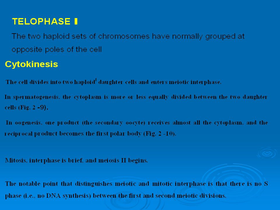
Telophase Nuclear membranes form Spindle disappears Division of cytoplasm occurs (cytokinesis)
")
27
Cytokinesis Cytoplasmic division occurs after nuclear division is complete. Two cells are formed.

28
The Human Karyotype

30
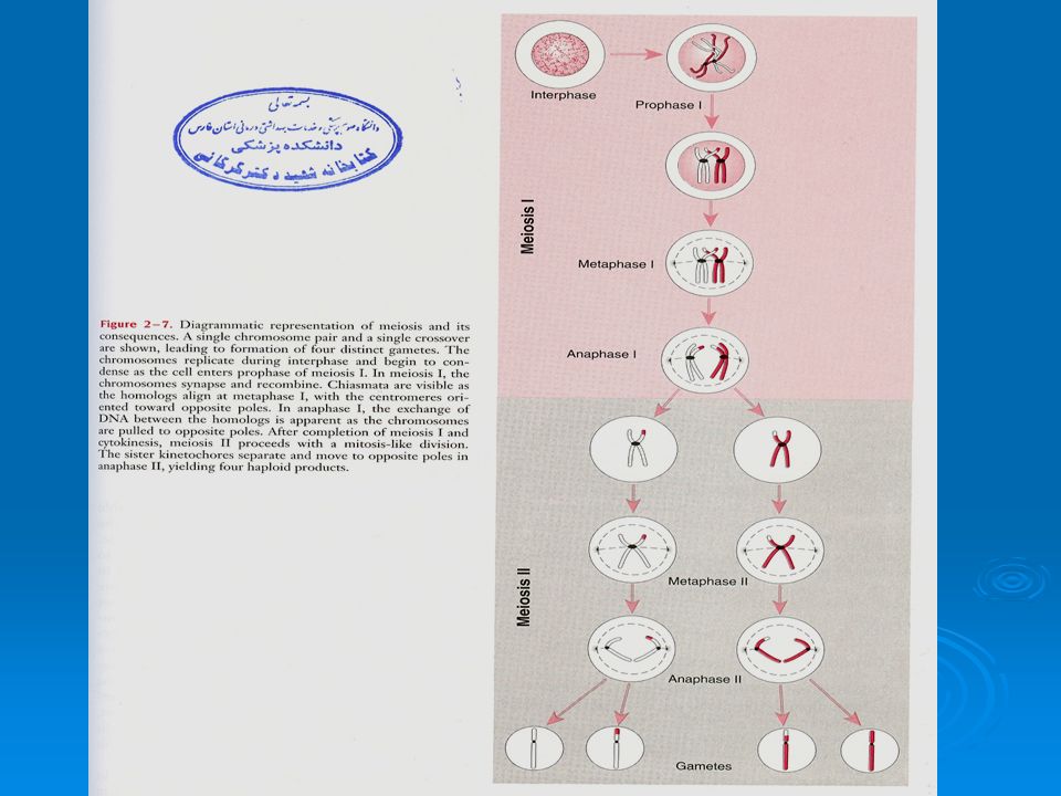
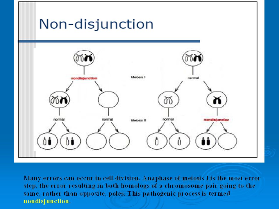
MEIOSIS - Meiosis is the type of cell division by which the diploid cells of the germline ( primary spermatocytes or primary oocytes, ) give rise to haploid gametes. -Meiosis consists of one round of DNA synthesis followed by two rounds of chromosome segregation and cell division

31
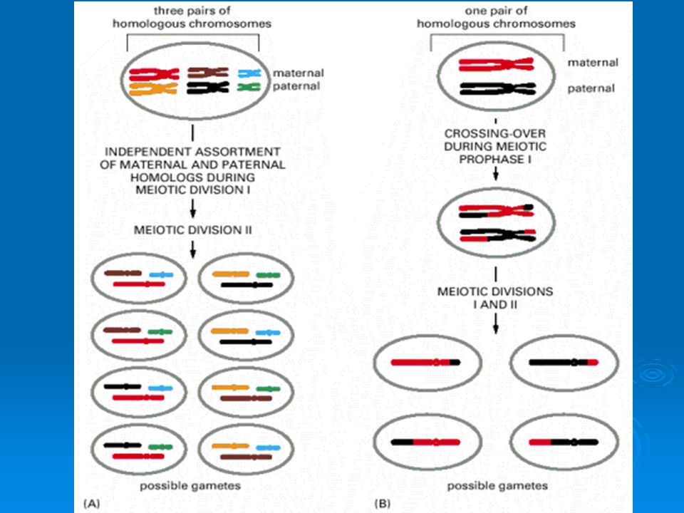
The two successive meiotic divisions are called meiosis I and meiosis II Meiosis I is also known as the reduction division because it is the division in which the chromosome number is reduced from diploid to haploid in which the chromosome number is reduced from diploid to haploid In Meiosis I occur genetic recombination (also called meiotic crossing over) occurs. thus ensuring that none of the gametes produced by meiosis is not identical to another. MEIOSIS

34
The First Meiotic Division (Meiosis I) PROPHASE 1 The prophase of meiosis I is a complicated process that differs from mitotic prophase in a number of ways, with important genetic from mitotic prophase in a number of ways, with important geneticconsequences. Several stages are defined: 1- Leptotene. 2- Zygotene 3- Pachytene 4- Diplotene 5- Diakinesis

35
The chromosomes, replicated during the preceding S phase and beginning to condense. and beginning to condense. two sister chromatids of each chromosome are so closely aligned that they cannot be distinguished. aligned that they cannot be distinguished.

36
homologous chromosomes begin to pair closely along their entire length (synapsis ). DNA sequences into alignment along the length of the entire chromosome.

37
The chromosomes become much more tightly coiled. Synapsis is complete, Each pair of homologs appears as a bivalent (tetrad four chromatids) Crossing over takes place
 Crossing over takes place.")
38
the synaptonemal complex disappears, the two components of each bivalent now begin to separate from each other. Although the homologous chromosomes separate, each of their centromeres remains intact. Eventually the two homologs of each bivalent are held together only at points called chiasmata (crosses),
,.")
39
In this stage, the chromosomes reach maximal condensation.

40
Metaphase I begins, as in mitosis

45
Meiosis II Meiosis II

47
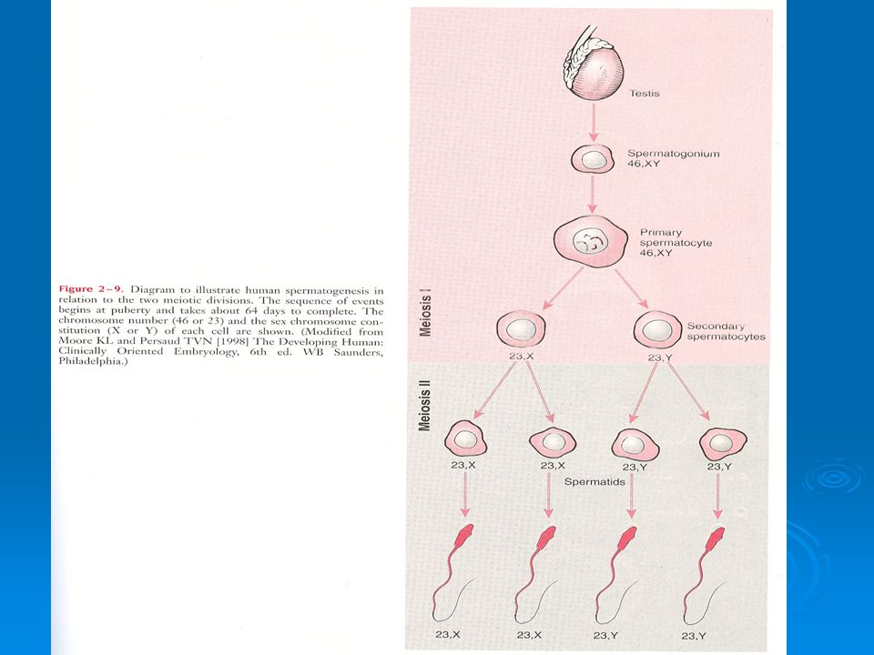
HUMAN GAMETOGENESIS AND FERTILIZATION Spermatogenesis

48
Oogenesis

Similar presentations


 eukaryotic cell of a sexually reproducing organism that result in four haploid (N)>")

 DIPLOID (2N) The condition of having two sets of chromosomes per nucleus The condition.>")


