Download presentation
Presentation is loading. Please wait.

1
Lab 4 & 5 Staining Technique
Practical Of Histopathology El-Hindi.M. & Abdelmoneim.A.

2
Objectives: 1. Understand the classification of Stains. 2. Understand basic tissue staining methods used in the clinical histology laboratory. 3. Identification the procedure of staining. 4. Understand special stains.

3
Overview: Most dyes used to visualize the membranes and organelles of the cell are water soluble. The embedded wax must therefore be removed prior to staining. Routine H&E staining and special stains play a critical role in tissue-based diagnosis or research.

4
Cont. By coloring otherwise transparent tissue sections, these stains allow highly trained pathologists and researchers to view, under a microscope, tissue morphology (structure) or to look for the presence or prevalence of particular cell types, structures or even microorganisms such as bacteria.
 or to look for the presence or prevalence of particular cell types, structures or even microorganisms such as bacteria.")
5
Cont. In the histopathology laboratory, the term “routine staining” refers to the hematoxylin and eosin stain (H&E) that is used “routinely” with all tissue specimens to reveal the underlying tissue structures and conditions. The term “special stains” has long been used to refer to a large number of alternative staining techniques that are used when the H&E does not provide all the information the pathologist or researcher needs.
 that is used routinely with all tissue specimens to reveal the underlying tissue structures and conditions. The term special stains has long been used to refer to a large number of alternative staining techniques that are used when the H&E does not provide all the information the pathologist or researcher needs.")
6
Classification of Stains
Acid stains Basic stains Neutral stains

7
Classification of Stains
Acid Dyes: In an acid dye the basic component is colored and the acid component is colorless. Acid dyes stain basic components e.g. eosin stains cytoplasm. The color imparted is shade of red. Basic Dyes: In a basic dye the acid component is colored and the basic component is colorless. Basic dyes stain acidic components e.g. basic fuchsin stains nucleus. The color imparted is shade of blue.

8
Cont. Neutral Dyes: When an acid dye is combined with a basic dye a neutral dye is formed. As it contains both colored radicals, it gives different colors to cytoplasm and nucleus simultaneously. This is the basis of Leishman stain (Blood Smear).
.")
9
Special stains When a specific components of tissue e.g. fibrous tissue, elastic tissue, nuclear material is to be stained, certain special stains are used which specifically stain that component tissue.

10
Types of special stains
1. PAS (Periodic Acid Schiff) stain: This stain demonstrates glycogen and neutral mucous substances, outlines basement membranes and reticulin and makes evident most types of fungi and parasites. 2. Alazarin Red: It is used to stain bone. 3. Sudan-Black: It is used for fat staining. 4. Masson’s Trichrome: It is used for differentiation of connective tissue elements. Reticular fibers, reticular fibres or reticulin is a type of fiber in connective tissue[1] composed of type III collagensecreted by reticular cells.[2] Reticular fibers crosslink to form a fine meshwork (reticulin). This network acts as a supporting mesh in soft tissues such as liver, bone marrow, and the tissues and organs of the lymphatic system.[3]
 stain: This stain demonstrates glycogen and neutral mucous substances, outlines basement membranes and reticulin and makes evident most types of fungi and parasites. 2. Alazarin Red: It is used to stain bone. 3. Sudan-Black: It is used for fat staining. 4. Masson’s Trichrome: It is used for differentiation of connective tissue elements. Reticular fibers, reticular fibres or reticulin is a type of fiber in connective tissue[1] composed of type III collagensecreted by reticular cells.[2] Reticular fibers crosslink to form a fine meshwork (reticulin). This network acts as a supporting mesh in soft tissues such as liver, bone marrow, and the tissues and organs of the lymphatic system.[3]")
11
Cont. 5. Papanicolaou’s stain: It is used to stain cells in cervical and sputum smear for cytology and to stain bone marrow and blood film. 6. Gymsa stain: it is used to stain blood film. 7. Phangeson stain: It is used to stain muscles. 8. Silver nitrate: It is used to stain skin human. 9. Mercury Brom Phenol Blue: It is used to stain total proteins.

12
Cont. 10. Stains for micro-organism: a. Gram-stain: Gram stain allows the separation of bacteria those that retain the crystal-violet-iodine complex (gram-positive) and those that are decolorized by alcohol treatment and counterstained by eosin, safranin or fuchsin. b. Ziehl_Neelsen stain: This stain detect acid fast bacilli. c. PAS stain: It is used for fungi, amoeba and Tricomonas. d. Modified Giemsa (2% Giemsa in water): Detects Helicobacter pylori.
 and those that are decolorized by alcohol treatment and counterstained by eosin, safranin or fuchsin. b. Ziehl_Neelsen stain: This stain detect acid fast bacilli. c. PAS stain: It is used for fungi, amoeba and Tricomonas. d. Modified Giemsa (2% Giemsa in water): Detects Helicobacter pylori.")
13
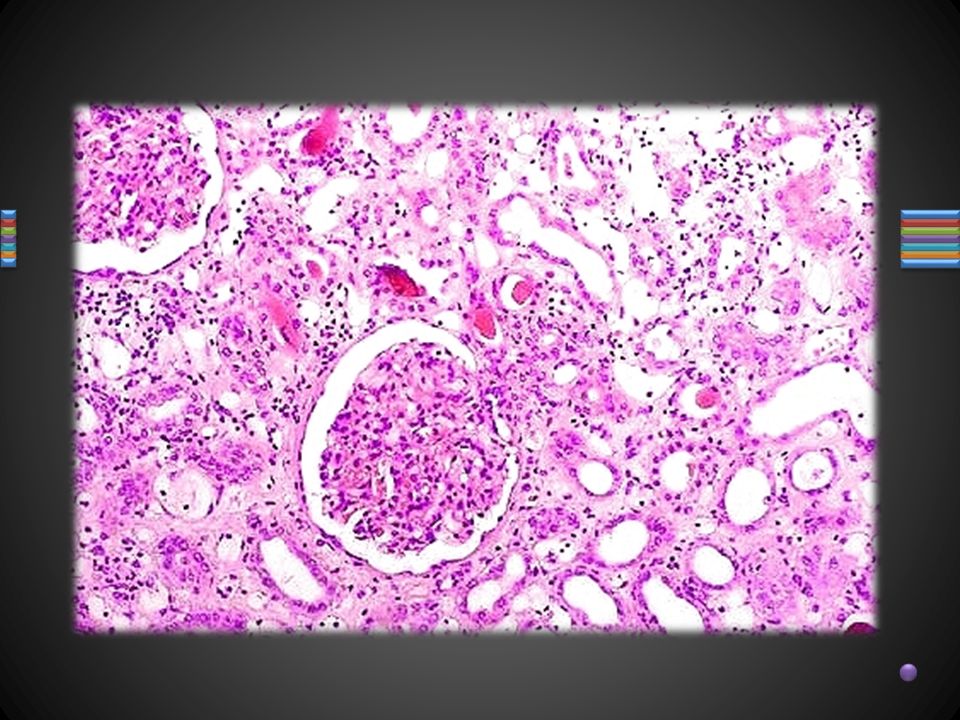
Hematoxylin and Eosin Stain
Hematoxylin staining requires the use of a mordant (most commonly aluminum salts) and stains the nuclear components of cells a dark blue. Hematoxylin is used in combination with eosin because eosin stains the cytoplasmic organelles varying shades of pink, red or orange. The combination of the two stains provides a broad range of morphological information about the section.
 and stains the nuclear components of cells a dark blue. Hematoxylin is used in combination with eosin because eosin stains the cytoplasmic organelles varying shades of pink, red or orange. The combination of the two stains provides a broad range of morphological information about the section.")
14
Frozen Section In this way tissue can be examined microscopically within 5-10 minutes of its removal from the body. It reduces the time of processing from 18 hours to 5 minutes. It has the disadvantage that only 8-16 micron thick section can be cut and finer details of tissue cannot be examined. Frozen section is performed on a machine called cryostat.

Similar presentations


 Abdelraheem BA>")


 or 1000000000.>")


 Staining. + Why stain tissue? Most tissues are colorless and unless one uses diffraction interference contrast microscopy,>")


