Download presentation
Presentation is loading. Please wait.

1
MEDULLARY THYROID CANCER
Dr. M. Sofi MD; FRCP (London); FRCPEdin; FRCSEdin
; FRCPEdin; FRCSEdin.")
2
Medullary thyroid cancer (MTC) is a form of thyroid carcinoma which originates from the parafollicular cells (C cells), which produce the hormone calcitonin. Medullary tumors are the third most common of all thyroid cancers. Sporadic, or isolated, MTC accounts for 75% of cases, and inherited MTC constitutes the rest. Inherited MTC occurs in association with multiple endocrine neoplasia (MEN) type 2A and 2B syndromes, but non-MEN familial MTC also occur Outcome depends on extent of disease, nature of tumor biology, and overall efficacy of surgical treatment. Advances in genetic testing in have revolutionized the management of this disease
 type 2A and 2B syndromes, but non-MEN familial MTC also occur. Outcome depends on extent of disease, nature of tumor biology, and overall efficacy of surgical treatment. Advances in genetic testing in have revolutionized the management of this disease.")
3
Characteristics of Medullary Thyroid Cancer
Occurs in 4 clinical settings and can be associated with other endocrine tumors More common in females than males (except for inherited cancers) Regional metastases (spread to neck lymph nodes) occurs early in the disease Spread to distant organs occurs late and can be to the: Liver, Bone Brain Adrenal medulla Usually originates in the upper central lobe of the thyroid
 Regional metastases (spread to neck lymph nodes) occurs early in the disease. Spread to distant organs occurs late and can be to the: Liver, Bone. Brain. Adrenal medulla. Usually originates in the upper central lobe of the thyroid.")
4
Characteristics of Medullary Thyroid Cancer
Poor prognostic factors include mean: Older than 50 years old Distant spread (metastases) In patients with other endocrine tumors due to MEN II-B syndrome. Residual disease (following surgery) or recurrence can be detected by measuring calcitonin hormone: Should be measured every 4 months for the first few years and then every 6 months for the rest of life
 In patients with other endocrine tumors due to MEN II-B syndrome. Residual disease (following surgery) or recurrence can be detected by measuring calcitonin hormone: Should be measured every 4 months for the first few years and then every 6 months for the rest of life.")
5
History A specific constellation of symptoms of medullary thyroid carcinoma (MTC) is not usually noted; however, one or more of the following symptoms may be observed: Patients may describe a lump at the base of the neck, which may interfere with swallowing or become more prominent Patients with locally advanced disease may present with hoarseness, dysphagia, and respiratory difficulty Although uncommon, patients may present with various paraneoplastic syndromes, including Cushing or carcinoid syndrome Diarrhea may occur from increased intestinal electrolyte secretion secondary to high plasma calcitonin levels Distant metastases (e.g., lung, liver, bone) may result in weight loss, lethargy, and bone pain
 is not usually noted; however, one or more of the following symptoms may be observed: Patients may describe a lump at the base of the neck, which. may interfere with swallowing or become more prominent. Patients with locally advanced disease may present with. hoarseness, dysphagia, and respiratory difficulty. Although uncommon, patients may present with various. paraneoplastic syndromes, including Cushing or carcinoid. syndrome. Diarrhea may occur from increased intestinal electrolyte. secretion secondary to high plasma calcitonin levels. Distant metastases (e.g., lung, liver, bone) may result in. weight loss, lethargy, and bone pain.")
6
Common symptom Neck lump: A single lump on the front of the neck is the most common symptom. It’s often discovered during a routine physical exam. Neck pain: Pain in the front of the neck may be related to the growth of a thyroid tumor. This pain can also extend to the ears. Hoarseness: If cancer has spread to that vocal cord, it can affect the quality of voice. Coughing: Thyroid cancer can sometimes cause a persistent cough. Trouble swallowing (dysphagia): If a thyroid tumor becomes large enough, it can press on the esophagus and make swallowing difficult. Shortness of breath (dyspnea): Similar to trouble swallowing, if a thyroid tumor is large enough it can push against the windpipe and interfere with breathing.
: If a thyroid tumor becomes large enough, it can press on the esophagus and make swallowing difficult. Shortness of breath (dyspnea): Similar to trouble swallowing, if a thyroid tumor is large enough it can push against the windpipe and interfere with breathing.")
7
Common symptom Severe diarrhea: The tumor produces high levels of a hormone-calcitonin, a prostaglandin that may cause severe diarrhea. Cushing syndrome: Although uncommon, patients may present with various paraneoplastic syndromes, including Cushing or carcinoid syndrome Facial flushing: A red face, neck, or chest paired with warm or burning sensations may occur Bone pain: People with medullary thyroid cancer may have bone pain if the cancer has spread to form bone lesions. Lethargy: Many people with advanced cancer may feel physically, emotionally, or mentally tired. Weight loss: Unusual weight loss is a symptom of advanced medullary thyroid cancer that has spread beyond the thyroid into other organs.

8
Physical Physical examination may demonstrate a dominant thyroid nodule at the base of the neck. Palpable cervical lymphadenopathy signifies disease that has progressed locally. Abdominal pain, jaundice, and rarely, bone tenderness may occur in patients with systemic metastases. Causes Medullary carcinoma of thethyroid (MTC) has a genetic association with multiple endocrine neoplasia (MEN) 2A and 2B; however, it is heritable by a non-MEN mode of transmission. Sporadic MTC occurs in 75% of patients, and familial MTC constitutes the other 25%. Mutations in RET can lead to MTC development in cells derived from neural crest tissue.
 has a genetic association with multiple endocrine neoplasia (MEN) 2A and 2B; however, it is heritable by a non-MEN mode of transmission. Sporadic MTC occurs in 75% of patients, and familial MTC constitutes the other 25%. Mutations in RET can lead to MTC development in cells derived from neural crest tissue.")
9
Classification of MTC Hereditary MTC (germline RET mutation present)
25 percent Sporadic MTC (no germline RET mutation identified) 75 percent • No somatic RET mutations found 35 percent • Somatic RET mutations found 65 percent - Exon 16, codon 918 60 percent - Exon 11, codon 630, 634 21 percent - Exon 10, codon 609, 620 9 percent - Exon 15, codon 891
 75 percent. • No somatic RET mutations found. 35 percent. • Somatic RET mutations found. 65 percent. - Exon 16, codon percent. - Exon 11, codon 630, percent. - Exon 10, codon 609, percent. - Exon 15, codon 891.")
10
Genetic screening in sporadic MTC
Germline RET testing should be requested in all patients with newly diagnosed sporadic MTC. Initial germline testing in patients sporadic MTC should include sequencing of exons 10, 11, and 13 through 16 of the RET gene. Sequencing of the remaining exons in the RET gene should be in patients highly suggestive of hereditary MTC who demonstrate no mutations in exons 10, 11, or 13 through 16 A much higher percentage (approximately 60 percent) of patients with sporadic MTC have somatic mutations in the RET gene within the tumor cells. These mutations are present only in the tumor cells and are not detected by standard genetic testing, ie, using leukocyte DNA. The presence of somatic RET mutations correlate with lymph node metastases, persistent disease, and lower survival.
 of patients with sporadic MTC have somatic mutations in the RET gene within the tumor cells. These mutations are present only in the tumor cells and are not detected by standard genetic testing, ie, using leukocyte DNA. The presence of somatic RET mutations correlate with lymph node metastases, persistent disease, and lower survival.")
11
Classification of multiple endocrine neoplasia type 2
Type 2A Type 2B MEN2A classical (medullary thyroid cancer, pheochromocytoma, primary hyperparathyroidism) Medullary thyroid cancer MEN2A with cutaneous lichen amyloidosis Pheochromocytoma MEN2A with Hirschsprung disease Other Familial medullary cancer without pheochromocytoma or parathyroid hyperplasia Mucosal neuromas Intestinal ganglioneuromas Marfanoid habitus
 Medullary thyroid cancer. MEN2A with cutaneous lichen amyloidosis. Pheochromocytoma. MEN2A with Hirschsprung disease. Other. Familial medullary cancer without pheochromocytoma or parathyroid hyperplasia. Mucosal neuromas. Intestinal ganglioneuromas. Marfanoid habitus.")
12
Differential diagnosis
Anaplastic Thyroid Carcinoma De Quervain Thyroiditis Follicular Thyroid Carcinoma Goiter Graves Disease Hyperthyroidism Intestinal Carcinoid Tumor Medullary Thyroid Carcinoma Papillary Thyroid Carcinoma Thyroid Lymphoma Thyroid Nodule Toxic Nodular Goiter Type 2 Multiple Endocrine Neoplasia

13
Laboratory Studies According to the American Thyroid Association, preoperative laboratory testing in patients with possible medullary thyroid carcinoma (MTC) has three purposes: To predict the extent of metastatic disease; this will determine the extent of preoperative imaging and may alter the surgical approach In patients with MEN 2, to identify primary hyperparathyroidism and/or pheochromocytoma —comorbid conditions that alter the surgical approach and surgical priorities To identify RET mutation carriers so that testing of appropriate family members can allow for early diagnosis and treatment of affected individuals
 has three purposes: To predict the extent of metastatic disease; this will determine the extent of preoperative imaging and may alter the surgical approach. In patients with MEN 2, to identify primary hyperparathyroidism and/or pheochromocytoma —comorbid conditions that alter the surgical approach and surgical priorities. To identify RET mutation carriers so that testing of appropriate family members can allow for early diagnosis and treatment of affected individuals.")
14
Calcitonin Calcitonin is the principal biochemical marker in MTC; it is used for detection, staging, postoperative management, and prognosis. The higher that the calcitonin levels are above normal, the greater the likelihood of MTC; basal levels of >100 pg/mL have been found to have 100% positive predictive value for MTC. Screening studies in patients with MEN 24-hour urinalysis for catecholamine metabolites (e.g., VMA, metanephrine) to rule out concomitant pheochromocytoma in patients with MEN type 2A or 2B. Pheochromocytoma must be treated before MTC. Obtain screening for the development of familial MTC in family members of patients with a history of MTC or MEN 2A or 2B
 to rule out concomitant pheochromocytoma in patients with MEN type 2A or 2B. Pheochromocytoma must be treated before MTC. Obtain screening for the development of familial MTC in family members of patients with a history of MTC or MEN 2A or 2B.")
15
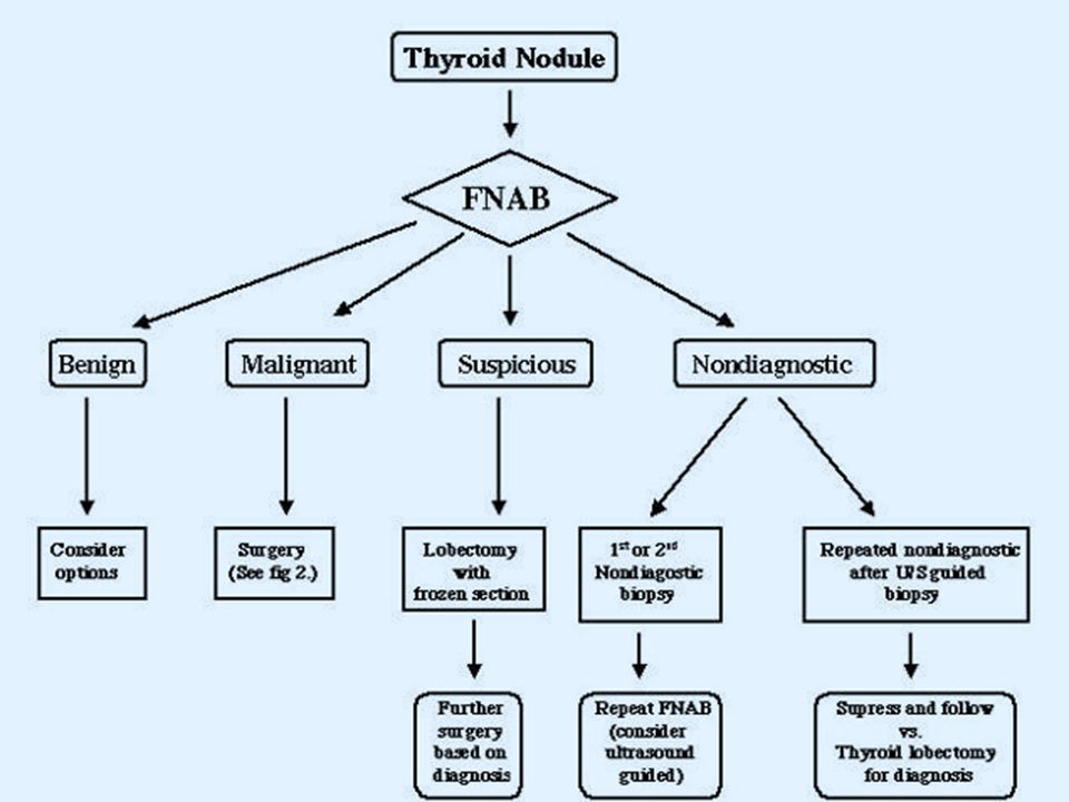
Imaging Studies Patients in whom medullary thyroid carcinoma (MTC) is diagnosed or suspected on the basis of fine needle aspiration findings or calcitonin levels should undergo preoperative ultrasonography to detect lymph node metastases. The study should include the superior mediastinum and the central and lateral neck compartments Patients with regional lymph node involvement or calcitonin levels >400 pg/mL should undergo preoperative CT scanning of the chest and neck, as well as 3-phase, contrast enhanced, multidetector liver CT or contrast-enhanced MRI imaging (MRI) to detect metastatic disease Fine-needle aspiration yields cytologic information, allowing diagnosis of MTC.
 to detect metastatic disease Fine-needle aspiration yields cytologic information, allowing diagnosis of MTC.")
16
Medullary thyroid carcinoma on ultrasound with typical small calcifications (arrows)
")
17
Fine needle aspirate with immunostaining for calcitonin in medullary cancer of the thyroid. The nuclei of the tumor cells are placed eccentrically and are larger and more pleomorphic than those of normal follicular cells. Immunocytologic staining for calcitonin is positive (brown staining which is best seen at the arrow). The background contains many red cells that nonspecifically take up the stain.
. The background contains many red cells that nonspecifically take up the stain..")
18
Surgical specimen showing typical histologic appearance of medullary cancer.

20
Treatment Surgery and radiation therapy have been the major treatments for medullary thyroid carcinoma. A total thyroidectomy with bilateral neck dissection is the gold standard for treating medullary thyroid cancer, and is the most definitive means of achieving a cure in patients without distant metastases or extensive nodal involvement.

21
Treatment Radiation External beam radiotherapy is recommended when there is a high risk of regional recurrence, even after optimum surgical treatment Unlike other differentiated thyroid carcinoma, there is no role for radioiodine treatment in medullary-type disease Protein kinase inhibitors Block the abnormal kinase proteins involved in the development and growth of medullary cancer cells, response in 10-30% Vandetanib the first drug approved by FDA for late-stage (metastatic) MTC in patients ineligible for surgery. Side effects of this drug include hypertension, nausea, diarrhea, cardiac electrical abnormalities, thrombotic or bleeding episodes.
 MTC in patients ineligible for surgery. Side effects of this drug include hypertension, nausea, diarrhea, cardiac electrical abnormalities, thrombotic or bleeding episodes.")
23
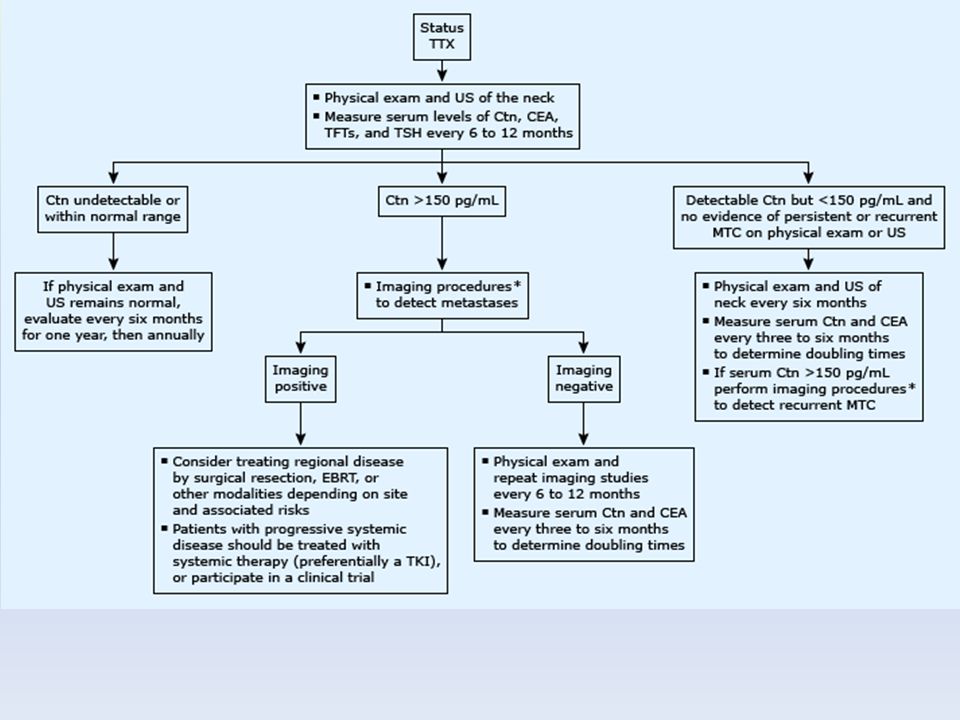
SUBSEQUENT MANAGEMENT — After thyroidectomy, it is important to evaluate patients to determine if surgery was curative. Measurement of serum calcitonin and carcinoembryonic antigen (CEA) are used to assess for cure. Subsequent management depends upon these values. Serum calcitonin and CEA measurement — Serum calcitonin and CEA should be measured two to three months after surgery to detect the presence of residual disease. Patients who have normal serum CEA and undetectable serum calcitonin values are considered biochemically cured and have the best prognosis. PROGNOSIS — Age and stage of disease at the time of diagnosis has been shown to be an important factor that influences prognosis: 5 and 10-year disease-free survival rates are higher among patients 40 years old or less (95 versus 65 %). Patients over age 40 years and (75 versus 50 %). 10-year survival rates for patients with stages I, II, III, and IV (MTC) are 100, 93, 71, and 21 % respectively.
 are used to assess for cure. Subsequent management depends upon these values. Serum calcitonin and CEA measurement — Serum calcitonin and CEA should be measured two to three months after. surgery to detect the presence of residual disease. Patients who have normal serum CEA and undetectable serum calcitonin. values are considered biochemically cured and have the best prognosis. PROGNOSIS — Age and stage of disease at the time of diagnosis has been shown to be an important factor that influences prognosis: 5 and 10-year disease-free survival rates are higher among patients 40. years old or less (95 versus 65 %). Patients over age 40 years and (75 versus 50 %). 10-year survival rates for patients with stages I, II, III, and IV (MTC) are. 100, 93, 71, and 21 % respectively.")
24
THANK YOU FOR YOUR ATTENTION

Similar presentations


 are derived from the diffuse neuroendocrine system, which is made up of.>")


>")

 Brooke Martin 3/20/08.>")


FRCSC(Surgical Oncology) Laparoscopic and Bariatric Surgery.>")
