Download presentation
Presentation is loading. Please wait.

2
The Special Senses

9
External Anatomy of the Eye

10
Lacrimal Apparatus of the Eye

11
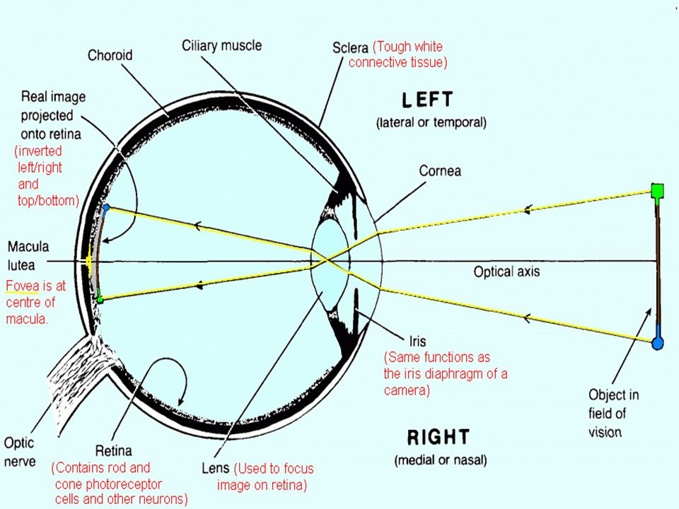
Anatomy of the Eyeball

12
Accessory structures of the Eye from a sagittal view

13
Detail view of the anterior anatomy of the eye

14
Photomicroscopic view of the Histology of the Eye showing the location of the central fovea

15
Intrinsic Eye Muscles and their response to light

16
The Visual Pathway

17
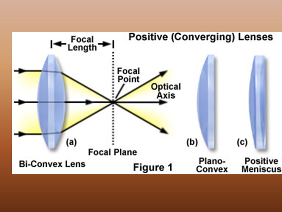
Light Refractory Pathway: 1.Bulbar Conjunctiva 2.Cornea 3.Aqueous Humor 4.Lens 5.Vitreous Humor 6.Ganglion Cell Layer 7.Inner Synaptic Layer 8.Bipolar Layer 9.Outer Synaptic Layer 10. Photoreceptor Layer

18
Abnormalities of The Eye: 1.Myopic - nearsighted 2.Hypermetropic - Farsighted 3.Presbyopia - age-related failure of lens to accommodate 4.Astigmatism - Distorted vision due to irregular-shaped lens or cornea 5.Color Blindness - genetic defect that causes dysfunction of cones

19
Accommodation of the Lens for near vision Ciliary muscles contract Ciliary body pulls forward and inward Tension on suspensory ligaments of lens is decreased Lens becomes thicker (rounder) due to its elasticity Pupils constricts
 due to its elasticity Pupils constricts")
20
Accommodation of the Lens for far vision Ciliary muscles relaxes Ciliary body returns to its resting state, backward and outward Tension on suspensory ligaments of lens is increased Lens becomes thinner (flatter) due to its elasticity Pupils dilate
 due to its elasticity Pupils dilate")
25
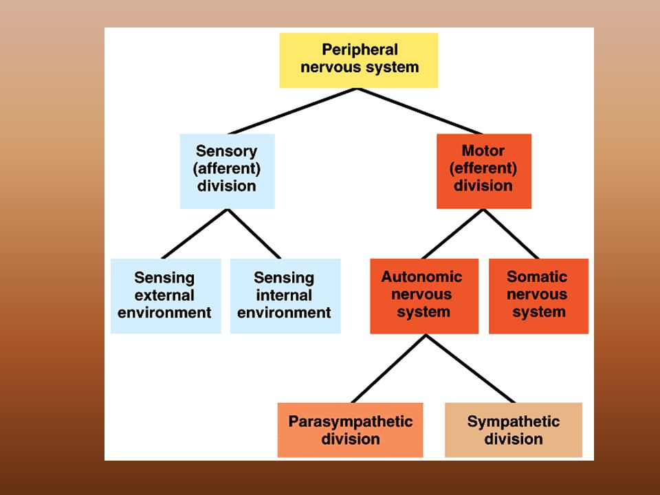
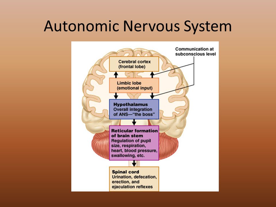
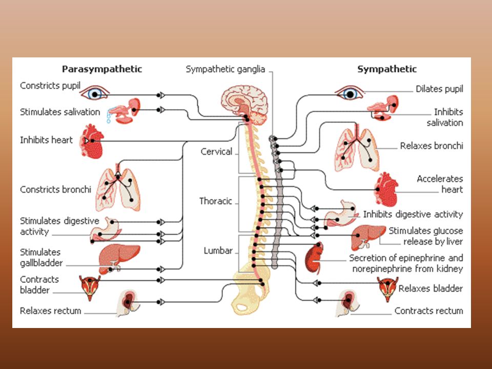
Autonomic Nervous System

Similar presentations


 1. Cornea 2. Sclera Middle Tunic (pg. 470-474) 3. Choroid Coat 4. Ciliary Body 5. Lens & Accommodation 6. Aqueous.>")


>")

 separated by the palpebral fissue Eyelashes Tarsal glands Lacrimal apparatus Vision Accessory structures.>")


>")


