Download presentation
Presentation is loading. Please wait.

1
Cell Culture Part 2

2
Recovery of Cells from Storage
Cells were stored in liquid nitrogen for a long time and need to be recovered for experimental use The vial should be immersed in a 37oC water bath and gently shaken until thawed The contents of the vial should be transferred into a 15 ml sterilized centrifuge tube containing about 10 ml pre-warmed complete medium and centrifuged at 1000 rpm for 5 min The supernatant should then be discarded and cell pellet should be resuspended in 15 ml complete medium Finally, the cell suspension is transferred to a sterile 75 cm2 tissue culture flask and incubated at 37oC in a humidified atmosphere of 95% air and 5% CO2

4
Subculturing Cells In order to maintain cell cultures in optimum conditions it is essential to keep cells in the log phase of growth Adherent cells will continue to grow in vitro until either: they have covered the surface available for growth or they have depleted the nutrients in the surrounding medium Cells kept for prolonged periods in the stationary phase of growth will lose plating efficiency and may become senescent, lose viability and other characteristics, or even die

5
Subculturing Adherent Cells
The frequency of subculture is dependent on a number of factors and will vary between cell lines, the factors include: inoculation density, growth rate, plating efficiency a measure of the number of colonies originating from single cells, and saturation density The maximum cell number attainable, under specified culture conditions, in a culture vessel This term is usually expressed as the number of cells per square centimeter in a monolayer culture or the number of cells per cubic centimeter in a suspension culture

6
Subculturing Adherent Cells

7
Subculturing Adherent Cells
Examine the condition of the cell monolayer using an inverted microscope and ensure that the cells are healthy and sub-confluent The medium is discarded from the vessel and the cells monolayer should be washed twice with warm PBS (magnesium & calcium free) The cells are detached using a warm solution of Trypsin/EDTA solution and the plate is left at 37oC for ~ 5 minutes or until the cells are detached with monitoring of cells under inverted microscope Cells should only be exposed to trypsin long enough to detach the cells Prolonged exposure can damage surface receptors on cells Sub-confluent Overgrown
 The cells are detached using a warm solution of Trypsin/EDTA solution and the plate is left at 37oC for ~ 5 minutes or until the cells are detached with monitoring of cells under inverted microscope. Cells should only be exposed to trypsin long enough to detach the cells. Prolonged exposure can damage surface receptors on cells. Sub-confluent. Overgrown.")
8
Subculturing Adherent Cells
Attached Detached Gently tap the flask with the palm of the hand a couple of times to release any remaining detached cells A warm complete medium is added to the cells to inactivate trypsin and the cells are pipetted several times to break clumps Remove the cell suspension from the flask and place in to a sterile container Centrifuge typically at 1000 rpm for 5 min to sediment the cells Pellet

9
Subculturing Adherent Cells
Pour off the supernatant from the container and resuspend the pellet in complete medium Perform a viable cell count and reseed a flask with an aliquot of cells at the required density The size of culture flask used depends on the number of cells required An appropriate volume of complete medium is added to the flask A cell count may not always be necessary if the cell line has a known split ratio Label each flask with cell line name, passage number, and date

10
Counting of Cells For the majority of manipulations using cell cultures, such as cytotoxicity tests, transfections, cell fusion techniques, cryopreservation and subculture routines it is necessary to quantify the number of cells prior to use Trypan blue is the most commonly used vital dye in microscopy for cell counting and to measure cells viability The principle of trypan blue is based on the fact that the chromophore is negatively charged and does not interact with the cell unless the membrane is damaged Equal volumes of cell suspension and 0.4% trypan blue are mixed by pipetting and left for 5 minutes at RT Prepare a clean hemocytometer chamber and fill it with cell suspension

11
Counting of Cells Count the number of cells in the four large corner squares: viable (seen as bright cells) and non-viable cells (stained blue) Use the following formula to calculate viable number of cells 1 2 4 3
 Use the following formula to calculate viable number of cells")
12
Subculturing Suspension Cells
Examine the cultures microscopically for signs of cell deterioration and high-density growth Cells in the exponential phase of growth will appear bright, round, and refractile, whereas dying cultures show cell lysis and shrunken cells Method 1: Remove a volume of the cells for a viable cell count Transfer the cells to a sterile container and centrifuge the cells at 1000 rpm for 5 min to form a pellet Discard the supernatant and resuspend the pellet with fresh media and split as desired

13
Subculturing Suspension Cells
Method 2: Take out required amount of cell suspension from the flask using pipette and place into new flask e.g. For 1:2 split from 20 ml of cell suspension take out 10 ml For 1:5 split from 20 ml of cell suspension take out 4 ml Add required amount of pre-warmed cell culture media to fresh flask. e.g. For 1:2 split from 20 ml add 10 ml fresh media to 10 ml cell suspension For 1:5 split from 20 ml add 16 ml fresh media to 4 ml cell suspension

14
Changing Media If cells have been growing well for a few days but are not yet confluent then they will require media changing to replenish nutrients and keep correct pH If there are a lot of cells in suspension (attached cell lines) or the media is starting to go orange rather than red then media should be changed as soon as possible To change media, warm up fresh culture media at 37oC in water bath or incubator for at least 30 mins Carefully pour of the media from the flask into a waste pot Immediately replace the media with fresh pre-warmed culture media and return to CO2 37oC incubator
 or the media is starting to go orange rather than red then media should be changed as soon as possible. To change media, warm up fresh culture media at 37oC in water bath or incubator for at least 30 mins. Carefully pour of the media from the flask into a waste pot. Immediately replace the media with fresh pre-warmed culture media and return to CO2 37oC incubator.")
15
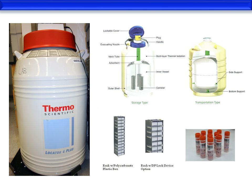
Storage of Cell Lines (Cryopreservation)
Because an established cell line is a valuable resource and its replacement is expensive and time consuming, it is vitally important that they are frozen and preserved for long-term storage If a cell line can be expanded sufficiently, preservation of cells by freezing will allow: secure stocks to be maintained without aging and protect them from problems of contamination incubator failure or medium and serum crises Ideally, 1×10⁶–1×10⁷ cells should be frozen in 10 ampoules

16
Factors Affectining Survival After Freezing
Factors favoring good survival after freezing and thawing are: High cell density at freezing (1×10⁶–1×10⁷ cells/ml) Presence of a preservative, such as glycerol or dimethyl sulfoxide (DMSO) at 5–10% Cryoprotective agents reduce the freezing point of the medium and also allow a slower cooling rate, greatly reducing the risk of ice crystal formation, which can damage cells and cause cell death Slow cooling, 1oC/min, down to −70oC and then rapid transfer to a liquid nitrogen freezer Rapid thawing Slow dilution, ∼20-fold, in medium to dilute out the preservative
 Presence of a preservative, such as glycerol or dimethyl sulfoxide (DMSO) at 5–10% Cryoprotective agents reduce the freezing point of the medium and also allow a slower cooling rate, greatly reducing the risk of ice crystal formation, which can damage cells and cause cell death. Slow cooling, 1oC/min, down to −70oC and then rapid transfer to a liquid nitrogen freezer. Rapid thawing. Slow dilution, ∼20-fold, in medium to dilute out the preservative.")
17
Factors Affectining Survival After Freezing
Reseeding at 2- to 5-fold the normal seeding concentration For example, if cells are frozen at 5×10⁶ cells in 1 ml of freezing medium with 10% DMSO and then thawed and diluted 1:20, the cell concentration will still be 2.5×10⁵ cells/ml at seeding, higher than the normal seeding concentration for most cell lines, and the DMSO concentration will be reduced to 0.5% which most cells will tolerate for 24 h Changing medium the following day (or as soon as all the cells have attached) to remove preservative
 to remove preservative.")
18
Quality Control All cell culture laboratories should be run according to Good Laboratory Practice Guidelines A number of organizations provide accreditation of clinical laboratories, and although most cell culture is probably done within research laboratories, similar principles should be applied The major issues are infection and contamination which cause inaccurate results

19
Culture Contamination
If a culture is contaminated this must be discovered as soon as possible, either to: Discard the culture before the contamination can spread to other cultures or to attempt decontamination The latter should only be used as a last option Decontamination is not always successful and can lead to the development of antibiotic-resistant organisms

20
Culture Contamination
Most bacterial, fungal, and yeast infections are readily detected by regular careful examination with the naked eye (e.g., by a change in the color of culture medium) and by using the microscope However, one of the most serious contaminations is mycoplasma, which is not visible by routine microscopy Any cell culture laboratory should have a mycoplasma screening program in operation Those collecting tissue for primary culture are particularly at risk
 and by using the microscope. However, one of the most serious contaminations is mycoplasma, which is not visible by routine microscopy. Any cell culture laboratory should have a mycoplasma screening program in operation. Those collecting tissue for primary culture are particularly at risk.")
21
Culture Contamination
The precautions that should be observed are as follows: Check frequently for contamination by looking for: a rapid change in pH (usually a fall, but some fungi can increase the pH) cloudiness in the medium Extracellular granularity under the microscope or any unidentified material floating in the medium If a contamination is detected, discard the flask unopened and autoclave If in doubt, remove a sample and examine by phase microscopy, Gram's stain, or standard microbiological techniques
 cloudiness in the medium. Extracellular granularity under the microscope. or any unidentified material floating in the medium. If a contamination is detected, discard the flask unopened and autoclave. If in doubt, remove a sample and examine by phase microscopy, Gram s stain, or standard microbiological techniques.")
22
Bacterial or Fungal Culture Contamination
Bacterial or fungal infections of cell cultures are usually obvious The phenol red, if present, will turn yellow as the infection uses up available nutrients and acidifies the medium Under the inverted microscope hyphae, yeast, or colonies of bacteria can be observed However, it is common practice to add antibiotics to cultures and this can mask low level infection for a considerable time

23
Mycoplasma Culture Contamination
The most common and most missed infection in cell culture laboratories is probably Mycoplasma Several species are involved and their effects are insidious The indicators that there might be a problem include: reduced growth rate, morphological changes, chromosomal aberration, and altered metabolism There are several ways of testing for Mycoplasma and many manufacturers provide kits It is possible to treat Mycoplasma infection with antibiotics, but avoidance is the best policy Most laboratories simply dispose of infected cultures and start again Fluorescence of the eukaryotic nuclei and the extranuclear prokaryotic DNA

24
Viruses Viral infection is also insidious, some cultures contain viruses Viruses are either integrated into their genome, or as endogenous non-lethal infections In many cases, these are not regarded as infections, and viral transformation of cells is an age-old method of producing continuous cell lines

25
Cross-Contamination If there are other cell lines in use in the laboratory, they can cross-contaminate even a primary culture, or misidentification can arise during subculture or recovery from the freezer Precautions must be taken to avoid cross-contamination: Do not handle more than one cell line at a time If this is impractical, do not have culture vessels and medium bottles for more than one cell line open at one time Never be tempted to use the same pipette or other device for different cell lines

26
Cross-Contamination Do not share media or other reagents among different cell lines Do not share media or reagents with other people Keep a panel of photographs of each cell line, at low and high densities, above the microscope, and consult this regularly when examining cells during maintenance This is particularly important if cells are handled over an extended period, and by more than one operator If continuous cell lines are in use in the laboratory, handle them after handing other, slower-growing, finite cell lines

Similar presentations


 (auxotroph) YPD with antibiotics.>")


