Download presentation
Presentation is loading. Please wait.

1
T. Lindenmaier D. Buchanan, S. McKay, I. Gyacskov,
Semi-Automated Segmentation of Three-dimensional Ultrasound Carotid Artery Plaque Volume T. Lindenmaier D. Buchanan, S. McKay, I. Gyacskov, A. Fenster and G. Parraga Imaging Research Laboratories, Robarts Research Institute, Department of Medical Biophysics, Biomedical Engineering Graduate Program, The University of Western Ontario, London, CANADA April 4, 2012

2
Overview Atherosclerosis and the carotid artery
Image acquisition using ultrasound Imaging phenotypes of carotid atherosclerosis Limitations of current methods Development of semi-automated measurement Application and Reproducibility

3
Motivation: Carotid Atherosclerosis
Libby, Peter. Inflammation in Atherosclerosis. Nature. December 2002.

4
ICA ECA CCA BF

5
Motivation: Atherosclerosis in Carotid Artery
Cerebrovascular disease accounts for 10% of all deaths worldwide1 1WHO, 2004

6
Ultrasound of Carotid Atherosclerosis
Intima-Media Thickness Courtesy of Christiane Mallet

7
Manual Planimetry Measurements
lumen-intima media-adventitia Longitudinal View Axial View

8
Intima-Media Thickness
Carotid Atherosclerosis: US Measurements Vessel Wall Volume Egger et al. J Ultrasound Med. (2008) Intima-Media Thickness Buchanan et al. Accepted to Ultrasound Med Biol. (2012) Total Plaque Volume Al-Shali et al. Atherosclerosis. (2005) Egger et al. Ultrasound Med Biol. (2007) Total Plaque Area Riccio et al. Cardiovascular Ultrasound. (2006)
 Intima-Media Thickness. Buchanan et al. Accepted to Ultrasound Med Biol. (2012) Total Plaque Volume. Al-Shali et al. Atherosclerosis. (2005) Egger et al. Ultrasound Med Biol. (2007) Total Plaque Area. Riccio et al. Cardiovascular Ultrasound. (2006)")
9
Manual Segmentation of 3DUS TPV
User sets axis of segmentation Measurements made in an axial view at 1mm inter-slice distance Inter-slice distance multiplied by segmentation area to calculate volume 100 500 400 300 200 600 V (mm3) CV (%) 5 10 15 20 Total Plaque Volume Adapted from Landry et al. Stroke. (2004)
 CV (%) Total Plaque Volume. Adapted from Landry et al. Stroke. (2004)")
10
Limitations of Current Measurements
Intima-Media Thickness (IMT) Narrow dynamic range (0.5mm to 1.0mm) 1-dimensional measurement No plaque Total Plaque Area (TPA) High inter-observer variability 2-dimensional measurement Not adequate to estimate 3D change with 2D measurement Total Plaque Volume (TPV) Long measurement time (slice-by-slice) Laborious
 Narrow dynamic range (0.5mm to 1.0mm) 1-dimensional measurement. No plaque. Total Plaque Area (TPA) High inter-observer variability. 2-dimensional measurement. Not adequate to estimate 3D change. with 2D measurement. Total Plaque Volume (TPV) Long measurement time (slice-by-slice) Laborious.")
11
Semi-automated TPV Measurement
Longitudinal View Axial View

12
Schematic of Longitudinal View
Semi-Automated TPV Measurement Contour 1 (longitudinal view) Contour 2 (axial view) Contour 3 (axial view) Contour 4 (axial view) Measurement View Schematic of Longitudinal View C1 C2 C3 C4 Min Z Max Z
 Contour 2. (axial view) Contour 3. (axial view) Contour 4. (axial view) Measurement View. Schematic of Longitudinal View. C1. C2. C3. C4. Min Z. Max Z.")
13
Semi-Automated TPV Measurement
C1 Min Z C2 C3 C4 Max Z x z y c b a P V = volume PFj = representative vertex Aj = area of triangle 1Van Gelder. Graphic Gems 5. (1995)
")
14
Methodology echogenic plaques from 17 subjects Selection of 23
2x5 rounds of semi - automated segmentation Images measured 5x with 5 Minutes between measurements Images measured 5x with 20 hours between measurements 1 round manual segmentation

15
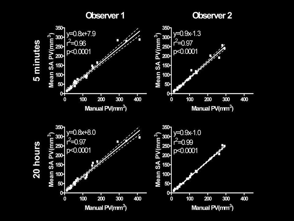
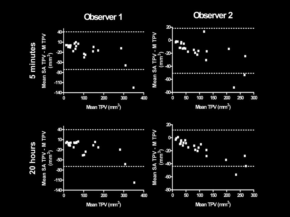
Manual vs. SA Measurements

16
Results Landry et al. Stroke. (2004)
")
19
Conclusion Intra-observer variability similar to manual measurement variability reported by Landry et al. (2004) High correlation between manual and SA for both time durations between measurements The manual TPV measurement can be replaced by the novel segmentation method. If adopted for clinical use, measurements could be generated right away (faster diagnosis).
.")
20
Acknowledgements Research Team Sandra Halko CRCC RPT Shayna McKay BSc
Andrew Wheatley BSc Miranda Kirby BSc Stephen Costella MESc Amir Owrangi MSc Trevor Szekeres MRT Sarah Svenningsen Lauren Villemaire Supervisory Committee Grace Parraga PhD Daniel Buchanan BSc Collaborators Aaron Fenster PhD FCCPM Igor Gyacskov 20

21
Thank you

Similar presentations


, RVT, MMM Director Non-invasive Cardiology and Echocardiography Professor of Medicine and Clinical Scholar Keck School of.>")


 Diagnostics Group 8 Laura Tanenbaum, Sam Tavakoli,>")

: A Reproducibility Study Mindy Columbus, Brian Wagner, Emma Barinas-Mitchell Department of Epidemiology, University.>")
 Bong-Soo Sohn School of Computer Science and Engineering Chung-Ang University.>")


