Download presentation
Presentation is loading. Please wait.

1
Diagnostic Slide Seminar Abdominal lesions Dr. Sandeep Mathur

2
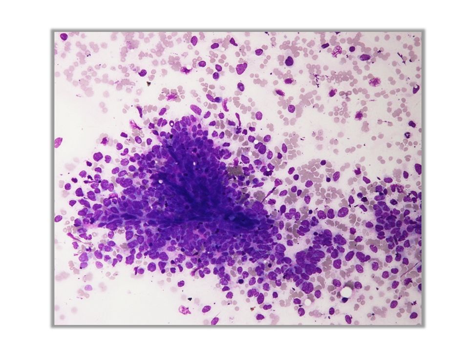
Case 1: FNAC from pancreatic SOL

3
Case 2: 24 year old female presented with pain abdomen for the last 3 years. Mass gradually increased and became palpable since 2 years. A mass in the abdomen, of size 15 x 20 cm, involving the left hypochondrium, left lumbar, left iliac fossa, hypogastrium, umbilical and right lumbar areas. USG guided FNA from Abdominal lump was performed.

4
Case 3: A 46 year old woman presented with increased menstrual bleeding for 6 months & pain in abdomen since 3 months. MRI revealed precaval lymph node with central necrosis & uterine leiomyoma with degenerative changes. CT guided fine needle aspiration from precaval lymph nodes was done.

6
Case 4: 54-year-old male patient presented with abdominal pain and recurrent lump in right iliac fossa.

Similar presentations


, Mumbai.>")


