Download presentation
Presentation is loading. Please wait.

1
Introduction to Pathology

2
What?

3
一、History of Pathology
Autopsy → Organ pathology (1761) LM → Cellular pathology (1854) Ultrastructural pathology with the application of EM (20 century 60s) Immunopathology, Molecular pathology, Genetic pathology, Quantitative pathology
 LM → Cellular pathology (1854) Ultrastructural pathology with the application. of EM (20 century 60s) Immunopathology, Molecular pathology, Genetic pathology, Quantitative pathology.")
4
二、The scope of pathology
1. The core of pathology: The four aspects of a disease process that form the core of pathology : (1) Etiology: causes of the disease (2) Pathogenesis: the mechanisms of its development (3) Morphologic changes: the structural alteration induced in the cells and organs of the body. (4) Clinical significance: the functional consequences of the morphologic changes.
 Etiology: causes of the disease. (2) Pathogenesis: the mechanisms of its development. (3) Morphologic changes: the structural alteration. induced in the cells and organs of the body. (4) Clinical significance: the functional. consequences of the morphologic changes.")
5
2. Classification: 3. Position: Autopsy Biopsy (1) Human pathology
Cytology (1) Human pathology (2) Experimental pathology 3. Position: It’s a bridging discipline involving both basic science and clinical practice
 Human pathology. (2) Experimental pathology. 3. Position: It’s a bridging discipline involving both. basic science and clinical practice.")
6
4. Text of Pathology: (1) General pathology: (2) Systemic pathology :
concerned with the basic reaction of cells and tissues to abnormal stimuli that underlie all diseases. (2) Systemic pathology : dedcribe the specific responses of specialized organs and tissues to defined stimuli.
 Systemic pathology : dedcribe the specific responses of specialized. organs and tissues to defined stimuli.")
7
三、Techniques of Pathology
1. Human pathology (1) Autopsy (2) Biopsy: surgical or diagnostic pathology (3) Cytology: smear, fine needle aspiration 2. Experimental pathology (1) Animal experiment: animal model (2) Tissue and cell culture
 Autopsy. (2) Biopsy: surgical or diagnostic pathology. (3) Cytology: smear, fine needle aspiration. 2. Experimental pathology. (1) Animal experiment: animal model. (2) Tissue and cell culture.")
9
四、Observation and New Technique of Morphology
(一)Gross appearance: size, shape weight color consistency surface edge, section
Gross appearance: size, shape. weight. color. consistency. surface. edge, section.")
10
(二)Histologic and cytologic observation:
most common and basic formalin fixed → HE (hematoxylin and eosin) stained Hemangioma of ventrical wall
 stained. Hemangioma of ventrical wall.")
11
(三)Histochemistry and cytochemistry
PAS→BM

12
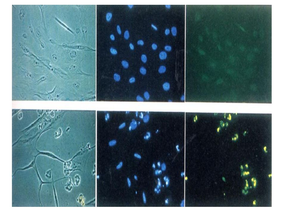
(四)Immunohistochemistry
1. Ag-Ab specific reaction 2. Applications (1) Location analysis cytokeratin→cell membrane (2) Clinical diagnosis and distinguishing diagnosis of tumor histogenesis
 Location analysis. cytokeratin→cell membrane. (2) Clinical diagnosis and distinguishing. diagnosis of tumor histogenesis.")
13
Leiomyosarcoma Actin (+)
")
14
(五)Ultrastructural observation
TEM (transmitting electron microscope) Filtering membrane
 Filtering membrane.")
15
SEM (scanning electron microscope)
Podocyte

16
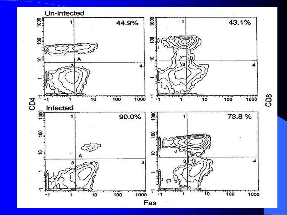
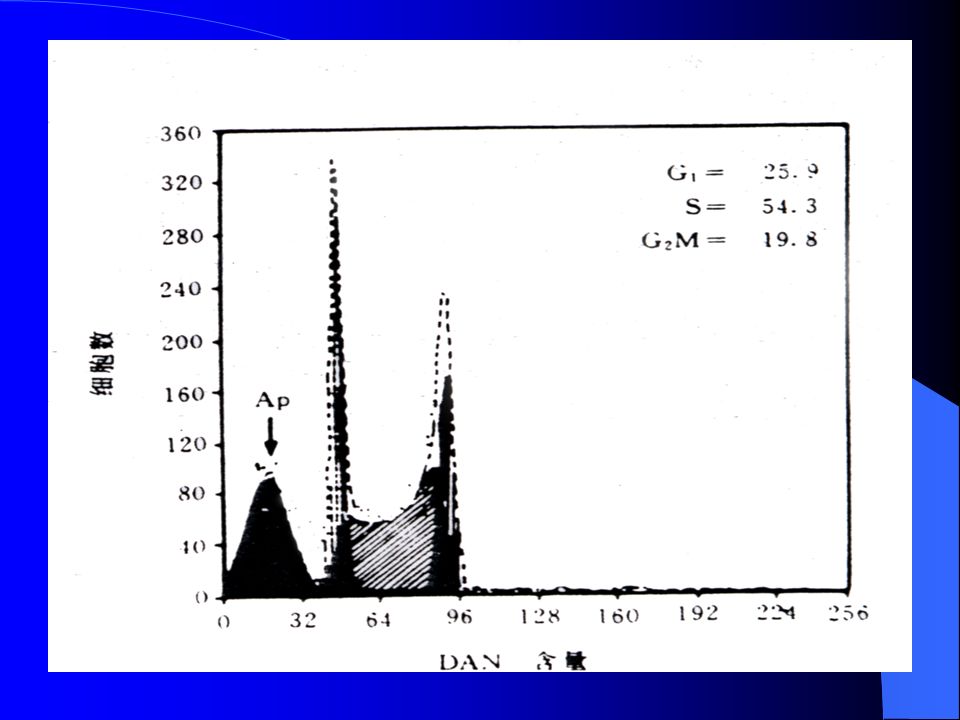
(六)Flow cytometry (FCM)
1. One kind of cells→quantitative 2. DNA ploidy analysis 3. Protein nucleus acid→quantitative analysis 4. Selection of collection of cells

19
(七)Image analysis (IA)
Nuclei: diameter; circumference; area; volume; morphology (八)Laser scanning confocal microscope (LSCM) aliving cell→observation in situ or development or quantitative
Laser scanning confocal. microscope (LSCM) aliving cell→observation in situ or. development or quantitative.")
20
(九)Molecular biology technique
1. Polymerase chain reaction (PCR) 2. DNA sequencing 3. Biochip technique (1) Gene chip (DNA chip) (2) Protein chip (protein microarray) (3) Tissue chip (tissue microarray)
 2. DNA sequencing. 3. Biochip technique. (1) Gene chip (DNA chip) (2) Protein chip (protein microarray) (3) Tissue chip (tissue microarray)")
21
Polymerase chain reaction (PCR)
")
Similar presentations


 Pathogenesis (mechanisms) pathologic changes: structural & functional.>")
