Download presentation
Presentation is loading. Please wait.

1
Structural Classification of the Nervous System
Central nervous system (CNS) Brain Spinal cord Peripheral nervous system (PNS) Nerves outside the brain and spinal cord
 Brain. Spinal cord. Peripheral nervous system (PNS) Nerves outside the brain and spinal cord.")
2
Functions of the Nervous System
Sensory input – gathering information To monitor changes occurring inside and outside the body Changes = stimuli Integration To process and interpret sensory input and decide if action is needed Motor output A response to integrated stimuli The response activates muscles or glands

4
Nervous Tissue: Neurons
Neurons = nerve cells Cells specialized to transmit messages Major regions of neurons Cell body – nucleus and metabolic center of the cell Processes – fibers that extend from the cell body

5
Neuron Anatomy Cell body – contains organelles. Neurons lack centrioles and are incapable of mitosis Nissl substance – specialized rough endoplasmic reticulum. Attached ribosomes. Function to synthesize vital protein molecules Neurofibrils – intermediate cytoskeleton that maintains cell shape Extensions outside the cell body Dendrites – short, branched receptive surfaces that conduct impulses toward the cell body Axons – long, usually singular and conduct impulses away from the cell body

6
Axons and Nerve Impulses
Axons end in axonal terminals Axonal terminals contain vesicles with neurotransmitters Axonal terminals are separated from the next neuron by a gap Synaptic cleft – gap between adjacent neurons Synapse – junction between nerves

7
Nervous Tissue: Support Cells (Neuroglia)
1. Astrocytes Abundant, star-shaped cells Brace neurons Form barrier between capillaries and neurons Control the chemical environment of the brain Figure 7.3a

8
2. Microglia 3. Ependymal cells Spider-like phagocytes
Dispose of debris 3. Ependymal cells Line cavities of the brain and spinal cord Circulate cerebrospinal fluid

9
Nervous Tissue: Support Cells
4. Oligodendrocytes Produce myelin sheath around nerve fibers in the central nervous system Figure 7.3d

10
Nervous Tissue: Support Cells
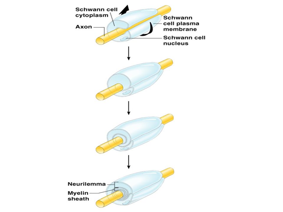
5. Satellite cells Protect neuron cell bodies 6. Schwann cells Form myelin sheath in the peripheral nervous system

12
Functional Classification of Neurons
Sensory (afferent) neurons Carry impulses from the sensory receptors Cutaneous sense organs Proprioceptors – detect stretch or tension Motor (efferent) neurons Carry impulses from the central nervous system
 neurons. Carry impulses from the sensory receptors. Cutaneous sense organs. Proprioceptors – detect stretch or tension. Motor (efferent) neurons. Carry impulses from the central nervous system.")
13
Interneurons (association neurons)
Found in neural pathways in the central nervous system Connect sensory and motor neurons

14
Structural Classification of Neurons
Multipolar neurons – many extensions from the cell body. Characteristic of Motor Neurons Figure 7.8a

15
Structural Classification of Neurons
Bipolar neurons – one axon and one dendrite. Found only associated with special senses. (Olfactory, vision) Figure 7.8b
 Figure 7.8b.")
16
Structural Classification of Neurons
Unipolar neurons – have a short single process leaving the cell body. Sensory Neurons Figure 7.8c

17
Nerve Impulses The surface of the nerve cell membrane is polarized due to unequal distribution of ions on either side of the membrane When nerve cells are at rest there is a greater concentration of Na+ ions outside the membrane and a greater concentration of K+ ions inside the membrane. (Rest Potential) This arrangement of ions gives the outside of the membrane a positive charge with respect to the inside.
 This arrangement of ions gives the outside of the membrane a positive charge with respect to the inside.")
19
A stimulus causes a change in permeability in a region of the membrane
Na+ ions rush into the cell and K+ ions rush out depolarizing the region of the membrane This region of depolarization is an Action Potential An action potential in one region stimulates adjacent regions to depolarize and the action potential moves away from the point of stimulus This moving action potential is a Nerve Impulse The membrane is repolarized in 1/1000 sec. by active transport

20
Continuation of the Nerve Impulse between Neurons
Impulses are able to cross the synapse to another nerve Neurotransmitter is released from a nerve’s axon terminal The dendrite of the next neuron has receptors that are stimulated by the neurotransmitter An action potential is started in the dendrite

22
Reflexes Reflex – automatic, unconscious, and involuntary responses to stimuli Simplest nerve pathways Autonomic Reflexes – involve contractions of smooth muscles Somatic Reflexes – involve contractions of skeletal muscles Help maintain homeostasis by controlling: body temperature, heart rate, breathing rate, blood pressure and digestive activities

23
Patellar Reflex – (knee-jerk reflex) Employs only two neurons
Patellar Reflex – (knee-jerk reflex) Employs only two neurons. Helps maintain posture Also swallowing, sneezing, coughing, vomiting are reflexes
 Employs only two neurons. Helps maintain posture. Also swallowing, sneezing, coughing, vomiting are reflexes.")
25
Withdrawl Reflex

26
Withdrawl Reflex 1. Receptor – sets up a nerve impulse when something painful is touched 2. Sensory Neuron – carries the impulse to the spinal cord 3. Interneuron – conducts the impulse from the sensory to a motor neuron 4. Motor Neuron – conducts the impulse to the effector 5. Effector – responds to stimulation by muscle contraction

Similar presentations


 from one part of the body to another. ◦ Major regions.>")


