Download presentation
Presentation is loading. Please wait.

1
ANATOMY PELVIC FLOOR

2
Landmarks Sacro-spinous lig. Sacro-tuberal lig. Arcus tendineus
Levator ani muscle Endopelvic fascia Sacro-uterine lig. Pubo-urethral lig. Recto-vaginal septum Vesico-vaginal septum Perineal body

3
Pelvic Floor Structure
Pelvic diaphragm Levator ani Coccygeus Perineal membrane Perineal body

4
Pelvic Floor structures

5
Levator Ani Critical component of pelvic support
Slow twitch fibers (I) Responsible for normal resting contraction of pelvic floor Keeps UGH closed Fast twitch fibers (II) Reflex contraction of muscle due to IAP Relaxation During, voiding, defeacation, parturition.
 Responsible for normal resting contraction of pelvic floor. Keeps UGH closed. Fast twitch fibers (II) Reflex contraction of muscle due to IAP. Relaxation. During, voiding, defeacation, parturition.")
6
Levator ani muscle M. pubo-coccygeus M. pubo-rectalis
- Pubo Vag. - Pubo Peri. - Pubo Analis (Attachement at intersphinc. Groove between int. Ext. Anal sphingter) Levator ani muscle OI M. pubo-rectalis (Forms U – sling behind ano-rectal junction & contributes to ano rectal angle) OI M. ilio-coccygeus (Post. Thinnest arises from ATLA & Is. Spines & join to form the iliococcy. Raphe & Lplate C C
 Levator ani muscle. OI. M. pubo-rectalis. (Forms U – sling behind ano-rectal junction & contributes to ano rectal angle) OI. M. ilio-coccygeus. (Post. Thinnest arises from ATLA & Is. Spines & join to form the iliococcy. Raphe & Lplate. C. C.")
7
Levator ani muscle M. pubo-rectalis M. pubo-coccygeus
M. ilio-coccygeus M. coccygeus

8
Levator ani muscle M. pubo-rectalis M. ilio-coccygeus

9
Levator Plate LP formed by union of Iliococcygeus muscle – supportive shelf

10
Levator Plate

11
Sagging of Levator Plate
Sagging of levator plate due to vertical inclination during valsalva & UGH

12
Pelvic Connective tissue
Ligament SSL, STL, Ant. Long Fascia Parietal (Collagen) allows attachment of muscle to boney pelvis. Visceral (Endo pelvic – Collagen, elastin, adipose) sub peritoneal perivascular CT, loose areolar tissue, allows expansion & contraction of pelvic organ & encases BV, lymphatic, nerves
 allows attachment of muscle to boney pelvis. Visceral (Endo pelvic – Collagen, elastin, adipose) sub peritoneal perivascular CT, loose areolar tissue, allows expansion & contraction of pelvic organ & encases BV, lymphatic, nerves.")
13
Sacro-spinous ligament (Coccygeous muscle overlies it)
")
14
Sacro-tuberal ligament

15
GSF – LSF – Sacro-spinous ligament Sacro-tuberal ligament
Piriformis, int. pud, inf & sup gluteal vessel, sciatic nerve & sacral. N plexus ( Neuro vasc. Injury during reconst. Surgery & Pud. N. block. LSF – Pudendal neuro vascular bundle & Obt. Int. tendon Sacro-spinous ligament Sacro-tuberal ligament

16
Pelvic Ligament Sacrospinous Sacrotuberous, & ant. Longitudnal Ligament – suture fixation sites in suspensory procedures. Coopers (IPL) Thickening of periosteum of pubic bone – Burch retropubic bladder neck suspension. Sacrospinous Ligament SSL fixation sutures penetrate the coccy - SSL complex & can lead to injury to L. Ani nerve
 Thickening of periosteum of pubic bone – Burch retropubic bladder neck suspension. Sacrospinous Ligament. SSL fixation sutures penetrate the coccy - SSL complex & can lead to injury to L. Ani nerve.")
17
Arcus tendineus

18
Parietal Fascia Arcus tendineus levator. Ani (ATLA)
Condensation of fascia overlying medial surface of OBT. INTER NUS muscle, point of origin of L. ANI muscle. Arcus tendineus fasciae pelvis (ATFP) Condensation of fascia over medial aspect of OI muscle & LA muscle, lateral point of attachment of anterior vag. Wall
 Condensation of fascia over medial aspect of OI muscle & LA muscle, lateral point of attachment of anterior vag. Wall.")
19
Delancey – levels of support
Lig. Sacro-uterinum

20
Delancey – levels of support
Level – I Suspensory Axis Supports upper 1/3 Vag. by USCL complex, apical support Leads to vault prolapse, Cx descent, cystocoele. Level – II Attachment Axis Middle 1/3 Vag. By L. Ani & ATFP Ant. Vag. Prolapse SUI Level – III Fusion Axis Strongest support Ant. – Urethra Lat.- Pubovag / PM, Post.- P. Body Distal rectocoele, perineal descent, Anal incontinence

21
Sacro-uterine ligament

22
Pubo-urethral ligament

23
Endopelvic fascia komplexes System

24
Perineal membrane (UGD)
Spans opening of ant. Pelvic outlet & supports distal vagina & Urethra Comp. Urethra, Urethro Vag. Sphinc. Muscle, ATFP Distal Vag. IS. PUBIC RAMI Perineal body

25
Urogenital triangle Anal triangle Symphysis Tuber ossis ischii
Os coccygis

26
Perineal Body Supports distal vag. & rectum – anatomical repair & reapproximation during episiotomy & perineal reconstructive surgery M. Bulbo cav. Sup. Trans Perineal. M Pubo Visc of L. ani RVS Peri. Membrane M. sphincter ani externus

28
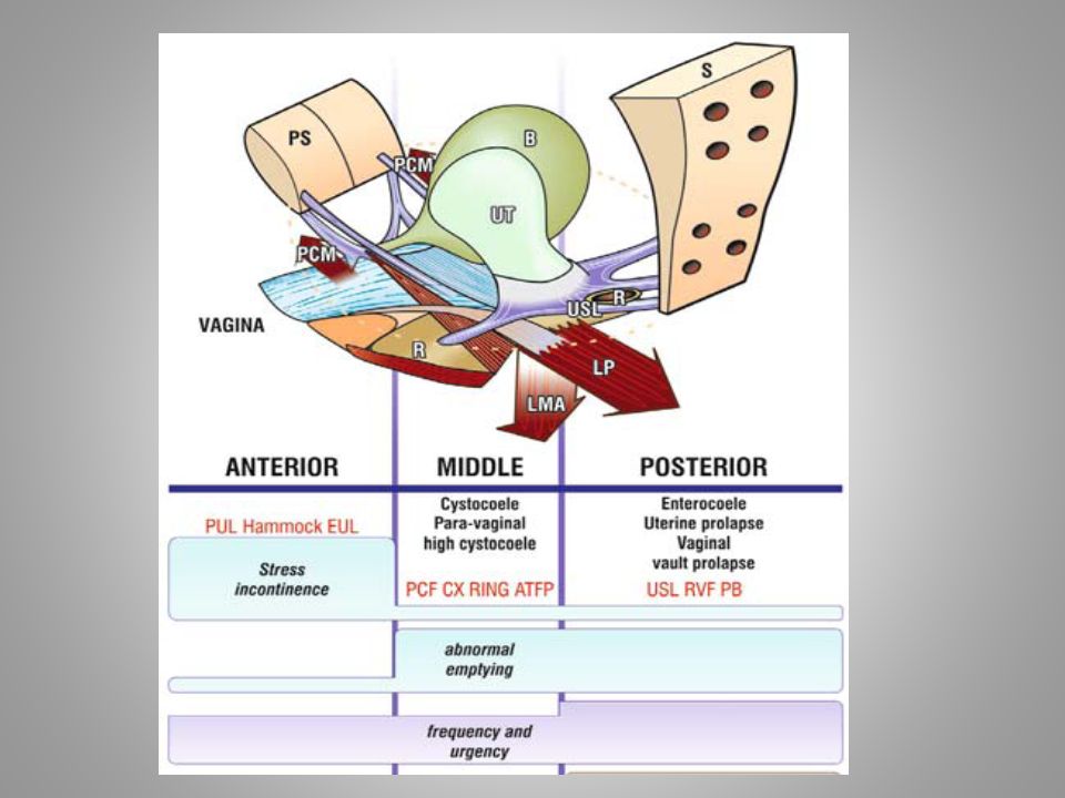
Zones Anterior Middle Posterior EUL / PUL – forms hammock
Pubo Cervical Fascia, ATFP, Cx ring Cardinal ligment, USL, RVS, PB Leads to incontinence Leads to paracolpium defect & cystocoele Leads to uterine prolapse Vault Prolapse, enterocoele.

29
Thank you

30
Lateral defect

31
Vesico-vaginal septum Recto-vaginal septum

32
Transobturator Landmarks
Adductor longus Urethra Obturator canal SAFE ENTRY ZONE of NEEDLE

33
Arcus tendineus fasciae pelvis
Arcus tendineus M. levator. ani

Similar presentations