Download presentation
Presentation is loading. Please wait.

1
Optimization of T-Cell Trapping in a Microfluidic Device Group #19 Jeff Chamberlain Matt Houston Eric Kim

2
MEMS- MicroElectroMechanical Systems Batch Fabrication Processes Cell Traps –High-throughput experimentation –Complex biochemical analysis –Single cell analysis –Reagent conservation –Quick environmental changes

3
Cell Trapping Basics Reagent #1 Reagent #2 Cells / Media Trap arrays:

4
Our Project Maximize trap efficiency by improving upon current trap designs. –Trap Efficiency : maximized number of traps with 1 cell/trap

5
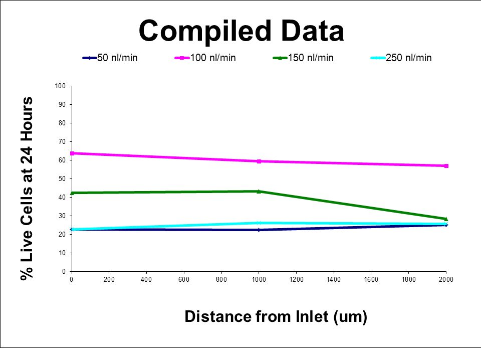
Cell Viability Results Flow Rate = 100 nl/min Flow Rate = 250 nl/min

7
SolidWorks ® Rendering of a Single Well

8
Picture of Well Array Square or rectangular shaped well of any depth Thousands of mirrored wells in one etching Front Surface Mirrors with high reflectivity Nearly orthogonal views of specimen 200 um

9
Background & Motivation Three Dimensional Image Information May Be Important for Biological Studies Chemotaxis Developmental Biology Cellular Division Pinocytic Loading Volumetric Measurements

10
Our System is Constructed From Silicon Wafer

11
Methods: Fabrication Silicon Wafer Silicon Wafer Grow SiO 2 Spin Coat Mask Layer Pattern with Photolithography Etch with HF Etch with HF Remove Photoresist Etch with KOH Coat with Platinum or Aluminum Cutaway View

12
SolidWorks ® Rendering of a Single Well

13
Results: Dictyostilium Extrusion A B C A B C Primary Image Reflection Reflection Reflection of the Reflection Reflection Objective

14
Coupling Microfluidics With the Pyramidal Wells Si Wafer Cross Section of One Well PDMS Flow PDMS Glass

15
Pyramidal Well Dimensions depth_min = 3.22728 cell_radius outside_dim = 3.48504 cell_radius Assuming: 0.5 base = 1.2 cell_radius

16
Micromirror Well Dimensions Cell TypeCell Dimensions (um)Minimum Required Depth (um) Outside Dimensions for Min Depth (um) T-cells5 diameter8.068217.4252 Jurkats10 diameter16.136434.8504 Dendritic Cells10-20 diameter16.1364 - 32.272834.8504 - 69.7008 Dicti10-20 diameter16.1364 - 32.272834.8504 - 69.7008 Myocyte15 by 10024.673754.9399 by 37.9399
Minimum Required Depth (um) Outside Dimensions for Min Depth (um) T-cells5 diameter Jurkats10 diameter Dendritic Cells10-20 diameter Dicti10-20 diameter Myocyte15 by by")
17
Future Directions Single Cell Design –Desire single cell per well for optimal imaging –Possibly controllably coupled with another As opposed to…

18
Future Directions ? Single Cell Design –Desire single cell per well for optimal imaging –Possibly controllably coupled with another

19
Future Directions Flow Modeling –Design a more efficient trapping system –Use flow data to design trap design that will keep cell in well ?

20
Future Directions PDMS-mirror bonding –Questions to whether the PDMS will bond to the mirror array –Use SU-80 and glass cover slip with a PDMS external flow system?

Similar presentations
>")
 Grey=Si, Blue=Silicon Dioxide, Red=Photoresist, Purple= Phosphorus.>")


![Fabrication of p-n junction in Si Silicon wafer [1-0-0] Type: N Dopant: P Resistivity: 10-20 Ω-cm Thickness: 505-545 µm.](/15/4784305/big_thumb.jpg "Fabrication of p-n junction in Si Silicon wafer [1-0-0] Type: N Dopant: P Resistivity: 10-20 Ω-cm Thickness: 505-545 µm.>")

 lecture04 Sherief Reda Division of Engineering, Brown University Spring 2008 [sources: Sedra/Prentice.>")


 Fabrication of photonic crystal structures on light emitting diodes by nanoimprint lithography.>")
