Download presentation
Presentation is loading. Please wait.

1
Human Anatomy and Physiology
Skin Human Anatomy and Physiology

2
Skin The skin is considered the largest organ of the body
Functions in thermoregulation, protection, metabolic functions and sensation. Two main regions, the epidermis, and the dermis The dermis is attached to an underlying hypodermis, also called subcutaneous connective tissue, which stores adipose tissue.

3
Epidermis The epidermis is the most superficial layer of the skin
The first barrier of protection from the invasion of foreign substances The epidermis is subdivided into five layers or strata, the stratum basale (bG), the stratum spinosum(SS), the stratum granulosum(SGR), the stratum lucidum(not seen in this photomicrograph) and the stratum corneum(SC) in which a keratinocyte gradually migates to the surface and is sloughed off in a process called desquamation.
, the stratum spinosum(SS), the stratum granulosum(SGR), the stratum lucidum(not seen in this photomicrograph) and the stratum corneum(SC) in which a keratinocyte gradually migates to the surface and is sloughed off in a process called desquamation.")
4
Stratum Germinatum The stratum basale (bG) provides the germinal cells necessary for the regeneration of the layers of the epidermis. These germinal cells are separated from the dermis by a thin layer of basement membrane. After a mitotic division a newly formed cell will undergo a progressive maturation called keratinization as its migrates to the surface.
 provides the germinal cells necessary for the regeneration of the layers of the epidermis. These germinal cells are separated from the dermis by a thin layer of basement membrane. After a mitotic division a newly formed cell will undergo a progressive maturation called keratinization as its migrates to the surface.")
5
Stratum Spinosum (SS) The cells that divide in the stratum basale soon begin to accumulate many desmosomes on their outer surface Stratum spinosum (SS)
")
6
Keratinization The progressive maturation of a keratinocyte is charcterized by the accumulation of keratin, called keratinization. The cells of the stratum basale (SB) accumlate dense basophilic keratohyalin granules (seen on the close-up view). These granules contain lipids, which along with the desmosomal connections, help to form a waterproof barrier that functions to prevent fluid loss from the body.
 accumlate dense basophilic keratohyalin granules (seen on the close-up view). These granules contain lipids, which along with the desmosomal connections, help to form a waterproof barrier that functions to prevent fluid loss from the body.")
7
Stratum Lucidum Epidermis varies in thickness throughout the body depending mainly on frictional forces and is thickest on the palms of the hands and soles of the feet. The stratum lucidum is normally only well seen in thick epidermis and represents a transition from the stratum basale to the stratum corneum.

8
Stratum Corneum A cell matures and accumulates keratin
The dead and dying cells filled with mature keratin form the stratum corneum (SC). The deeper cells of the stratum corneum retain their desmosomal junctions, but as they are pushed to the surface by newly forming cells of the stratum basale (BG), the dead cells gradually break apart and are lost, a process called desquamation.
. The deeper cells of the stratum corneum retain their desmosomal junctions, but as they are pushed to the surface by newly forming cells of the stratum basale (BG), the dead cells gradually break apart and are lost, a process called desquamation.")
9
Dermis The dermis (D) assumes the important functions of thermoregulation and supports the vascular network to supply the avascular epidermis with nutrients. Two zones, a papillary dermis and a reticular layer. The dermis contains mostly fibroblasts which secrete collagen, elastin and ground substance that give the support and elasticity of the skin. Also present are immune cells that are involved in defense against foreign invaders passing through the epidermis
 assumes the important functions of thermoregulation and supports the vascular network to supply the avascular epidermis with nutrients. Two zones, a papillary dermis and a reticular layer. The dermis contains mostly fibroblasts which secrete collagen, elastin and ground substance that give the support and elasticity of the skin. Also present are immune cells that are involved in defense against foreign invaders passing through the epidermis.")
10
Papillary Dermis The papillary dermis (PD) contains vascular networks that have two important functions. support the avascular epidermis with vital nutrients secondly to provide a network for thermoregulation. The vasculature is organized so that by increasing or decreasing blood flow, heat can either be conserved or dissipated. The vasculature interdigitates in areas called dermal papillae (DP). The papillary dermis also contains the free sensory nerve endings and structures called Meissner’s corpuscles( the free sensory nerve endings in) highly sensitive areas.
. The papillary dermis also contains the free sensory nerve endings and structures called. Meissner’s corpuscles( the free sensory nerve endings in) highly sensitive areas.")
11
Reticular Layer The reticular layer of the dermis (RD) consists of dense connective tissue The reticular layer houses important epithelial derived structures such as glands and hair follicles. Pacinian corpuscles are also found here. They are deep pressure receptors derived from nervous tissue.

12
Serous Membrane Serous membrane contains epithelial tissue.
Found in body cavities not open to exterior. Two layers Visceral Parietal Named for location Thoracic: pluera Abdominal: peritoneum Cardiac: pericardium

13
Serous membrane Serous membrane Visceral pleura Parietal pleura
Visceral peritoneum Parietal peritoneum Visceral pericardium Parietal pericardium

14
Mucous membrane Epithelium resting on loose C.T. (lamina propria)
Lines cavities that are open to exterior Respiratory ciliated columnar Digestive simple columnar Reproductive Urinary no mucus

15
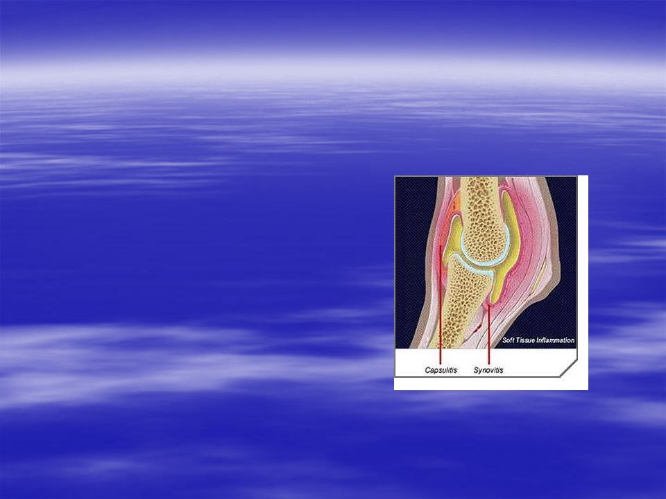
Synovial membrane Connective tissue Line fibrous capsule around joint
Line bursae Line tendon sheath

Similar presentations


 Largest organ of the body (15% of body weight) Skin thickness variable, normally 1-2 mm Protection –chemical barrier (waterproof)>")
epidermis –stratified squamous epithelium cells –constantly replaced from inside out –entirely from ectoderm.>")


 Skin derivatives>")
