Download presentation
Presentation is loading. Please wait.

1
Histology of Central Nervous System Dr. Sama ul Haque

2
Objectives Describe the microscopic anatomy of gray matter & white matter Describe the microscopic anatomy of neuroglial cells of CNS Describe the microscopic anatomy of spinal cord. Discuss the microscopic anatomy of Cerebral cortex. Discuss the microscopic anatomy of Cerebellum. Describe the microscopic anatomy Meninges

3
Gray matter & White matter Gray matter Contains abundant neuronal cell bodies, dendrites, the initial unmyelinated portions of axons, & neuroglial cells. White matter Contains myelinated fibers & neuroglial cells.

4
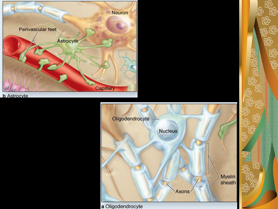
Neuroglial Cells of CNS They are nonneuronal, supportive cells 4 types of cells can be seen: 1. Astrocyte Form part blood-barrier 2 types: fibrous astrocytes protoplasmic astrocytes 2. Oligodendrocytes Form myelin sheath around axons in the CNS

6
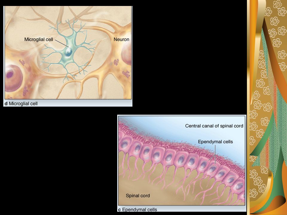
3. Microglia Are small cells with complex shapes. Phagocytotic cells. 4. Ependymal Cells Line the ventricles of the brain and the central canal of the spinal cord low columnar cilated epithelial cells

8
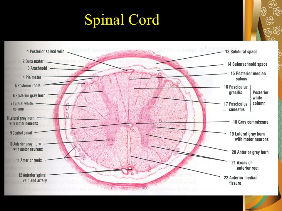
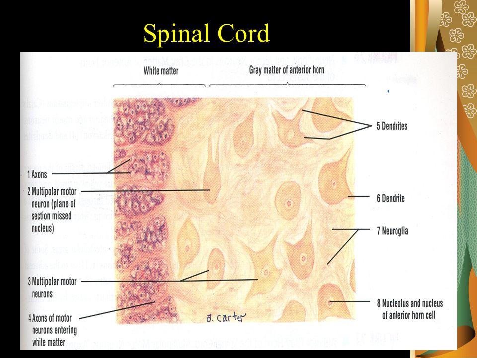
Spinal Cord

11
(Motor Neurons) Spinal Cord
 Spinal Cord")
12
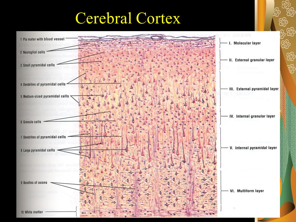
Gray Matter I.Molecular Layer (Plexiform Layer): Pia Mater and Neuroglial cells II.External Granular layer: Small pyramidal cells III.External Pyramidal layer: Medium-sized pyramidal cells IV.Internal Granular layer (Ganglionic Layer): Small granule cells Cerebral Cortex
: Pia Mater and Neuroglial cells II.External Granular layer: Small pyramidal cells III.External Pyramidal layer: Medium-sized pyramidal cells IV.Internal Granular layer (Ganglionic Layer): Small granule cells Cerebral Cortex")
13
Gray Matter V. Internal Pyramidal layer: Largest pyramidal cells (Betz Cells) VI. Multiform layer (Layer of polymorphic cells): Fusiform cells Granule cells Stellate cells Cells of Martinotti Cerebral Cortex
: Fusiform cells Granule cells Stellate cells Cells of Martinotti Cerebral Cortex.")
14
White Matter Bundles of Axons Cerebral Cortex

16
Cerebral Cortex (Layer V)
")
17
T.S of Cerebellum

18
Gray Matter I.Molecular Layer: Stellate cells Basket cells Neuroglial cells II.Purkinje cell layer: Purkinje cells: Flask shaped Arranged in a single layer having Thick dendrites. Cerebellum

19
Gray Matter III. Granular layer: Small Granule cells with dark stained nuclei Golgi type II cells Glomeruli Cerebellum

20
Cerebellar Cortex

21
Meninges Are connective tissue covering the brain & spinal cord Three meningial layers are distinguished: 1. Dura is thick external layer consisting of dense C.T 2. Arachanoid(arachnoeides, spiderweblike) has two components: (1) Sheet of connective tissue in contact with the dura mater (2) System of loosely arranged trabeculae containing fibroblasts and collagen.
 has two components: (1) Sheet of connective tissue in contact with the dura mater (2) System of loosely arranged trabeculae containing fibroblasts and collagen..")
22
3. Pia is a delicate & loose connective tissue containing many blood vessels.

23
Thank You

Similar presentations


 It is phylogenetically newest and structurally most complex. Neocortex is.>")


 Anatomy Support structures –(bone) –meninges –cerebrospinal fluid (CSF) Protective structure –blood-brain barrier General.>")


>")


 2008, 2005 by Mosby, Inc., an affiliate of Elsevier Inc. All rights reserved. Copyright © 2005, Elsevier, Inc. All rights reserved. Slide.>")
